Chapter 1 | Introduction to Special Stains - Dako
Chapter 1 | Introduction to Special Stains - Dako
Chapter 1 | Introduction to Special Stains - Dako

Create successful ePaper yourself
Turn your PDF publications into a flip-book with our unique Google optimized e-Paper software.
<strong>Introduction</strong> <strong>to</strong> <strong>Special</strong> <strong>Stains</strong> <strong>Introduction</strong> <strong>to</strong> <strong>Special</strong> <strong>Stains</strong><br />
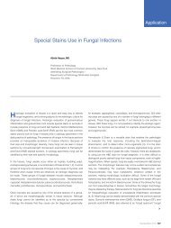
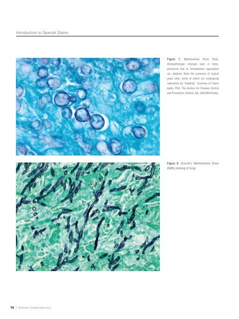
Figure 7. Methenamine Silver Stain.<br />
His<strong>to</strong>pathologic changes seen in his<strong>to</strong>plasmosis<br />
due <strong>to</strong> His<strong>to</strong>plasma capsulatum<br />
var. duboisii. Note the presence of typical<br />
yeast cells, some of which are undergoing<br />
replication by “budding”. Courtesy of Libero<br />
Ajello, PhD, The Centers for Disease Control<br />
and Prevention, Atlanta, GA, USA/Wikimedia.<br />
Figure 8. Grocott’s Methenamine Silver<br />
(GMS) staining of fungi.<br />
Figure 9. His<strong>to</strong>pathology of Treponema<br />
pallidum spirochetes using a modified<br />
Steiner Silver Stain. Image credit: Dr.<br />
Edwin P. Ewing, Jr., The Centers for<br />
Disease Control and Prevention, Atlanta,<br />
GA, USA/Wikimedia.<br />
Figure 10. Helicobacter stained with<br />
Warthin-Starry, <strong>Dako</strong> Code AR181. The<br />
arrow points <strong>to</strong> some black H. pylori<br />
organisms in yellow mucus.<br />
14 | special stains and H & e special stains and H & e | 15