Chapter 1 | Introduction to Special Stains - Dako
Chapter 1 | Introduction to Special Stains - Dako
Chapter 1 | Introduction to Special Stains - Dako

You also want an ePaper? Increase the reach of your titles
YUMPU automatically turns print PDFs into web optimized ePapers that Google loves.
<strong>Introduction</strong> <strong>to</strong> <strong>Special</strong> <strong>Stains</strong> <strong>Introduction</strong> <strong>to</strong> <strong>Special</strong> <strong>Stains</strong><br />
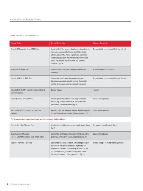
Table 2. Commonly used special stains.<br />
<strong>Special</strong> Stain Clinical Application Staining Specificity<br />
Grocott’s Methenamine Silver (GMS) Stain Useful in identifying a variety of pathogenic fungi, including<br />
Aspergillus fumigatus, Blas<strong>to</strong>myces dermatitidis, Candida<br />
albicans, Coccidioides immitis, Cryp<strong>to</strong>coccus neoformans,<br />
His<strong>to</strong>plasma capsulatum, Nocardia asteroids, Pneumocystis<br />
carinii, Pneumocystis Jiroveci (human) and Sporothrix<br />
schenckii; Fig. 6-8<br />
Mayer’s Mucicarmine Stain Detects encapsulated yeast-like fungus Cryp<strong>to</strong>coccus<br />
neofarmans<br />
Periodic Acid-Schiff (PAS) Stain Used for the identification of Aspergillus fumigatus,<br />
Blas<strong>to</strong>myces dermatitidis, Candida albicans, Coccidioides<br />
immitis, Cryp<strong>to</strong>coccus neofarmans, Sporothrix schenckii<br />
Sayeed’s Stain (Schiff’s reagent, 0.5% periodic acid,<br />
Mayer’s hemalum)<br />
Detects H. pylori H. pylori<br />
Steiner & Steiner Staining Method Detects spirochetes and legionella, and pneumophila<br />
bacteria, e.g., Borrelia burgdorferi, H. pylori, Legionella<br />
pneumophila, Treponema pallidum; Fig. 9<br />
Warthin-Starry Stain (these are reduced silver<br />
methods)<br />
For demonstrating connective tissue, muscle, collagen, lipid and fibrin<br />
Identifies Alipia feles, Bar<strong>to</strong>nella henselae, Borrelia burgdorferi,<br />
H. pylori, Legionella pneumophila, Treponema pallidum; Fig. 10<br />
Gomori’s One-Step Trichrome Stain Used for distinguishing collagen and smooth muscle fibers;<br />
Fig.11<br />
Jones’ Basement Membrane<br />
Periodic Schiff-Methenamine Silver (PASM) Stain<br />
Used for the identification of basement membranes (of the<br />
glomerulus in the kidney or in tissue samples); Fig. 12<br />
Masson’s Trichrome Stain (TRI) Used for distinguishing cells from surrounding connective<br />
tissue which has several variants and is probably the<br />
trichrome most used in his<strong>to</strong>pathology. Black nuclei, red<br />
cy<strong>to</strong>plasm (including muscle), blue or green collagen<br />
(including fine fibers), cartilage and mucus; Fig. 13<br />
Polysaccharide components of the fungal cell wall<br />
Polysaccharides on the capsule<br />
Polysaccharide components of the fungal cell wall<br />
<strong>Stains</strong> whole organisms<br />
<strong>Stains</strong> whole organisms<br />
Collagen and smooth muscle fibers<br />
Basement membranes<br />
Muscle, collagen fibers, fibrin and erythrocytes<br />
<strong>Special</strong> Stain Clinical Application Staining Specificity<br />
Russel-Movat Pentachrome Stain Used for simultaneous demonstration of muscle, elastic<br />
fibers, collagen/reticular fibers, ground substance and<br />
fibrinoid in tissues<br />
Oil Red O and Sudan Black B <strong>Stains</strong> Used for staining lipids in frozen sections and some<br />
lipoproteins on paraffin sections<br />
Orcein Stain Used for staining elastic fibers Elastic fibers<br />
Lendrum’s Method (Picro-Mallory Stain) Fibrin Fibrin<br />
Phosphotungstic Acid-Hema<strong>to</strong>xylin (PTAH) Stain Used for demonstrating striated muscle fibers<br />
Also used <strong>to</strong> stain abnormal neuroglia (reactive astrocy<strong>to</strong>sis)<br />
Silver methods for reticulum and<br />
basement membranes<br />
(e.g., Reticulin/ Nuclear Fast Red Stain)<br />
Used for the identification of reticulin fibers in tissue<br />
samples; Fig.14<br />
Muscle, elastic fibers, collagen/reticular fibers<br />
Lipids, including triglycerides (which necessarily are<br />
neutral). Oil Red O stains only the most hydrophobic<br />
lipids (triglycerides and cholesterol esters). Sudan<br />
Black B stains these and also phospholipids and<br />
sphingomyelins, which are less hydrophobic<br />
Muscle fibers, collagen<br />
Reticulin (collagen with high level of hexosylation,<br />
including Type IV)<br />
6 | special stains and H & e special stains and H & e | 7<br />
Verhoeff Stain<br />
Van Gieson Stain<br />
For detecting nucleic acids<br />
Used for the identification of elastic laminae and fibers<br />
in tissues; Fig.15<br />
The Verhoeven Stain is specific for elastic fibers.<br />
The Van Gieson Stain is specific for collagen.<br />
Verhoeff’s iron-hema<strong>to</strong>xylin stains elastin and<br />
nuclei black. Van Gieson’s picro-fuchsine gives<br />
yellow cy<strong>to</strong>plasm and red collagen fibers<br />
Ethyl Green-Pyronine Stain Used for differential demonstration of DNA and RNA A buffered mixture of the two dyes gives blue-green<br />
DNA and red RNA (rRNA in cy<strong>to</strong>plasm, nucleoli)<br />
Feulgen Stain Used for the identification of chromosomal material or<br />
deoxyribonucleic acid (DNA in paraffin-embedded tissue<br />
or cell specimens); Fig.16<br />
Deoxyribonucleic acid (DNA)