Chapter 1 | Introduction to Special Stains - Dako
Chapter 1 | Introduction to Special Stains - Dako
Chapter 1 | Introduction to Special Stains - Dako

Create successful ePaper yourself
Turn your PDF publications into a flip-book with our unique Google optimized e-Paper software.
<strong>Introduction</strong> <strong>to</strong> <strong>Special</strong> <strong>Stains</strong> <strong>Introduction</strong> <strong>to</strong> <strong>Special</strong> <strong>Stains</strong><br />
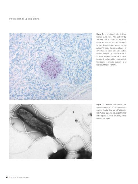
Figure 3. Lung stained with Acid-Fast<br />
Bacteria (AFB) Stain, <strong>Dako</strong> Code AR162.<br />
This AFB stain is suitable for the visualization<br />
of acid-fast bacteria belonging<br />
<strong>to</strong> the Mycobacterium genus on the<br />
Artisan Staining System. Application of<br />
carbol-fuchsin stains acid-fast bacteria<br />
fuchsia, followed by decolorization of<br />
all tissue elements except the acid-fast<br />
bacteria. A methylene blue counterstain is<br />
then applied <strong>to</strong> impart a blue color <strong>to</strong> all<br />
background tissue elements.<br />
Figure 4a. Electron micrograph (EM)<br />
(negative staining) of H. pylori possessing<br />
multiple flagella. Courtesy of Wikimedia.<br />
Prof. Yutaka Tsutsumi, MD, Department of<br />
Pathology, Fujita Health University School<br />
of Medicine, Japan.<br />
Sheathed polar flagella<br />
Body of the Bacteria<br />
Figure 4b. Illustration of S-shaped H. pylori with four sheathed polar flagella. The majority of helicobacters possess this basic morphology of an S-shape with polar,<br />
sheathed flagella, though variations in size and the number of spirals are seen in a number of other species. These bacteria are usually around 0.5 × 5 μm, and the<br />
S-shaped morphology has been correlated with maximum in vitro motility. Thin sections of H. pylori revealed through an electron microscope show an outer and inner<br />
membrane separated by the periplasm of approximately 30 nm thickness (see EM picture above). The dense cy<strong>to</strong>plasm contains nucleoid material and ribosomes<br />
(Source: Jani O’Rourke and Günter Bode. Morphology and Ultrastructure of Helicobacter pylori. Physiology and Genetics. Eds. Harry L. T. Mobley, George L. Mendz, and<br />
Stuart L. Hazell. ASM Press. 2001). Illustration by Rashmil Saxena, BFA, HT(ASCP) CM .<br />
10 | special stains and H & e special stains and H & e | 11