Chapter 1 | Introduction to Special Stains - Dako
Chapter 1 | Introduction to Special Stains - Dako
Chapter 1 | Introduction to Special Stains - Dako

Create successful ePaper yourself
Turn your PDF publications into a flip-book with our unique Google optimized e-Paper software.
<strong>Introduction</strong> <strong>to</strong> <strong>Special</strong> <strong>Stains</strong> <strong>Introduction</strong> <strong>to</strong> <strong>Special</strong> <strong>Stains</strong><br />
Meissner’s<br />
corpuscle<br />
Oil gland<br />
Sweat gland<br />
Epidermis<br />
Dermis<br />
Hypodermis<br />
Hair shaft<br />
Arrec<strong>to</strong>r pili<br />
Hair follicle<br />
Hair root<br />
Pacinian<br />
corpuscle<br />
Vein<br />
Artery<br />
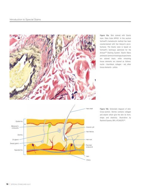
Figure 15a. Skin stained with Elastic<br />
stain, <strong>Dako</strong> Code AR163. In this section<br />
Verhoeff’s hema<strong>to</strong>xylin method has been<br />
counterstained with Van Gieson’s picrofuchsine.<br />
The Elastic stain is based on<br />
Verhoeff’s technique optimized for the<br />
Artisan Staining System. Elastin fibers<br />
and elastic lamina in his<strong>to</strong>logical specimens<br />
are stained black, while remaining<br />
tissue elements are stained as follows:<br />
nuclei - blue/black, collagen - red, other<br />
tissue elements - yellow.<br />
Figure 15b. Schematic diagram of skin:<br />
Cross-section. Dermis contains collagen<br />
and elastin which give the skin its form,<br />
shape and elasticity. Illustration by<br />
Rashmil Saxena, BFA, HT(ASCP) CM .<br />
Figure 16. Breast tissue stained with<br />
Feulgen, <strong>Dako</strong> Code AR174. The Feulgen<br />
stain is used <strong>to</strong> demonstrate DNA in<br />
tissue sections. RNA is not stained<br />
by this procedure. The DNA is stained<br />
magenta with Schiff’s reagent. The stained<br />
DNA is contrasted against a light green<br />
counterstain <strong>to</strong> allow better visualization<br />
by light microscopy or image analysis.<br />
Figure 17. Amyloid stained with Congo<br />
Red, <strong>Dako</strong> Code AR161. The Congo<br />
Red stain is used <strong>to</strong> detect amyloid, an<br />
abnormal protein product that can be<br />
found in various pathologic conditions.<br />
This stain is based on Benhold’s and<br />
demonstrates amyloid in pink <strong>to</strong> dark<br />
salmon with light microscopy or the<br />
characteristic “apple-green birefringence’’<br />
with polarized light. Mayer’s hema<strong>to</strong>xylin<br />
is used as a counterstain. The preferred<br />
method for visualization of amyloid is<br />
under polarized light.<br />
18 | special stains and H & e special stains and H & e | 19