ER/PR pharmDx™ Interpretation Manual - Dako
ER/PR pharmDx™ Interpretation Manual - Dako
ER/PR pharmDx™ Interpretation Manual - Dako

Create successful ePaper yourself
Turn your PDF publications into a flip-book with our unique Google optimized e-Paper software.
PATHOLOGY<br />
<strong>ER</strong>/<strong>PR</strong> pharmDx TM <strong>Interpretation</strong> <strong>Manual</strong><br />
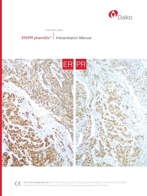
<strong>ER</strong> <strong>PR</strong><br />
For In Vitro Diagnostic Use. FDA 510(k) cleared as an aid in identifying patients eligible for treatment with anti-hormonal<br />
or aromatase inhibitor therapies as well as an aid in the prognosis and management of breast cancer.
Table of Contents<br />
Introduction 2<br />
<strong>ER</strong>/<strong>PR</strong> pharmDx overview 3<br />
the <strong>ER</strong>/<strong>PR</strong> pharmDx Kit 4<br />
<strong>ER</strong>/<strong>PR</strong> Expression Rates 5<br />
Concordance studies 6<br />
n Concordance of Immunohistochemistry<br />
(IHC) to ligand-binding Assay<br />
n <strong>ER</strong>/<strong>PR</strong> pharmDx tM Concordance to the<br />
Allred Procedure<br />
n Determination of the Cut-off IHC score<br />
<strong>ER</strong>/<strong>PR</strong> Expression in normal tissue 9<br />
<strong>ER</strong>/<strong>PR</strong> pharmDx training Checklist 10<br />
Quality Control 11<br />
Control slides for <strong>ER</strong>/<strong>PR</strong> pharmDx <br />
Validation 12<br />
Examples of Acceptable and<br />
Unacceptable staining of<br />
<strong>ER</strong>/<strong>PR</strong> pharmDx Control slides 13<br />
<strong>ER</strong>/<strong>PR</strong> pharmDx Results:<br />
Evaluation and Reporting 14<br />
<strong>ER</strong>/<strong>PR</strong> pharmDx scoring system 15<br />
Image Guide for Allred scoring<br />
for <strong>ER</strong>/<strong>PR</strong> pharmDx 16<br />
n Estrogen Receptor<br />
n Progesterone Receptor<br />
Additional Images 18<br />
n Estrogen Receptor<br />
n Progesterone Receptor<br />
Additional specimens stained<br />
with <strong>ER</strong>/<strong>PR</strong> pharmDx 20<br />
Artifacts and Various Factors<br />
Affecting staining 21<br />
n Epitope Retrieval Artifacts<br />
n background staining<br />
<strong>ER</strong>/<strong>PR</strong> pharmDx <br />
Pathology Report Form 22<br />
References 23<br />
<strong>ER</strong>/<strong>PR</strong> pharmDx <strong>Interpretation</strong> <strong>Manual</strong><br />
tAblE oF ContEnts<br />
1
IntRoDUCtIon<br />
Introduction<br />
Welcome to the <strong>ER</strong>/<strong>PR</strong> pharmDx <br />
<strong>Interpretation</strong> <strong>Manual</strong><br />
this guide for pathologists includes key technical<br />
histological staining and interpretation tips applicable<br />
when using the <strong>ER</strong>/<strong>PR</strong> pharmDx tM kit. Utilization of the<br />
suggestions that follow will ensure that your laboratory<br />
achieves the quality results expected from <strong>ER</strong>/<strong>PR</strong><br />
pharmDx tM .<br />
2 <strong>ER</strong>/<strong>PR</strong> pharmDx <strong>Interpretation</strong> <strong>Manual</strong><br />
the <strong>ER</strong>/<strong>PR</strong> pharmDx tM <strong>Interpretation</strong> <strong>Manual</strong> objectives<br />
are simple:<br />
n to ensure that the <strong>ER</strong>/<strong>PR</strong> pharmDx tM assay is being<br />
performed consistent with <strong>Dako</strong> recommendations for<br />
optimal results.<br />
n to encourage reproducible results by introducing a<br />
standard approach to staining and interpretation.<br />
n to provide pathologists with a tool to allow consistent<br />
interpretation of <strong>ER</strong>/<strong>PR</strong> pharmDx tM kit results to<br />
appropriately guide patient management for breast<br />
cancer therapy.<br />
n to facilitate troubleshooting of the <strong>ER</strong>/<strong>PR</strong> pharmDx tM<br />
Kit, if problems occur.
<strong>ER</strong>/<strong>PR</strong> pharmDx tM overview<br />
the <strong>ER</strong>/<strong>PR</strong> pharmDx tM Kit is a semi-quantitative<br />
immunohistochemical (IHC) assay to identify estrogen<br />
receptor (<strong>ER</strong>) and progesterone receptor (<strong>PR</strong>) expression<br />
in normal and neoplastic tissues, formalin-fixed and<br />
paraffin-embedded for histological evaluation. <strong>ER</strong>/<strong>PR</strong><br />
pharmDx tM specifically detects the <strong>ER</strong> alpha protein as<br />
well as the <strong>PR</strong> protein located in the nuclei of <strong>ER</strong> and<br />
<strong>PR</strong>-expressing cells, respectively.<br />
Investigations into the biological mechanisms for breast<br />
cancer have found that the growth rate is dependent on<br />
the presence of estrogen or progesterone or both in most<br />
breast cancers. thus, estrogen receptor and proges-<br />
terone receptor status in breast cancer is considered to<br />
be a validated prognostic and predictive factor for patient<br />
management for anti-hormonal therapy. 1-5<br />
<strong>ER</strong>/<strong>PR</strong> pharmDx tM is indicated as an aid in identifying<br />
patients eligible for treatment with anti-hormonal or<br />
aromatase inhibitor therapies, as well as an aid in the<br />
prognosis and management of breast cancer.<br />
<strong>ER</strong>/<strong>PR</strong> pharmDx Provides the Basis<br />
for Reliable <strong>ER</strong> and <strong>PR</strong> Assessment<br />
n FDA 510(k) cleared, standard, reproducible assay.<br />
n new, highly specific <strong>ER</strong> antibody cocktail and <strong>PR</strong><br />
antibody with demonstrated sensitivity and specificity.<br />
n optimized protocol with clinically validated scoring<br />
system for the determination of <strong>ER</strong>/<strong>PR</strong> status<br />
applicable in the management of breast cancer<br />
patients. 5-9<br />
n Concordance demonstrated between <strong>ER</strong>/<strong>PR</strong><br />
pharmDx tM and an established reference method<br />
with positive/negative cut-off IHC score calibrated<br />
using samples with known biochemical and clinical<br />
response data. 10<br />
n Verified cut-off IHC score for positivity for<br />
<strong>ER</strong>/<strong>PR</strong> pharmDx tM .<br />
n FDA clearance and confidence in test sensitivity and<br />
specificity lessens the burden of extensive validation<br />
by laboratory staff.<br />
<strong>ER</strong>/<strong>PR</strong> pharmDx <strong>Interpretation</strong> <strong>Manual</strong><br />
oV<strong>ER</strong>VIEw<br />
3
tHE <strong>ER</strong>/<strong>PR</strong> pharmDx KIt<br />
the <strong>ER</strong>/<strong>PR</strong> pharmDx Kit<br />
the <strong>ER</strong>/<strong>PR</strong> pharmDx tM Kit is a semi-quantitative IHC<br />
assay to identify <strong>ER</strong> and <strong>PR</strong> expression in normal and<br />
neoplastic tissues, formalin-fixed and paraffin-embedded<br />
for histological evaluation. <strong>ER</strong>/<strong>PR</strong> pharmDx tM specifically<br />
detects <strong>ER</strong> alpha protein as well as the <strong>PR</strong> protein located<br />
in the nuclei of <strong>ER</strong> and <strong>PR</strong>-expressing cells, respectively.<br />
Following incubation of the primary monoclonal antibody<br />
to human <strong>ER</strong> or <strong>PR</strong> proteins or the negative Control<br />
Reagent, this validated protocol employs a ready-to-use<br />
visualization reagent based on dextran technology.<br />
this reagent consists of both secondary goat anti-mouse<br />
antibody molecules and horseradish peroxidase<br />
Two <strong>ER</strong>/<strong>PR</strong> pharmDx Kit Configurations are Available<br />
K4071 <strong>ER</strong>/<strong>PR</strong> pharmDx Kit for the <strong>Dako</strong> Autostainer 50 tests<br />
sK310 <strong>ER</strong>/<strong>PR</strong> pharmDx Kit for Automated Link Platforms 50 tests<br />
The <strong>ER</strong>/<strong>PR</strong> pharmDx Kit Includes:<br />
n <strong>ER</strong>/<strong>PR</strong> pharmDx TM Mouse Anti-Human <strong>ER</strong> Antibody<br />
4 <strong>ER</strong>/<strong>PR</strong> pharmDx <strong>Interpretation</strong> <strong>Manual</strong><br />
Cocktail <strong>ER</strong> Mouse Monoclonal Antibody Cocktail<br />
(Clones 1D5 and <strong>ER</strong>-2-123)<br />
n <strong>ER</strong>/<strong>PR</strong> pharmDx TM Mouse Anti-Human <strong>PR</strong> Antibody<br />
<strong>PR</strong> Mouse Monoclonal Antibody (Clone PgR 1294)<br />
n <strong>ER</strong>/<strong>PR</strong> pharmDx TM Negative Control Reagent<br />
negative Control Reagent (Cocktail of Mouse IgG 1<br />
and Mouse IgG 2a )<br />
n <strong>ER</strong>/<strong>PR</strong> pharmDx TM Control Slides<br />
Each slide contains two pelleted, formalin-fixed,<br />
paraffin-embedded cell lines representing negative (0)<br />
and moderate levels of <strong>ER</strong> or <strong>PR</strong> protein expression<br />
(dependent on primary antibody applied to slide).<br />
n <strong>ER</strong>/<strong>PR</strong> pharmDx TM Epitope Retrieval Solution (10x)<br />
n <strong>ER</strong>/<strong>PR</strong> pharmDx TM Peroxidase-Blocking Reagent<br />
n <strong>ER</strong>/<strong>PR</strong> pharmDx TM Visualization Reagent<br />
n <strong>ER</strong>/<strong>PR</strong> pharmDx TM DAB+ Substrate-Chromogen<br />
n Wash Buffer (10x)<br />
molecules linked to a common dextran polymer back-<br />
bone. Enzymatic conversion of the subsequently added<br />
chromogen results in formation of a visible reaction<br />
product at the antigen site. the specimens may then be<br />
counterstained and coverslipped. Results are interpreted<br />
using a light microscope. Control slides containing two<br />
formalin-fixed, paraffin-embedded human cell lines are<br />
provided for quality control of the kit reagent performance.<br />
A minimum of four slides per patient sample is required:<br />
one slide for tumor presence, one slide for <strong>ER</strong> protein<br />
evaluation, one slide for <strong>PR</strong> protein evaluation and one<br />
slide for negative Control Reagent.<br />
Materials Required, but not Supplied:<br />
n Calibrated pressure cooker with the capability of<br />
reaching and maintaining a temperature of 125 °C<br />
for 5 minutes<br />
n Hematoxylin (Code sK308 or s3301)<br />
It is essential that laboratories strictly adhere to utilization<br />
of the reagents and protocol specified for use with <strong>ER</strong>/<strong>PR</strong><br />
pharmDx tM to ensure consistent, reproducible results. All<br />
reagents are formulated specifically for use with this test.
<strong>ER</strong>/<strong>PR</strong> Expression Rates<br />
Historical studies have shown that <strong>ER</strong>/<strong>PR</strong> status is correlated with untreated outcome,<br />
i.e. prognostic for well-differentiated invasive breast cancer, and especially correlated with<br />
response to anti-hormonal therapy. As shown in table 1 below, a phenotype of <strong>ER</strong> and<br />
<strong>PR</strong> expression offers a more accurate prediction of a patient’s response to therapy. thus,<br />
estrogen receptor and progesterone receptor test results in breast cancer specimens are<br />
considered to be a validated prognostic and predictive factor for patient management for<br />
anti-hormonal therapy. 1-5<br />
Table 1. Percent of Incidence and Response Rate of Estrogen<br />
Receptor / Progesterone Receptor Expression Phenotypes<br />
Phenotype Incidence (%) Response Rate (%)<br />
<strong>ER</strong>+ / <strong>PR</strong>+ 58 77<br />
<strong>ER</strong>+ / <strong>PR</strong>– 23 27<br />
<strong>ER</strong>– / <strong>PR</strong>+ 4 46<br />
<strong>ER</strong>– / <strong>PR</strong>– 15 11<br />
6, 7<br />
<strong>ER</strong>, estrogen receptor; <strong>PR</strong>, progesterone receptor; patients with advanced breast cancer receiving anti-hormonal therapy.<br />
Figure 1<br />
Kit Procedure<br />
Step 1<br />
Epitope Retrieval in pressure<br />
cooker. Incubate 5 minutes at<br />
125 °C.<br />
Step 2<br />
Application of <strong>ER</strong>/<strong>PR</strong> pharmDx TM<br />
Peroxidase-Blocking Reagent.<br />
Incubate 5 minutes.<br />
Step 3<br />
Application of Primary Antibody.<br />
Incubate 30 minutes.<br />
Step 4<br />
Application of <strong>ER</strong>/<strong>PR</strong> pharmDx TM<br />
Visualization Reagent.<br />
Incubate 30 minutes.<br />
Step 5<br />
Application of <strong>ER</strong>/<strong>PR</strong> pharmDx TM<br />
DAB+ Substrate-Chromogen.<br />
Incubate 10 minutes.<br />
<strong>ER</strong>/<strong>PR</strong> pharmDx <strong>Interpretation</strong> <strong>Manual</strong><br />
5
ConCoRDAnCE stUDIEs<br />
Concordance studies<br />
<strong>ER</strong>/<strong>PR</strong> pharmDx tM was developed to provide a<br />
reproducible test system concordant to a previously<br />
validated reference IHC method and utilizes a clinically<br />
validated scoring system for the determination of <strong>ER</strong>/<strong>PR</strong><br />
status applicable in the management of breast cancer<br />
patients. 5-9<br />
6 <strong>ER</strong>/<strong>PR</strong> pharmDx <strong>Interpretation</strong> <strong>Manual</strong><br />
Concordance of Immunohistochemistry<br />
(IHC) to Ligand-Binding Assay<br />
Concordance was performed between a reference IHC<br />
method (Allred procedure) and a ligand-binding assay.<br />
As shown below, a strong correlation for estrogen and<br />
progesterone receptor expression between ligand-<br />
binding assay (lbA) and immunohistochemistry (IHC)<br />
has been demonstrated and validated (see Figures 2<br />
and 3). 6<br />
Figure 2<br />
Kaplan-Meier curves of DFS comparing IHC and LBA methods of assessing <strong>ER</strong> in the subset of patients receiving endocrine therapy. Used with permission of DC Allred, M.D. 6<br />
Figure 3<br />
Kaplan-Meier curves of DFS comparing IHC and LBA methods of assessing <strong>PR</strong> in the subset of patients receiving endocrine therapy. Used with permission of DC Allred, M.D. 6
<strong>ER</strong>/<strong>PR</strong> pharmDx Concordance to the Allred Procedure<br />
(Reference IHC Method)<br />
A pilot study comparing different assay procedures and<br />
antibodies to the Allred procedure was performed on a<br />
set of 20 tissues. the assay procedure and antibodies<br />
that produced the most similar testing results to the Allred<br />
procedure were selected for further testing. 10<br />
the validity of the assay procedure and antibody<br />
selection/dilution was tested on a set of specimens<br />
assembled in tissue arrays. testing consisted of staining<br />
of specimens using the Allred procedure as the reference<br />
method, compared to the <strong>ER</strong>/<strong>PR</strong> pharmDx tM staining<br />
procedure, with all specimens graded and interpreted<br />
using the Allred scoring method. Distributions of staining<br />
results for positive/negative determination are presented<br />
in tables 2 and 3 for <strong>ER</strong> and <strong>PR</strong>, respectively. Concor-<br />
dance to the Allred method for positive/negative hormone<br />
receptor result was 99% for both receptors. 10<br />
Table 2. <strong>Dako</strong> <strong>ER</strong> pharmDx TM Test Results Compared to Allred Procedure <strong>ER</strong> Test Results<br />
<strong>Dako</strong> <strong>ER</strong> Test Result<br />
Allred Procedure <strong>ER</strong> Test Result<br />
Positive Negative Total<br />
Positive 158 0 158<br />
Negative 2 52 54<br />
Total 160 52 212<br />
Positive agreement = 158/160 = 0.9875<br />
Negative agreement = 52/52 = 1.0<br />
Concordance was 210/212=0.9906. The Kappa statistic was calculated as 0.9748, with a 95% CI of 0.9402-1.0095.<br />
Table 3. <strong>Dako</strong> <strong>PR</strong> pharmDx TM Test Results Compared to Allred Procedure <strong>PR</strong> Test Results<br />
<strong>Dako</strong> <strong>PR</strong> Test Result<br />
Allred Procedure <strong>PR</strong> Test Result<br />
Positive Negative Total<br />
Positive 128 0 128<br />
Negative 2 74 76<br />
Total 130 74 204<br />
Positive agreement = 128/130 = 0.9846<br />
Negative agreement = 74/74 = 1.0<br />
Concordance was 202/204=0.9902. The Kappa statistic was calculated as 0.9789, with a 95% CI of 0.9498-1.0080.<br />
<strong>ER</strong>/<strong>PR</strong> pharmDx <strong>Interpretation</strong> <strong>Manual</strong><br />
ConCoRDAnCE stUDIEs<br />
7
DEt<strong>ER</strong>MInAtIon oF CUt-oFF<br />
Determination of Cut-Off IHC Score (Positive/Negative)<br />
the interpretation of results, i.e. definitions of “positive”<br />
and “negative,” has been established by calibration to<br />
clinical outcome. the Allred score provides a scoring<br />
system which incorporates not only the proportion of<br />
cells stained, but also the intensity of these cells.<br />
8 <strong>ER</strong>/<strong>PR</strong> pharmDx <strong>Interpretation</strong> <strong>Manual</strong><br />
on the basis of responses from patients receiving any<br />
adjuvant therapy, an optimal cut-off IHC score (>2) for<br />
predicting patient improved outcome was established<br />
(see Figures 4 and 5).<br />
Figure 4<br />
Univariate Disease-Free Survival (DFS) curves for all possible <strong>ER</strong> IHC scores in patients receiving adjuvant therapy.<br />
Reprinted with permission from the American Society of Clinical Oncology.<br />
Figure 5<br />
Univariate Disease-Free Survival (DFS) curves for all possible <strong>PR</strong> IHC scores in patients receiving adjuvant therapy.<br />
Reprinted with permission from Modern Pathology, 2004 Macmillian Publishers Ltd. 9
<strong>ER</strong>/<strong>PR</strong> Expression in normal tissue<br />
Table 4. Evaluation of Normal Tissue Staining by <strong>Dako</strong> <strong>ER</strong>/<strong>PR</strong> pharmDx TM<br />
Tissue Type (# Tested) Staining Intensity<br />
tissue Element stained <strong>ER</strong> <strong>PR</strong><br />
Adrenal (3) none none<br />
Bone Marrow (3) none none<br />
Breast (3)<br />
Ductal epithelial cells 3,3,3 3,3,3<br />
Brain/Cerebellum (3) none none<br />
Brain/Cerebrum (3) none none<br />
Cervix (3)<br />
basal epithelium 2,3,3 none<br />
stromal cells 1,3,3 2,3,3<br />
Colon (3) none none<br />
Esophagus (3) none none<br />
Heart (3) none none<br />
Kidney (3) none none<br />
Liver (3) none none<br />
Lung (3) none none<br />
Mesothelial Cells (3) none none<br />
Ovary (3)<br />
surface epithelium 0,0,2 0,3,3<br />
stromal cells none 0,3,3<br />
Pancreas (3)<br />
Islet cells* none 1,3,3<br />
Parathyroid (3) none none<br />
Peripheral Nerve (3) none none<br />
Pituitary (3)<br />
Pituicytes 0,1,3 1,2,3*<br />
Prostate (3)<br />
stromal cells 1,1,2 0,2,2<br />
Salivary Gland (3) none none<br />
Skeletal Muscle (3) none none<br />
Skin (3) none none<br />
Small Intestine (3)<br />
Muscularis propria none 0,0,2<br />
Spleen (3) none none<br />
Stomach (3) none none<br />
Testis (3) none none<br />
Thymus (3) none none<br />
Thyroid (3) none none<br />
Tonsil (3) none none<br />
Uterus (3)<br />
Endometrial glands 2,3,3 3,3,3 *<br />
Endometrial stroma 2,2,3 3,3,3<br />
Myometrium 2,3,3 3,3,3<br />
All slides were graded for intensity only on a 0-3 scale.<br />
Nuclear staining.<br />
* Nuclear and cytoplasmic staining.<br />
<strong>ER</strong>/<strong>PR</strong> pharmDx <strong>Interpretation</strong> <strong>Manual</strong><br />
<strong>ER</strong>/<strong>PR</strong> Ex<strong>PR</strong>EssIon<br />
9
tRAInInG CHECKlIst<br />
Table 5. <strong>ER</strong>/<strong>PR</strong> pharmDx TM Training Checklist<br />
Institution<br />
trained by Date<br />
Person trained/title<br />
<strong>Dako</strong> Autostainer or Automated link Platform staining Run<br />
software Version Instrument serial number<br />
Companion Products Yes No<br />
Calibrated pressure cooker<br />
Hematoxylin (Code sK308 or s3301)<br />
<strong>Dako</strong> Autostainer or Automated Link Yes No<br />
Platform Procedure<br />
Control slides and kit stored at 2-8 °C?<br />
Cell line control slides and all reagents<br />
equilibrated to room temperature (20-25 °C)<br />
prior to starting assay?<br />
tissues formalin-fixed?<br />
specimens stained within two months of<br />
sectioning when stored at room temperature?<br />
Clearing solutions changed after 200 slides?<br />
Deparaffinization and rehydration<br />
protocol followed?<br />
<strong>ER</strong>/<strong>PR</strong> pharmDx tM Epitope Retrieval solution<br />
prepared properly?<br />
Prepare sufficient quantity of Epitope Retrieval Solution 10x,<br />
by diluting 1:10 with reagent-quality water, deionized or<br />
distilled water.<br />
wash buffer prepared properly?<br />
Prepare sufficient quantity of Wash Buffer 10x,<br />
by diluting 1:10 with reagent-quality<br />
water, deionized or distilled water.<br />
Distilled or deionized water (not tap water)<br />
used for water washes after last alcohol<br />
bath in deparaffinization?<br />
Appropriate epitope retrieval temperature and<br />
incubation time (125 °C for 5 minutes) in<br />
a calibrated pressure cooker?<br />
Progressive hematoxylin counterstain used?<br />
<strong>Dako</strong> Autostainer Procedure Yes No<br />
slides placed in wash buffer for a minimum<br />
of 5 minutes before loading onto<br />
the Autostainer?<br />
Appropriate protocol template used?<br />
was the Autostainer programming reviewed<br />
for accuracy?<br />
<strong>ER</strong>/<strong>PR</strong> pharmDx tM DAb+ substrate-Chromogen<br />
prepared properly?<br />
Add 11 drops of DAB+ Chromogen to one vial (11 mL)<br />
of DAB+ Substrate Buffer and mix.<br />
10 <strong>ER</strong>/<strong>PR</strong> pharmDx <strong>Interpretation</strong> <strong>Manual</strong><br />
Automated Link Platform Procedure Yes No<br />
slides placed in wash buffer for a minimum<br />
of 5 minutes before loading onto the<br />
Automated link Platform?<br />
Appropriate protocol template used?<br />
<strong>ER</strong>/<strong>PR</strong> pharmDx tM DAb+ substrate-Chromogen<br />
prepared properly?<br />
Add 20 µL of <strong>ER</strong>/<strong>PR</strong> pharmDx TM DAB+ Chromogen<br />
to each 1 mL of <strong>ER</strong>/<strong>PR</strong> pharmDx TM DAB+ Substrate<br />
Buffer and mix.<br />
Instrumentation / Equipment Yes No<br />
Is regular preventive maintenance performed<br />
on the pressure cooker and the <strong>Dako</strong> Autostainer<br />
or Automated link Platform?<br />
Is the pressure cooker properly calibrated?<br />
Do you have all the necessary equipment and<br />
reagents to perform the <strong>ER</strong>/<strong>PR</strong> pharmDx tM assay<br />
according to protocol?<br />
If not, specify what is missing in comments below.<br />
If you answered NO to any of the above, you have deviated<br />
from protocol and should consult with your <strong>Dako</strong> technical<br />
support Representative for assistance.<br />
Additional observations or comments:
Quality Control<br />
the first quality control step for interpretation is the<br />
evaluation of the <strong>ER</strong>/<strong>PR</strong> pharmDx tM Control slides. Each<br />
of the supplied control slides contains two pelleted,<br />
formalin-fixed, paraffin-embedded human cell lines: one<br />
positive and one negative with <strong>ER</strong> and <strong>PR</strong> antibodies.<br />
two control slides should be run in each staining<br />
procedure, one incubated with the <strong>ER</strong> antibody cocktail<br />
and one incubated with the <strong>PR</strong> antibody. the evaluation of<br />
the <strong>Dako</strong> supplied control slides indicates the validity of<br />
the staining run. the control slides should not be used to<br />
aid in interpretation of patient results. If either of the con-<br />
trol cell lines have staining results outside the acceptable<br />
criteria, results from all of the test slides stained simulta-<br />
neously within the same run should be considered invalid<br />
and the test should be repeated.<br />
tissue controls should be fresh biopsy/surgical specimens<br />
fixed, processed and embedded as soon as possible in<br />
the same manner as the patient sample(s). Positive tissue<br />
controls are indicative of correctly prepared tissues and<br />
proper staining techniques. one positive tissue control<br />
for each set of test conditions should be included in each<br />
staining run. Endocervix is recommended as a control<br />
tissue that contains both <strong>ER</strong> and <strong>PR</strong> expressing cells.<br />
the specimens used for the positive tissue controls<br />
should give weak positive staining so that subtle changes<br />
in the primary antibody sensitivity can be detected. the<br />
control slides supplied with this system or specimens<br />
processed differently from the patient sample(s) validate<br />
reagent performance only and do not verify tissue<br />
preparation.<br />
Known positive tissue controls should only be utilized<br />
for monitoring the correct performance of processed<br />
tissues and test reagents, not as an aid in formulating a<br />
specific diagnosis of patient samples. If the positive tissue<br />
controls fail to demonstrate appropriate positive staining,<br />
results with the test specimens should be considered<br />
invalid and the test should be repeated.<br />
Use a negative control tissue (known to be <strong>ER</strong> and <strong>PR</strong><br />
negative) fixed, processed and embedded in the same<br />
manner as the patient sample(s) with each staining<br />
run to verify the specificity of the primary antibody and<br />
to indicate unintended cross-reactivity to cells/cellular<br />
components. the variety of different cell types present in<br />
most tissue sections offers internal negative control sites.<br />
If specific staining occurs in the negative control tissue,<br />
results with the patient specimens should be considered<br />
invalid and the test should be repeated.<br />
<strong>ER</strong>/<strong>PR</strong> pharmDx <strong>Interpretation</strong> <strong>Manual</strong><br />
QUAlIty ContRol<br />
11
ContRol slIDEs<br />
Control slides for <strong>ER</strong>/<strong>PR</strong> pharmDx tM Validation<br />
Estrogen Receptor<br />
Figure 6<br />
CAMA-1 positive cell line control stained with <strong>ER</strong> from <strong>ER</strong>/<strong>PR</strong> pharmDx TM Kit;<br />
40x magnification<br />
Progesterone Receptor<br />
Figure 8<br />
CAMA-1 positive cell line control stained with <strong>PR</strong> from <strong>ER</strong>/<strong>PR</strong> pharmDx TM Kit;<br />
40x magnification<br />
Table 6. Acceptance Criteria for Positive Cell Line Controls<br />
(Negative cell lines should exhibit no nuclear staining)<br />
12 <strong>ER</strong>/<strong>PR</strong> pharmDx <strong>Interpretation</strong> <strong>Manual</strong><br />
<strong>ER</strong> <strong>PR</strong><br />
Intensity Score (based on 0-3 scale) 2-3 2-3<br />
Figure 7<br />
HT-29 negative cell line control stained with <strong>ER</strong> from <strong>ER</strong>/<strong>PR</strong> pharmDx TM Kit;<br />
40x magnification<br />
Figure 9<br />
HT-29 negative cell line control stained with <strong>PR</strong> from <strong>ER</strong>/<strong>PR</strong> pharmDx TM Kit;<br />
40x magnification
Examples of Acceptable and Unacceptable staining with <strong>ER</strong>/<strong>PR</strong> pharmDx tM<br />
Control slides<br />
Estrogen Receptor (CAMA-1)<br />
Figure 10<br />
Unacceptable staining.<br />
Unacceptable weak staining intensity (too light);<br />
Weak staining may result in false-negative results;<br />
40x magnification<br />
Progesterone Receptor (CAMA-1)<br />
Figure 13<br />
Unacceptable staining.<br />
Unacceptable weak staining intensity (too light);<br />
Weak staining may result in false-negative results;<br />
40x magnification<br />
Figure 11<br />
Acceptable staining;<br />
40x magnification<br />
Figure 14<br />
Acceptable staining;<br />
40x magnification<br />
Figure 12<br />
Unacceptable staining.<br />
Unacceptable strong staining intensity (too dark);<br />
Excessively strong staining may result in<br />
false-positive results;<br />
40x magnification<br />
Figure 15<br />
Unacceptable staining.<br />
Unacceptable strong staining intensity (too dark);<br />
Excessively strong staining may result in<br />
false-positive results;<br />
40x magnification<br />
<strong>ER</strong>/<strong>PR</strong> pharmDx <strong>Interpretation</strong> <strong>Manual</strong><br />
ContRol slIDEs<br />
13
EVAlUAtIon & REPoRtInG<br />
<strong>ER</strong>/<strong>PR</strong> pharmDx tM Results: Evaluation and Reporting<br />
Slide Evaluation Should be Performed by<br />
a Pathologist Using a Light Microscope<br />
<strong>ER</strong>/<strong>PR</strong> pharmDx tM stains cell nuclei when using<br />
anti-<strong>ER</strong> and anti-<strong>PR</strong>. the immunostaining pattern in<br />
breast cancer is normally heterogeneous. scoring is<br />
based on examination of all tumor cells on the slide.<br />
Figure 16<br />
Allred Scoring Guidelines for <strong>ER</strong>/<strong>PR</strong> pharmDx<br />
<br />
TM<br />
Scoring Guidelines (“Allred Score”) modified and used with the permission of D.C. Allred, M.D. 6<br />
<br />
<br />
<br />
<br />
<br />
<br />
14 <strong>ER</strong>/<strong>PR</strong> pharmDx <strong>Interpretation</strong> <strong>Manual</strong><br />
<br />
<br />
<br />
n A Proportion score (Ps) is assigned representing the<br />
proportion of tumor cells with positive nuclear staining.<br />
n An Intensity score (Is) is assigned representing the<br />
AV<strong>ER</strong>AGE staining intensity of all positive tumor cells.<br />
n A total score (ts) is the sum of Ps plus Is (ranging<br />
from 0, 2–8). A positive result for both <strong>ER</strong> and <strong>PR</strong> is<br />
defined as ts ≥ 3, which was validated in numerous<br />
large clinical studies. 5-9
<strong>ER</strong>/<strong>PR</strong> pharmDx tM scoring system<br />
Table 7. Allred Scoring Guidelines<br />
ProPortion Score (PS)* PS obServation intenSity Score (iS)** iS obServation<br />
0 None 0 None<br />
1 > 0 to 1 /100 1 Weak<br />
2 > 1 /100 to 1 /10 2 Intermediate<br />
3 > 1 /10 to 1 /3 3 Strong<br />
4 > 1 /3 to 2 /3<br />
5 > 2 /3 to 1<br />
Total Score = PS + IS<br />
Each Proportion Score encompasses a range represented by a whole number.<br />
total Score (tS)*** interPretation<br />
0, 2 Negative<br />
≥ 3 Positive<br />
* Proportion of tumor cells with positive nuclear staining<br />
** Average intensity of all positive tumor cells<br />
*** Sum of Proportion Score (PS) and Intensity Score (IS)<br />
Figure 17<br />
<strong>ER</strong>/<strong>PR</strong> pharmDx TM Positive/Negative Results<br />
No Staining<br />
Negative Result<br />
(TS = 0)<br />
<strong>ER</strong>/<strong>PR</strong> Staining of Tumor Cells<br />
Weak<br />
Staining<br />
Negative Result<br />
(TS = 2)<br />
Any Positive Staining<br />
≤1% Positive >1% Positive<br />
Intermediate to<br />
Strong Staining<br />
Positive Result<br />
(TS = 3-4)<br />
Weak to Strong<br />
Staining<br />
Positive Result<br />
(TS = 3-8)<br />
<strong>ER</strong>/<strong>PR</strong> pharmDx <strong>Interpretation</strong> <strong>Manual</strong><br />
sCoRInG systEM<br />
15
IMAGE GUIDE<br />
Image Guide for Allred scoring for <strong>ER</strong>/<strong>PR</strong> pharmDx tM<br />
Estrogen Receptor (40x magnification)<br />
Figure 18<br />
Breast cancer (PS 0) + (IS 0) = TS 0 → Negative<br />
Figure 20<br />
Breast cancer (PS 3) + (IS 1) = TS 4 → Positive<br />
Figure 22<br />
Breast cancer (PS 5) + (IS 2) = TS 7 → Positive<br />
PS = Proportion Score IS = Intensity Score TS = Total Score<br />
16 <strong>ER</strong>/<strong>PR</strong> pharmDx <strong>Interpretation</strong> <strong>Manual</strong><br />
Figure 19<br />
Breast cancer (PS 1) + (IS 1) = TS 2 → Negative<br />
Figure 21<br />
Breast cancer (PS 5) + (IS 1) = TS 6 → Positive<br />
Figure 23<br />
Breast cancer (PS 5) + (IS 3) = TS 8 → Positive
Progesterone Receptor (40x magnification)<br />
Figure 24<br />
Breast cancer (PS 0) + (IS 0) = TS 0 → Negative<br />
Figure 26<br />
Breast cancer (PS 1) + (IS 2) = TS 3 → Positive<br />
Figure 28<br />
Breast cancer (PS 4) + (IS 1) = TS 5 → Positive<br />
PS = Proportion Score IS = Intensity Score TS = Total Score<br />
Figure 25<br />
Breast cancer (PS 1) + (IS 1) = TS 2 → Negative<br />
Figure 27<br />
Breast cancer (PS 3) + (IS 1) = TS 4 → Positive<br />
Figure 29<br />
Breast cancer (PS 5) + (IS 3) = TS 8 → Positive<br />
<strong>ER</strong>/<strong>PR</strong> pharmDx <strong>Interpretation</strong> <strong>Manual</strong><br />
IMAGE GUIDE<br />
17
IMAGE GUIDE<br />
Additional Images<br />
Estrogen Receptor<br />
Figure 30<br />
Breast cancer (PS 3) + (IS 3) = TS 6<br />
10x magnification 10x magnification<br />
20x magnification 20x magnification<br />
40x magnification<br />
PS = Proportion Score IS = Intensity Score TS = Total Score<br />
18 <strong>ER</strong>/<strong>PR</strong> pharmDx <strong>Interpretation</strong> <strong>Manual</strong><br />
Figure 31<br />
Breast cancer (PS 4) + (IS 2) = TS 6<br />
40x magnification
Progesterone Receptor<br />
Figure 32<br />
Breast cancer (PS 3) + (IS 3) = TS 6<br />
10x magnification 10x magnification<br />
20x magnification 20x magnification<br />
40x magnification<br />
PS = Proportion Score IS = Intensity Score TS = Total Score<br />
Figure 33<br />
Breast cancer (PS 3) + (IS 1) = TS 4<br />
40x magnification<br />
IMAGE GUIDE<br />
<strong>ER</strong>/<strong>PR</strong> pharmDx <strong>Interpretation</strong> <strong>Manual</strong> 19
IMAGE GUIDE<br />
Additional specimens stained with <strong>ER</strong>/<strong>PR</strong> pharmDx tM<br />
Breast Cancer/Normal Breast Staining<br />
Figure 34<br />
Breast cancer with normal breast (internal control) stained with <strong>ER</strong>;<br />
(PS 3) + (IS 3) = TS 6; 20x magnification<br />
Endocervix Staining<br />
Figure 36<br />
Endocervix stained with <strong>ER</strong>; 20x magnification<br />
Nuclear/Cytoplasmic Staining<br />
Figure 38<br />
Breast cancer stained with <strong>ER</strong>; Example of nuclear/cytoplasmic staining;<br />
(PS 4) + (IS 3) = TS 7; 40x magnification<br />
20 <strong>ER</strong>/<strong>PR</strong> pharmDx <strong>Interpretation</strong> <strong>Manual</strong><br />
Figure 35<br />
Breast cancer with normal breast (internal control) stained with <strong>PR</strong>;<br />
(PS 2) + (IS 1) = TS 3; 20x magnification<br />
Figure 37<br />
Endocervix stained with <strong>PR</strong>; 20x magnification<br />
PS = Proportion Score IS = Intensity Score TS = Total Score
Artifacts and Various Factors Affecting staining<br />
Epitope Retrieval Artifacts<br />
<strong>ER</strong>/<strong>PR</strong> pharmDx tM includes pre-treatment by means of<br />
epitope retrieval in a pressure cooker. tissue sections<br />
may occasionally be harmed by epitope retrieval,<br />
causing disruption of cell membranes and overall tissue<br />
architecture. breast tissue commonly contains fat which<br />
can be easily disrupted. the use of positively charged<br />
slides may improve the adherence of tissue.<br />
Breast Cancer with Adipocytes<br />
Figure 39<br />
Breast cancer stained with <strong>ER</strong>; Example of breast carcinoma with adipocytes<br />
(PS 2) + (IS 2) = TS 4; 40x magnification<br />
Background Staining<br />
background staining is defined as diffuse, non-specific<br />
staining of a specimen. It can be caused by several<br />
factors. these factors include, but are not limited to,<br />
fixation and processing of the specimen, incomplete<br />
removal of paraffin from sections prior to staining, and<br />
incomplete rinsing of slides.<br />
Unacceptable Background<br />
Figure 40<br />
Breast cancer stained with <strong>ER</strong>; Example of unacceptable background staining<br />
Invalid test; 40x magnification<br />
<strong>ER</strong>/<strong>PR</strong> pharmDx <strong>Interpretation</strong> <strong>Manual</strong><br />
ARtIFACts<br />
21
Table 8. <strong>ER</strong>/<strong>PR</strong> pharmDx TM Pathology Report Form<br />
Patient name _____________________________________________ Collection Date _____________________________________<br />
ordering Physician ________________________________________ Acceptance Date ___________________________________<br />
ordering Facility __________________________________________ Report Date ________________________________________<br />
Medical Record # __________________________________________ lab Reference # ___________________________________<br />
specimen ID # ____________________________________________ Patient Gender _____________________________________<br />
Date of birth ______________________________________________<br />
Description<br />
Deparaffinized tissue and appropriate control tissue sections are stained using the FDA 510(k) cleared<br />
<strong>Dako</strong> <strong>ER</strong>/<strong>PR</strong> pharmDx tM Immunohistochemistry Kit.<br />
A positive result is based on nuclear staining within the tumor and defined as a total score of ≥3 using<br />
the Allred scoring Guidelines for <strong>ER</strong>/<strong>PR</strong> pharmDx tM .<br />
Patient Result<br />
Estrogen Receptor<br />
Progesterone Receptor<br />
<strong>ER</strong>/<strong>PR</strong> pharmDx tM is indicated as an aid in identifying patients eligible for treatment with anti-hormonal<br />
or aromatase inhibitor therapies, as well as an aid in the prognosis and management of breast cancer.<br />
Allred Scoring Guidelines<br />
22 <strong>ER</strong>/<strong>PR</strong> pharmDx <strong>Interpretation</strong> <strong>Manual</strong><br />
ProPortion Score (PS)* PS obServation intenSity Score (iS)** iS obServation<br />
0 None 0 None<br />
1 > 0 to 1 /100 1 Weak<br />
2 > 1 /100 to 1 /10 2 Intermediate<br />
3 > 1 /10 to 1 /3 3 Strong<br />
4 > 1 /3 to 2 /3<br />
5 > 2 /3 to 1<br />
Total Score = PS + IS<br />
Each Proportion Score encompasses a range represented by a whole number.<br />
total Score (tS)*** interPretation<br />
0, 2 Negative<br />
≥ 3 Positive<br />
* Proportion of tumor cells with positive nuclear staining<br />
** Average intensity of all positive tumor cells<br />
*** Sum of Proportion Score (PS) and Intensity Score (IS)<br />
PoSITIvE NEgATIvE<br />
PLEASE PHOTOCOPY FOR YOUR USE
References<br />
1. Elledge RM, Fuqua sAw. Estrogen and progesterone receptors.<br />
In: Harris, et al, editors. Diseases of the breast. Philadelphia: lippincott,<br />
williams & wilkins; 2000. p. 471-3.<br />
2. Fitzgibbons Pl, Page Dl, weaver D, thor AD, Allred DC, Clark GM,<br />
et al. Prognostic factors in breast cancer. College of American<br />
Pathologists Consensus statement 1999. Arch Pathol lab Med.<br />
2000;124:966-78.<br />
3. bardou VJ, Arpino G, Elledge RM, osborne CK, Clark GM. Progesterone<br />
receptor status significantly improves outcome prediction over estrogen<br />
receptor status alone for adjuvant endocrine therapy in two large breast<br />
cancer databases. J Clin oncol. 2003;21:1973-9.<br />
4. Rhodes A, Jasani b, balaton AJ, Miller KD. Immunohistochemical<br />
demonstration of oestrogen and progesterone receptors: correlation<br />
of standards achieved on in house tumours with that achieved on<br />
external quality assessment material in over 150 laboratories from<br />
26 countries. J Clin Pathol. 2000;53:292-301.<br />
5. Elledge RM, Green s, Pugh R, Allred DC, Clark GM, Hill J, et al.<br />
Estrogen receptor (<strong>ER</strong>) and progesterone receptor (PgR), by<br />
ligand-binding assay compared with <strong>ER</strong>, PgR and ps2, by immuno-<br />
histochemistry in predicting response to tamoxifen in metastatic<br />
breast cancer: a southwest oncology Group study. Int J Cancer.<br />
2000;89:111-7.<br />
6. Allred DC, Harvey JM, berardo M, Clark GM. Prognostic and predictive<br />
factors in breast cancer by immunohistochemical analysis. Mod Pathol.<br />
1998;11:155-68.<br />
7. McGuire wl, Chamness GC, Fuqua sA. Estrogen receptor variants<br />
in clinical breast cancer. Mol Endocrinol. 1991;5:1571-7.<br />
8. Harvey JM, Clark GM, osborne CK, Allred DC. Estrogen receptor status<br />
by immunohistochemistry is superior to the ligand-binding assay for<br />
predicting response to adjuvant endocrine therapy in breast cancer.<br />
J Clin oncol. 1999;17:1474-81.<br />
9. Mohsin sK, weiss H, Havighurst t, Clark GM, berardo M, Roanh lD,<br />
et al. Progesterone receptor by immunohistochemistry and clinical<br />
outcome in breast cancer: a validation study. Modern Pathol.<br />
2004;17:1545-54.<br />
10. Phillips t, Murray G, wakamiya K, Askaa J, Huang D, welcher R, Pii K,<br />
Allred DC. Development of standard estrogen and progesterone<br />
receptor immunohistochemical assays for selection of patients for<br />
antihormonal therapy. Appl Immunohistochem Mol Morphol.<br />
2007;15:325-331.<br />
<strong>ER</strong>/<strong>PR</strong> pharmDx <strong>Interpretation</strong> <strong>Manual</strong><br />
REF<strong>ER</strong>EnCEs<br />
23
notEs<br />
notes<br />
____________________________________________________________________<br />
____________________________________________________________________<br />
____________________________________________________________________<br />
____________________________________________________________________<br />
____________________________________________________________________<br />
____________________________________________________________________<br />
____________________________________________________________________<br />
____________________________________________________________________<br />
____________________________________________________________________<br />
____________________________________________________________________<br />
____________________________________________________________________<br />
____________________________________________________________________<br />
____________________________________________________________________<br />
____________________________________________________________________<br />
____________________________________________________________________<br />
____________________________________________________________________<br />
____________________________________________________________________<br />
____________________________________________________________________<br />
____________________________________________________________________<br />
____________________________________________________________________<br />
____________________________________________________________________<br />
____________________________________________________________________<br />
____________________________________________________________________<br />
____________________________________________________________________<br />
____________________________________________________________________<br />
____________________________________________________________________<br />
24 <strong>ER</strong>/<strong>PR</strong> pharmDx <strong>Interpretation</strong> <strong>Manual</strong>
Corporate Headquarters<br />
Denmark<br />
+45 44 85 95 00<br />
www.dako.com<br />
Distributors in more<br />
than 50 countries<br />
Australia<br />
+61 2 9316 4633<br />
Austria<br />
+43 1 408 4334 50<br />
Belgium<br />
+32 016 38 72 20<br />
Canada<br />
+1 905 858 8510<br />
Denmark<br />
+45 44 85 97 56<br />
France<br />
+33 1 30 50 00 50<br />
Germany<br />
+49 40 69 69 470<br />
Ireland<br />
+353 1 479 0568<br />
Italy<br />
+39 02 58 078 1<br />
Japan<br />
+81 3 5802 7211<br />
The Netherlands<br />
+31 20 42 11 100<br />
Norway<br />
+47 23 14 05 40<br />
Poland<br />
+48 58 661 1879<br />
Spain<br />
+34 93 499 05 06<br />
Sweden<br />
+46 8 556 20 600<br />
Switzerland<br />
+41 41 760 11 66<br />
United Kingdom<br />
+44 (0)1 353 66 99 11<br />
United States of America<br />
+1 805 566 6655<br />
28252 01NOV07