Cysteine protease inhibitors as chemotherapy - Proceedings of the ...
Cysteine protease inhibitors as chemotherapy - Proceedings of the ...
Cysteine protease inhibitors as chemotherapy - Proceedings of the ...

You also want an ePaper? Increase the reach of your titles
YUMPU automatically turns print PDFs into web optimized ePapers that Google loves.
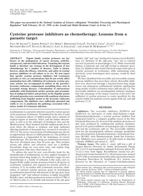
Proc. Natl. Acad. Sci. USA<br />
Vol. 96, pp. 11015–11022, September 1999<br />
Colloquium Paper<br />
This paper w<strong>as</strong> presented at <strong>the</strong> National Academy <strong>of</strong> Sciences colloquium ‘‘Proteolytic Processing and Physiological<br />
Regulation’’ held February 20–21, 1999, at <strong>the</strong> Arnold and Mabel Beckman Center in Irvine, CA.<br />
<strong>Cysteine</strong> <strong>prote<strong>as</strong>e</strong> <strong>inhibitors</strong> <strong>as</strong> <strong>chemo<strong>the</strong>rapy</strong>: Lessons from a<br />
par<strong>as</strong>ite target<br />
PAUL M. SELZER*†, SABINE PINGEL‡, IVY HSIEH*, BERNHARD UGELE§, VICTOR J. CHAN*, JUAN C. ENGEL*,<br />
MATTHEW BOGYO, DAVID G. RUSSELL � ,JUDY A. SAKANARI*, AND JAMES H. MCKERROW* , ** ,††<br />
Departments <strong>of</strong> *Pathology, **Pharmaceutical Chemistry, Biochemistry, and ‡ Medicine, University <strong>of</strong> California, San Francisco, CA 94143; � W<strong>as</strong>hington<br />
University, St. Louis, MO 63110; and § I. Frauenklinik, Klinikum Innenstadt, Ludwig-Maximilians Universität München, 80337 Munich, Germany<br />
ABSTRACT Papain family cysteine <strong>prote<strong>as</strong>e</strong>s are key<br />
factors in <strong>the</strong> pathogenesis <strong>of</strong> cancer inv<strong>as</strong>ion, arthritis,<br />
osteoporosis, and microbial infections. Targeting this enzyme<br />
family is <strong>the</strong>refore one strategy in <strong>the</strong> development <strong>of</strong> new<br />
<strong>chemo<strong>the</strong>rapy</strong> for a number <strong>of</strong> dise<strong>as</strong>es. Little is known,<br />
however, about <strong>the</strong> efficacy, selectivity, and safety <strong>of</strong> cysteine<br />
<strong>prote<strong>as</strong>e</strong> <strong>inhibitors</strong> in cell culture or in vivo. We now report<br />
that specific cysteine <strong>prote<strong>as</strong>e</strong> <strong>inhibitors</strong> kill Leishmania<br />
par<strong>as</strong>ites in vitro, at concentrations that do not overtly affect<br />
mammalian host cells. Inhibition <strong>of</strong> Leishmania cysteine <strong>prote<strong>as</strong>e</strong><br />
activity w<strong>as</strong> accompanied by defects in <strong>the</strong> par<strong>as</strong>ite’s<br />
lysosome�endosome compartment resembling those seen in<br />
lysosomal storage dise<strong>as</strong>es. Colocalization <strong>of</strong> anti-<strong>prote<strong>as</strong>e</strong><br />
antibodies with biotinylated surface proteins and accumulation<br />
<strong>of</strong> undigested debris and <strong>prote<strong>as</strong>e</strong> in <strong>the</strong> flagellar pocket<br />
<strong>of</strong> treated par<strong>as</strong>ites were consistent with a pathway <strong>of</strong> <strong>prote<strong>as</strong>e</strong><br />
trafficking from flagellar pocket to <strong>the</strong> lysosome�endosome<br />
compartment. The <strong>inhibitors</strong> were sufficiently absorbed and<br />
stable in vivo to ameliorate <strong>the</strong> pathology <strong>as</strong>sociated with a<br />
mouse model <strong>of</strong> Leishmania infection.<br />
Leishmani<strong>as</strong>is is a par<strong>as</strong>itic infection caused by various species<br />
<strong>of</strong> <strong>the</strong> protozoan Leishmania. Transmitted by <strong>the</strong> bite <strong>of</strong> sand<br />
flies, Leishmania infects 12 million people and is endemic in<br />
tropical regions <strong>of</strong> America, Africa, and <strong>the</strong> Indian subcontinent,<br />
<strong>as</strong> well <strong>as</strong> in <strong>the</strong> subtropics <strong>of</strong> Sou<strong>the</strong><strong>as</strong>t Asia and <strong>the</strong><br />
Mediterranean. Three hundred and fifty million people live in<br />
are<strong>as</strong> where <strong>the</strong> dise<strong>as</strong>e is common, and large epidemics<br />
affecting hundreds <strong>of</strong> thousands have occurred <strong>as</strong> recently <strong>as</strong><br />
1991 (1). The severe visceral form <strong>of</strong> leishmani<strong>as</strong>is may also be<br />
an opportunistic dise<strong>as</strong>e in AIDS patients (1). The problem <strong>of</strong><br />
leishmani<strong>as</strong>is is compounded by <strong>the</strong> inadequacy <strong>of</strong> current<br />
<strong>chemo<strong>the</strong>rapy</strong>. The first-line drugs are antimonial derivatives<br />
that were developed more than 40 years ago. They produce<br />
serious side effects, and refractory c<strong>as</strong>es are a problem.<br />
Second-line drugs are even more toxic, and require long,<br />
repeated doses with close observation (1).<br />
To address <strong>the</strong> need for new, cost-effective leads for <strong>the</strong><br />
<strong>chemo<strong>the</strong>rapy</strong> <strong>of</strong> leishmani<strong>as</strong>is, we have applied strategies <strong>of</strong><br />
structure-b<strong>as</strong>ed drug design (2). An attractive target for new<br />
<strong>chemo<strong>the</strong>rapy</strong> is a family <strong>of</strong> ca<strong>the</strong>psin L-like (cpL) and ca<strong>the</strong>psin<br />
B-like (cpB) cysteine <strong>prote<strong>as</strong>e</strong>s found in all species <strong>of</strong><br />
Leishmania examined, and required for par<strong>as</strong>ite growth or<br />
virulence (3–5). In studies with Leishmania mexicana, elimination<br />
<strong>of</strong> selected cysteine <strong>prote<strong>as</strong>e</strong> genes by homologous<br />
recombination showed that null mutants <strong>of</strong> <strong>the</strong> cpL gene array<br />
designated ‘‘cpb’’ had reduced virulence in highly susceptible<br />
BALB�c mice, and <strong>the</strong>y produced no lesions at all in C57BL�6<br />
or CBA�Ca mice (3, 4). Double null mutants <strong>of</strong> <strong>the</strong> cpL gene<br />
PNAS is available online at www.pn<strong>as</strong>.org.<br />
11015<br />
families ‘‘cpb’’ and ‘‘cpa’’ produced no lesions even in BALB�c<br />
mice (3). Deletion <strong>of</strong> <strong>the</strong> cpB gene ‘‘cpc’’ led to reduced<br />
survival <strong>of</strong> par<strong>as</strong>ites in macrophages (3, 6). While structurally<br />
distinct, Leishmania cpL and cpB overlap in substrate specificity<br />
(2). Inhibitors that would effectively target both types <strong>of</strong><br />
cysteine <strong>prote<strong>as</strong>e</strong>s in Leishmania, while maintaining some<br />
selectivity versus homologous host enzymes, would be ideal<br />
drug leads.<br />
We have identified both reversible and irreversible cysteine<br />
<strong>prote<strong>as</strong>e</strong> <strong>inhibitors</strong> that meet <strong>the</strong>se criteria. Reversible <strong>inhibitors</strong><br />
were discovered through a structure-b<strong>as</strong>ed drug design<br />
screen and subsequent combinatorial syn<strong>the</strong>tic optimization<br />
using models <strong>of</strong> both Leishmania major cpB and cpL (2). The<br />
irreversible <strong>inhibitors</strong> are pseudopeptide substrate analogues<br />
that take advantage <strong>of</strong> <strong>the</strong> unique reactivity <strong>of</strong> <strong>the</strong> active site<br />
sulfhydryl <strong>of</strong> cysteine <strong>prote<strong>as</strong>e</strong>s to confer specificity for this<br />
enzyme family but maintain activity against both cpL and cpB<br />
<strong>prote<strong>as</strong>e</strong>s (7, 8).<br />
METHODS<br />
Inhibitors. The reversible <strong>inhibitors</strong> ZLIII43A and<br />
ZLIII115A are derivatives <strong>of</strong> oxalic bis[(2-hydroxy-1-naphthyl)methylene]hydrazide,<br />
a cysteine <strong>prote<strong>as</strong>e</strong> inhibitor lead<br />
compound found in a computer graphics screen <strong>of</strong> <strong>the</strong> Fine<br />
Chemicals Directory (9). Several <strong>of</strong> <strong>the</strong> syn<strong>the</strong>tic derivatives <strong>of</strong><br />
that lead, produced by combinatorial syn<strong>the</strong>tic chemistry,<br />
proved to be potent <strong>inhibitors</strong> <strong>of</strong> homologous cpLs <strong>of</strong> malaria<br />
(10) and <strong>the</strong> Leishmania cpB (2). The irreversible inhibitor<br />
used w<strong>as</strong> <strong>the</strong> pseudopeptide substrate analogue morpholine<br />
urea-phenylalanine-homophenylalanine-vinylsulfonyl-benzene<br />
(K11002, Arris Pharmaceuticals, South San Francisco,<br />
CA). Inhibitors were prepared <strong>as</strong> 20 mM stocks in dimethyl<br />
sulfoxide (DMSO) and stored at �20°C.<br />
Prote<strong>as</strong>e Assays. The native L. major cpB w<strong>as</strong> a gift <strong>of</strong><br />
Jacques Bouvier (Novartis, St. Aubin, Switzerland). Papain<br />
(EC 3.4.22.2) and mammalian ca<strong>the</strong>psin B (bovine spleen; EC<br />
3.4.22.1) were from Sigma. Recombinant cruzain w<strong>as</strong> produced<br />
<strong>as</strong> previously described (11). All <strong>prote<strong>as</strong>e</strong>s were <strong>as</strong>sayed<br />
at 25°C with an automated microtiter plate spectr<strong>of</strong>luorometer<br />
(Labsystem FluoroScan II; Northbrook, IL). Activity w<strong>as</strong><br />
detected by <strong>the</strong> liberation <strong>of</strong> 7-amino-4-methylcoumarin<br />
Abbreviations: cpL, ca<strong>the</strong>psin L-like cysteine <strong>prote<strong>as</strong>e</strong>; cpB, ca<strong>the</strong>psin<br />
B-like cysteine <strong>prote<strong>as</strong>e</strong>; AMC, 7-amino-4-methylcoumarin.<br />
† To whom reprint requests may be addressed at present address:<br />
Hoechst Roussel Vet GmbH, Research Pharmaceuticals, Building H<br />
811, D-65926 Frankfurt�Main, Germany. E-mail: PSelzer@<br />
hrvet.com.<br />
†† To whom reprint requests may be addressed at: Department <strong>of</strong><br />
Pathology, University <strong>of</strong> California, Tropical Dise<strong>as</strong>e Research<br />
Unit, VAMC, 4150 Clement Street 113B, San Francisco, CA 94121.<br />
E-mail: jmck@cgl.ucsf.edu.
11016 Colloquium Paper: Selzer et al. Proc. Natl. Acad. Sci. USA 96 (1999)<br />
(AMC) (excitation wavelength � 355 nm and emission wavelength<br />
� 460 nm) from <strong>the</strong> syn<strong>the</strong>tic peptide substrate Z-Phe-<br />
Arg-AMC (Z � benzyloxycarbonyl) (Enzyme Systems Products,<br />
Livermore, CA). The enzyme concentrations were determined<br />
by active site titration. Reversible <strong>inhibitors</strong> at<br />
various concentrations were preincubated with <strong>the</strong> respective<br />
enzyme for 5 min before <strong>the</strong> reaction w<strong>as</strong> started by adding <strong>the</strong><br />
substrate. Enzyme activities were expressed in percent <strong>of</strong><br />
residual activity compared with an uninhibited control, and<br />
were plotted versus incre<strong>as</strong>ing inhibitor concentrations to<br />
calculate <strong>the</strong> IC50. Assay conditions were <strong>as</strong> follows: L. major<br />
cpB: 100 mM sodium acetate at pH 5.5, 10 mM dithiothreitol<br />
(DTT), 1 mM EDTA, 0.1% Triton X-100, 50 �M Z-Phe-Arg-<br />
AMC final concentration (from a 10 mM stock solution in<br />
DMSO); Km � 7 �M. Papain and mammalian ca<strong>the</strong>psin B: 100<br />
mM sodium acetate at pH 5.5, 10 mM DTT, 100 �M Z-Phe-<br />
Arg-AMC final concentration; Km � 50 �M and 110 �M,<br />
respectively. Cruzain: The <strong>as</strong>say conditions were <strong>the</strong> same <strong>as</strong><br />
for papain except that <strong>the</strong> substrate concentration w<strong>as</strong> 20 �M;<br />
Km � 1 �M. Km values were determined by nonlinear regression<br />
using <strong>the</strong> s<strong>of</strong>tware ULTRAFIT (Bios<strong>of</strong>t, Ferguson, MO).<br />
Irreversible <strong>inhibitors</strong> were <strong>as</strong>sayed in a time-b<strong>as</strong>ed inactivation<br />
<strong>as</strong>say. The inactivation process w<strong>as</strong> b<strong>as</strong>ed on <strong>the</strong> following<br />
scheme<br />
k1 E � I L|;<br />
k �1<br />
kinact EI O¡<br />
E-I<br />
where E � enzyme, I � inhibitor, EI � noncovalent enzyme<br />
inhibitor complex, E-I � inactivated enzyme, k1 and k�1 �<br />
noncovalent rate constants (Ki � k�1�k1), and kinact � firstorder<br />
inactivation constant. The values <strong>of</strong> Ki and kinact were<br />
determined from progress curves in <strong>the</strong> presence <strong>of</strong> substrate<br />
and inhibitor. These curves were fit to a first-order equation<br />
(ULTRAFIT) to produce kobs (observed inactivation constant)<br />
values, where kobs � kinact[I]�Ki, app � [I], where Ki, app �<br />
apparent Ki). Plotting 1�kobs versus 1�[I] gives <strong>the</strong> values for Ki,<br />
app and kinact. Taking <strong>the</strong> substrate into consideration, <strong>the</strong> true<br />
Ki w<strong>as</strong> calculated by Ki � Ki, app�(1 � [S]�Km). At le<strong>as</strong>t six<br />
different inhibitor concentrations were determined in duplicate<br />
for a minimum <strong>of</strong> three independent experiments. The<br />
reaction w<strong>as</strong> started by adding <strong>the</strong> enzyme, and <strong>the</strong> timedependent<br />
inactivation w<strong>as</strong> monitored. Enzyme (E) and substrate<br />
(S) concentrations: L. major cpB, E � 1–2 nM, S � 2.5<br />
�M; cruzain, E � 5 nM, S � 5 �M; papain, E � 6 nM, S �<br />
15 �M; and cpB, E � 10 nM, S � 10 �M.<br />
Cell Culture Assays. L. major prom<strong>as</strong>tigotes LV39(MRHO�<br />
SU�59�P) were grown at 27°C in 5 ml (25-cm 2 cell culture<br />
fl<strong>as</strong>k; Costar, Cambridge, MA) <strong>of</strong> RPMI medium 1640 containing<br />
10% (vol�vol) heat-inactivated fetal bovine serum<br />
(FBS) and 20% brain heart infusion tryptose. Par<strong>as</strong>ites were<br />
maintained in <strong>the</strong> exponential growth ph<strong>as</strong>e by p<strong>as</strong>sing <strong>the</strong>m<br />
twice a week. For inhibitor studies, 10 6 cells per ml were<br />
inoculated in new cultures, and cell growth w<strong>as</strong> determined by<br />
counting <strong>the</strong> par<strong>as</strong>ites with a Neubauer hemocytometer (A.O.<br />
Instruments, Buffalo, NY). The mouse macrophage cell line<br />
J774 w<strong>as</strong> maintained in 75-cm 2 cell culture fl<strong>as</strong>ks (Costar) at<br />
37°C in RPMI medium 1640 containing 5% FBS (12 ml total<br />
volume) and p<strong>as</strong>sed once a week. Irradiated J774 cells (10 min,<br />
2,700 rad, 24 h before infection) were cultured on gl<strong>as</strong>s<br />
coverslips in six-well cluster plates (Costar) and infected with<br />
stationary-ph<strong>as</strong>e prom<strong>as</strong>tigotes in a ratio <strong>of</strong> 1:10 for 12 h. After<br />
<strong>the</strong> infected macrophage monolayers had been w<strong>as</strong>hed three<br />
times with RPMI 1640, <strong>inhibitors</strong> were added to <strong>the</strong> culture<br />
and plates were incubated for 5 days at 32°C in a 5% CO2�95%<br />
air atmosphere. To determine <strong>the</strong> number <strong>of</strong> am<strong>as</strong>tigotes per<br />
macrophage, cells were fixed in 100% methanol and stained<br />
with Giemsa stain. At le<strong>as</strong>t 200 macrophages per experiment<br />
were examined to monitor <strong>the</strong> effect <strong>of</strong> <strong>the</strong> <strong>inhibitors</strong>. Inhibitors<br />
dissolved in DMSO were from 20 mM stock solutions.<br />
DMSO concentrations up to 0.5% showed no effect on prom<strong>as</strong>tigotes,<br />
am<strong>as</strong>tigotes, or J774 cells.<br />
Electron Microscopy and ImmunoGold Localization. One<br />
to 5 � 10 8 prom<strong>as</strong>tigote par<strong>as</strong>ites, treated or untreated, were<br />
w<strong>as</strong>hed twice with PBS (4°C, 10 min, 3,000 rpm in a Beckman<br />
Accuspin-FR centrifuge). Cells were fixed in 0.1 M sodium<br />
cacodylate buffer at pH 7.4 containing 1.5% glutaraldehyde<br />
(0.25% for ImmunoGold labeling) and 1% sucrose. Epon<br />
embedding, LR white embedding, and thin sectioning were<br />
performed according to standard protocols (12–14).<br />
For ImmunoGold labeling, a polyclonal antiserum raised<br />
against <strong>the</strong> native L. major cpB w<strong>as</strong> used in a 1:20 or 1:100<br />
dilution, followed by a secondary antibody conjugated with<br />
10-nm gold particles (goat antibody to rabbit IgG, 1:50,<br />
Amersham Life Sciences). Serum from <strong>the</strong> rabbit before<br />
immunization, BSA, and bovine serum were used for specificity<br />
controls. Photographs were taken with a Zeiss EM10C.<br />
Alternatively, prom<strong>as</strong>tigotes <strong>of</strong> L. major were surface labeled<br />
with 500 �g�ml N-hydroxysuccinimide-biotin in PBS<br />
(pH 7.6) for 20 min on ice. The cells were w<strong>as</strong>hed and placed<br />
in medium at 25°C for 60 min. They were fixed in 200 mM Pipes<br />
with 4% paraformaldehyde, frozen, and processed for immunoelectron<br />
microscopy <strong>as</strong> described previously (15, 16). The<br />
thawed cryosections were probed with streptavidin (1 �g�ml),<br />
followed by mouse monoclonal anti-streptavidin and rabbit<br />
antibody to L. major ca<strong>the</strong>psin B. The antibodies were revealed<br />
by 12-nm gold-conjugated goat anti-mouse IgG and 18-nm<br />
gold-conjugated goat anti-rabbit IgG (Jackson ImmunoResearch).<br />
Prom<strong>as</strong>tigote Extracts and Western Blot Analysis. Five �<br />
10 9 prom<strong>as</strong>tigotes were w<strong>as</strong>hed twice with PBS at pH 7.4. Cells<br />
were sonicated (Sonic Dismembranator 300, Fisher Scientific)<br />
on ice (three � 10 sec, relative output 0.6) and adjusted with<br />
sodium acetate buffer at pH 5.5 to 1 � 10 9 cells per ml.<br />
Aliquots were stored at �20°C for 4 months without any loss<br />
<strong>of</strong> cysteine <strong>prote<strong>as</strong>e</strong> activity. Samples <strong>of</strong> 100 �l were solubilized<br />
by adding 20 �l <strong>of</strong> 6-fold concentrated Laemmli buffer.<br />
Samples were subjected to SDS�10% PAGE and transferred to<br />
nitrocellulose sheets. The immunoblots were incubated in<br />
2.5% (wt�vol) blocking reagent (Boehringer Mannheim) in<br />
100 mM maleic acid buffer (pH 7.5) for 60 min at room<br />
temperature, and <strong>the</strong>n incubated overnight at 4°C with a rabbit<br />
polyclonal antiserum raised against L. major cpB or L. mexicana<br />
cpL that had been diluted 1:1000 or 1:500, respectively,<br />
in 100 mM Tris�HCl, pH 7.5, with 0.05% Tween 20 and 1%<br />
FCS. After incubation with horseradish peroxid<strong>as</strong>econjugated<br />
secondary antibodies (1:3000; goat anti-rabbit IgG;<br />
Gibco BRL Life Technologies) for 60 min at room temperature,<br />
<strong>the</strong> blots were developed using ECL (Amersham Life<br />
Science). For active site labeling <strong>of</strong> cysteine <strong>prote<strong>as</strong>e</strong>s, prom<strong>as</strong>tigote<br />
extracts were incubated ei<strong>the</strong>r with 50 �M 14 Clabeled<br />
K11002 for 15 min at room temperature or with<br />
125 I-labeled p-nitrophenyl-derivatized E-64 and vinyl sulfone<br />
<strong>as</strong> previously described (17). Samples <strong>of</strong> 100 �l were subjected<br />
to SDS�PAGE and analyzed by fluorography.<br />
Animal Model <strong>of</strong> Infection. All procedures were approved by<br />
<strong>the</strong> University <strong>of</strong> California, San Francisco Committee on<br />
Table 1. Inhibition <strong>of</strong> cysteine <strong>prote<strong>as</strong>e</strong>s with reversible <strong>inhibitors</strong><br />
IC50, �M<br />
Enzyme<br />
ZLIII115A ZLIII43A<br />
L. major cpB 10 2<br />
Cruzain 10 5<br />
Papain �50 10<br />
Mammalian ca<strong>the</strong>psin B 20 20<br />
See Prote<strong>as</strong>e Assays for details <strong>of</strong> <strong>as</strong>say used.
Colloquium Paper: Selzer et al. Proc. Natl. Acad. Sci. USA 96 (1999) 11017<br />
Table 2. Inhibition <strong>of</strong> cysteine <strong>prote<strong>as</strong>e</strong>s with K11002<br />
Enzyme kinact, s �1 Ki, �M kinact�Ki, s �1 �M �1<br />
L. major cpB 0.021 � 0.0014 0.205 � 0.077 107,000 � 32,000<br />
Cruzain 0.064 � 0.027 0.17 � 0.074 383,000 � 27,000<br />
Papain 0.072 � 0.009 0.261 � 0.025 275,000 � 10,000<br />
Mammalian ca<strong>the</strong>psin B 0.014 � 0.001 9.8 � 2.3 1,400 � 250<br />
Note <strong>the</strong> similar kinact values versus <strong>the</strong> differences in <strong>the</strong> Ki values that are mainly responsible for <strong>the</strong><br />
divergence <strong>of</strong> <strong>the</strong> second-order rate constants. See Prote<strong>as</strong>e Assays for details <strong>of</strong> <strong>as</strong>say used.<br />
Animal Research. Two � 10 5 metacyclic L. major (WHOM�<br />
IR�173) were obtained by peanut agglutinin selection <strong>as</strong><br />
previously described (18) and were injected into each hind<br />
footpad <strong>of</strong> female BALB�c mice (18–20 g, Simonsen Laboratories,<br />
Gilroy, CA). Strain WHOM�IR�173 w<strong>as</strong> used because<br />
<strong>of</strong> its higher virulence in mice compared with strain<br />
LV39(MRHO�SU�59�P). Compounds were dissolved in<br />
100% DMSO and stored at �20°C. The final concentrations<br />
were adjusted with sterile water to give a 70:30 (vol�vol)<br />
DMSO�H2O mixture. Twenty-four hours after infection, mice<br />
were treated with 100 �l <strong>of</strong> K11002 or ZLIII115A (100 mg�kg<br />
per day, every day, 4 weeks, intraperitoneal) in ei<strong>the</strong>r a single<br />
dose or split into two treatments per day. Each set tested<br />
consisted <strong>of</strong> five mice, including an untreated control, a<br />
DMSO-treated control, <strong>as</strong> well <strong>as</strong> uninfected mice treated with<br />
<strong>the</strong> appropriate compound. To monitor <strong>the</strong> course <strong>of</strong> <strong>the</strong><br />
infection, <strong>the</strong> thickness <strong>of</strong> <strong>the</strong> footpads w<strong>as</strong> me<strong>as</strong>ured once a<br />
week by a standard method using a metric caliper (dial<br />
thickness gauge no. 7305; Mitutoyo, Kaw<strong>as</strong>aki, Japan) (19). To<br />
quantify par<strong>as</strong>ite burden, whole footpad histology <strong>as</strong> well <strong>as</strong><br />
limited par<strong>as</strong>ite dilution <strong>as</strong>says from footpad tissues (20) were<br />
performed at <strong>the</strong> end <strong>of</strong> <strong>the</strong> experiments. To investigate side<br />
effects <strong>of</strong> <strong>the</strong> compounds, mouse liver tissue w<strong>as</strong> embedded in<br />
paraffin and 5-�m sections were stained with hematoxylin.<br />
Sections <strong>of</strong> treated tissues were <strong>the</strong>n compared with control<br />
liver tissue.<br />
Cytokine Assays. IL-4 and IFN-� were <strong>as</strong>sayed in inhibitortreated<br />
and untreated mice by monoclonal-b<strong>as</strong>ed ELISA and<br />
normalized to standard controls <strong>as</strong> previously described (20).<br />
RESULTS<br />
The reversible hydrazide <strong>inhibitors</strong>, ZLIII115A and ZLIII43A,<br />
were tested against <strong>the</strong> L. major cpB, cruzain (<strong>the</strong> major cpL<br />
<strong>of</strong> Trypanosoma cruzi), papain, and mammalian ca<strong>the</strong>psin B.<br />
There w<strong>as</strong> 2- to 10-fold higher inhibitory activity toward <strong>the</strong> L.<br />
major cpB versus <strong>the</strong> plant or mammalian <strong>prote<strong>as</strong>e</strong>s (Table 1).<br />
In <strong>the</strong> c<strong>as</strong>e <strong>of</strong> <strong>the</strong> irreversible pseudopeptide inhibitor K11002<br />
(7), <strong>the</strong> first-order inactivation constant (kinact) w<strong>as</strong> similar for<br />
<strong>the</strong> four enzymes. However, differences <strong>of</strong> up to 100-fold were<br />
observed for <strong>the</strong> second-order rate constants (kinact�Ki) (Table<br />
2). The differences in activity <strong>of</strong> <strong>the</strong> mammalian ca<strong>the</strong>psin B<br />
versus <strong>the</strong> plant or par<strong>as</strong>ite <strong>prote<strong>as</strong>e</strong>s are mainly due to<br />
differences in Ki. The L. major cpB, cruzain, and papain were<br />
inhibited to a similar extent. This similarity is consistent with<br />
<strong>the</strong> paradoxical ca<strong>the</strong>psin L-like substrate preference <strong>of</strong> <strong>the</strong> L.<br />
major cpB, due to a single amino acid modification in <strong>the</strong> S2<br />
binding pocket (2).<br />
The three <strong>inhibitors</strong> were next tested in cell cultures <strong>of</strong> L.<br />
major prom<strong>as</strong>tigotes, <strong>the</strong> extracellular stage <strong>of</strong> <strong>the</strong> par<strong>as</strong>ite.<br />
Inhibitors were added to replicating Leishmania <strong>as</strong> a single<br />
dose, and cell growth w<strong>as</strong> monitored for 3 days. Both <strong>the</strong><br />
irreversible and <strong>the</strong> reversible <strong>inhibitors</strong> blocked replication <strong>of</strong><br />
<strong>the</strong> par<strong>as</strong>ite. Concentrations <strong>of</strong> 5 �M inhibited par<strong>as</strong>ite growth<br />
10-fold, where<strong>as</strong> 20 �M and 50 �M completely inhibited cell<br />
growth (Fig. 1). Exchanging <strong>the</strong> medium every day for a total<br />
<strong>of</strong> 3 days, <strong>the</strong>reby keeping inhibitor concentrations stable (20<br />
�M and 50 �M), led to death <strong>of</strong> <strong>the</strong> par<strong>as</strong>ites. After <strong>the</strong> fourth<br />
day <strong>of</strong> this latter experiment, <strong>the</strong> medium w<strong>as</strong> replaced with<br />
fresh medium without inhibitor, and <strong>the</strong> fl<strong>as</strong>ks were again kept<br />
under culture conditions. Even after 10 days no par<strong>as</strong>ites could<br />
be detected, indicating a complete cure <strong>of</strong> <strong>the</strong> Leishmania<br />
culture by <strong>the</strong> cysteine <strong>prote<strong>as</strong>e</strong> <strong>inhibitors</strong>.<br />
To confirm that <strong>the</strong> <strong>inhibitors</strong> could access <strong>the</strong> intracellular<br />
cysteine <strong>prote<strong>as</strong>e</strong>s <strong>of</strong> L. major, prom<strong>as</strong>tigote par<strong>as</strong>ites were<br />
harvested after treatment with 50 �M K11002 for 24 h and<br />
extracted. The residual cysteine <strong>prote<strong>as</strong>e</strong> activity <strong>as</strong> me<strong>as</strong>ured<br />
with <strong>the</strong> fluorogenic substrate Z-Phe-Arg-AMC (which detects<br />
both L. major cpL and cpB activity) in K11002-treated<br />
cells w<strong>as</strong> 20% � 7% (n � 5) relative to <strong>the</strong> control par<strong>as</strong>ites.<br />
Targeting <strong>of</strong> ca<strong>the</strong>psins by K11002 w<strong>as</strong> also confirmed by<br />
‘‘tagging’’ <strong>the</strong> target <strong>prote<strong>as</strong>e</strong>s with radioactively labeled inhibitor<br />
and identification <strong>of</strong> protein species by parallel Western<br />
blot (Fig. 2). The predominant cysteine <strong>prote<strong>as</strong>e</strong> in L. major<br />
prom<strong>as</strong>tigotes is cpB (21), and this species w<strong>as</strong> <strong>the</strong> predominant,<br />
but not exclusive, target <strong>of</strong> <strong>the</strong> vinyl sulfone inhibitor.<br />
The mature catalytic domains <strong>of</strong> L. major ca<strong>the</strong>psins B and L<br />
FIG. 1. Effects <strong>of</strong> K11002 (A) and ZLIII115A (B) on <strong>the</strong> growth <strong>of</strong> L. major prom<strong>as</strong>tigotes. The compounds were added at time point 0, and<br />
cell growth w<strong>as</strong> monitored for 3 days. ZLIII43A showed very similar inhibition pr<strong>of</strong>iles (2). F, Control; ‚, 5�M; E, 20�M; and �, 50�M. Points<br />
are means <strong>of</strong> three independent experiments.
11018 Colloquium Paper: Selzer et al. Proc. Natl. Acad. Sci. USA 96 (1999)<br />
FIG. 2. Inhibitor targets and Western blot analysis <strong>of</strong> prom<strong>as</strong>tigote lysates. Proteins from whole prom<strong>as</strong>tigote extracts (Lanes 1 and 2), or whole<br />
prom<strong>as</strong>tigote were separated by SDS�PAGE and blotted to nitrocellulose membrane. Lane 1 w<strong>as</strong> probed with a rabbit antiserum raised against<br />
a cpL from L. mexicana (a gift <strong>of</strong> Jeremy Mottram, University <strong>of</strong> Gl<strong>as</strong>gow); lane 2 w<strong>as</strong> probed with a rabbit antiserum raised against cpB from<br />
L. major. Extracts in lane 3 were incubated with 125 I-labeled E-64, an epoxide cysteine <strong>prote<strong>as</strong>e</strong> inhibitor. Extracts in lane 4 were labeled with<br />
125 I-vinyl sulfone <strong>as</strong> described previously (17). Note <strong>the</strong> predominant labeling <strong>of</strong> <strong>the</strong> mature (catalytic domain) cpB in lanes 3 and 4 by both <strong>inhibitors</strong>.<br />
The vinyl sulfone also binds to <strong>the</strong> less abundant mature CpL. Both <strong>inhibitors</strong> label higher molecular weight <strong>prote<strong>as</strong>e</strong> precursors, which can be<br />
tentatively identified <strong>as</strong> active intermediates in <strong>prote<strong>as</strong>e</strong> processing (Int.) by Western blotting (lanes 1 and 2) and reexpression <strong>of</strong> specific <strong>prote<strong>as</strong>e</strong><br />
genes in <strong>prote<strong>as</strong>e</strong>-null organisms (Sanya Sanderson and Jeremy Mottram, personal communication). The ‘‘complex’’ band is presumably an<br />
aggregate <strong>of</strong> <strong>prote<strong>as</strong>e</strong> with itself or with a carrier protein.<br />
were labeled, <strong>as</strong> were intermediates in <strong>prote<strong>as</strong>e</strong> processing<br />
(Fig. 2).<br />
To determine effects <strong>of</strong> <strong>the</strong> <strong>inhibitors</strong> on Leishmania am<strong>as</strong>tigotes,<br />
<strong>the</strong> stage <strong>of</strong> <strong>the</strong> par<strong>as</strong>ite that resides within mammalian<br />
host cells, irradiated macrophages (J774 cells) were infected<br />
with prom<strong>as</strong>tigote stationary-ph<strong>as</strong>e Leishmania. After 12 h,<br />
50% <strong>of</strong> <strong>the</strong> macrophages were infected with 1 to 4 par<strong>as</strong>ites per<br />
host cell. Cells were <strong>the</strong>n treated with a single dose (40 �M)<br />
<strong>of</strong> inhibitor and cultured for ano<strong>the</strong>r 5 days. At day 5, 85% <strong>of</strong><br />
untreated J774 cells carried more than 9 par<strong>as</strong>ites. This<br />
observation confirms that par<strong>as</strong>ites replicate within <strong>the</strong> host<br />
cells and infect new macrophages. Replication <strong>of</strong> par<strong>as</strong>ites w<strong>as</strong><br />
decre<strong>as</strong>ed in cultures treated with <strong>the</strong> pseudopeptide inhibitor<br />
or <strong>the</strong> hydrazide <strong>inhibitors</strong>, and few if any new macrophages<br />
were infected after treatment (Table 3). Host cell morphology<br />
w<strong>as</strong> not affected by <strong>the</strong> treatment, and nonirradiated, inhibitor-treated<br />
macrophages had no difference in growth rate<br />
compared with untreated cells.<br />
The hydrazide <strong>inhibitors</strong> and <strong>the</strong> pseudopeptide inhibitor<br />
produced very similar effects on <strong>the</strong> organelle structure within<br />
<strong>the</strong> par<strong>as</strong>ites. After 24 h <strong>of</strong> treatment, myelin figures, undigested<br />
cell debris, dense bodies, and multivesicular bodies<br />
Table 3. Treatment <strong>of</strong> am<strong>as</strong>tigote par<strong>as</strong>ites<br />
Compound<br />
(40 �M)<br />
%<strong>of</strong><br />
host cells<br />
infected<br />
% <strong>of</strong> host cells with<br />
given number <strong>of</strong> am<strong>as</strong>tigotes<br />
1–4 5–9 �10<br />
Control 12 h 48 � 9 48 � 9 — —<br />
Control 5 d 85 � 7 25 � 5 22 � 5 38 � 7<br />
ZLIII115A 5 d 58 � 9 58 � 9 — —<br />
ZLIII43A 5 d 56 � 12 56 � 12 — —<br />
K11002 5 d 65 � 7 58 � 2 7 � 9 —<br />
Irradiated J774 host cell macrophages were infected with L. major<br />
prom<strong>as</strong>tigotes for 12 h to allow infection <strong>of</strong> <strong>the</strong> cells and development <strong>of</strong><br />
am<strong>as</strong>tigote par<strong>as</strong>ites. After 12 h, 50% <strong>of</strong> <strong>the</strong> macrophages were infected.<br />
The established infection w<strong>as</strong> <strong>the</strong>n treated with a single dose <strong>of</strong> <strong>the</strong><br />
hydrazide (ZL-) or <strong>the</strong> vinyl sulfone (K11002) <strong>prote<strong>as</strong>e</strong> <strong>inhibitors</strong>. After<br />
5 days at 32°C, untreated (control) cells were highly infected, where<strong>as</strong><br />
treated cells remained essentially unchanged with respect to <strong>the</strong> initial<br />
(already established) infection. Numbers are expressed in percent and are<br />
means � SD <strong>of</strong> three independent experiments.
FIG. 3. Electron micrographs <strong>of</strong> Epon-embedded L. major prom<strong>as</strong>tigotes.<br />
Par<strong>as</strong>ites were untreated (A) or treated for 24 h with 50<br />
�M K11002 (B and C) or50�M ZLIII115A (D). Treatment <strong>of</strong> <strong>the</strong><br />
par<strong>as</strong>ite with ei<strong>the</strong>r inhibitor had very similar effects, resulting in <strong>the</strong><br />
appearance <strong>of</strong> diverse multivesicular and dense bodies (arrowheads),<br />
lipid inclusions (arrows), and myelin figures (<strong>as</strong>terisk). n, Nucleus; g,<br />
Golgi apparatus; f, flagellar pocket; m, mitochondrion; k, kinetopl<strong>as</strong>t.<br />
(Bars � 10 �m.)<br />
appeared within <strong>the</strong> abnormally dilated par<strong>as</strong>ite lysosomes and<br />
<strong>the</strong> flagellar pocket (Fig. 3), <strong>the</strong> site where endocytosis and<br />
exocytosis takes place (22). These abnormalities resemble<br />
alterations seen in lysosomal storage dise<strong>as</strong>es caused by <strong>the</strong><br />
deficiency or absence <strong>of</strong> specific lysosomal hydrol<strong>as</strong>es (23).<br />
The nucleus and <strong>the</strong> Golgi apparatus were not affected, but<br />
some cells showed dilated mitochondria. In <strong>the</strong>se latter cells<br />
<strong>the</strong> kinetopl<strong>as</strong>t DNA w<strong>as</strong> no longer condensed but appeared<br />
in diffuse patches. No effects on treated mammalian host cells<br />
at <strong>the</strong> light microscopic or ultr<strong>as</strong>tructural level were observed.<br />
C<br />
Colloquium Paper: Selzer et al. Proc. Natl. Acad. Sci. USA 96 (1999) 11019<br />
A B<br />
E F<br />
D<br />
To localize <strong>the</strong> Leishmania cysteine <strong>prote<strong>as</strong>e</strong>s within <strong>the</strong><br />
par<strong>as</strong>ite cell, ImmunoGold electron microscopic analysis using<br />
a L. major cpB-specific antiserum w<strong>as</strong> performed. In untreated<br />
cells <strong>the</strong> gold label appeared only in lysosomes (Fig. 4 C and<br />
D). Treated cells were more heavily labeled in <strong>the</strong> dilated<br />
lysosome�endosome compartment and in <strong>the</strong> flagellar pocket<br />
(Fig. 4 E and F). Apparently empty flagellar pockets were also<br />
heavily labeled in treated par<strong>as</strong>ites, but not in untreated<br />
par<strong>as</strong>ites. To confirm target <strong>prote<strong>as</strong>e</strong> localization at <strong>the</strong> site <strong>of</strong><br />
inhibitor-induced abnormalities, untreated prom<strong>as</strong>tigotes<br />
were surface-labeled with N-hydroxysuccinimide-biotin and<br />
placed back in culture to facilitate internalization <strong>of</strong> labeled<br />
proteins. This method allows visualization <strong>of</strong> <strong>the</strong> endosomal�<br />
lysosomal network <strong>of</strong> <strong>the</strong> cells. ImmunoGold electron microscopy<br />
<strong>of</strong> <strong>the</strong>se par<strong>as</strong>ites revealed an abundance <strong>of</strong> ca<strong>the</strong>psin B<br />
in <strong>the</strong> flagellar pocket and in vesicles subtending that structure<br />
(Fig. 4 A and B). Some <strong>of</strong> <strong>the</strong>se vesicles contained biotinylated<br />
proteins, indicating that <strong>the</strong>y are endosomes or lysosomes,<br />
where<strong>as</strong> o<strong>the</strong>rs contained only ca<strong>the</strong>psin B, suggesting that<br />
<strong>the</strong>y may be secretory vesicles.<br />
Because <strong>of</strong> <strong>the</strong> selective arrest <strong>of</strong> par<strong>as</strong>ite versus host cell<br />
growth by <strong>inhibitors</strong> added to cultures, <strong>the</strong> efficacy <strong>of</strong> <strong>the</strong><br />
cysteine <strong>prote<strong>as</strong>e</strong> <strong>inhibitors</strong> in vivo w<strong>as</strong> evaluated in Leishmania-infected<br />
BALB�c mice. Twenty-four hours after infection,<br />
mice received intraperitoneal injections <strong>of</strong> K11002 or<br />
ZLIII115A dissolved in DMSO�H2O (70:30). By 2 weeks,<br />
control mice had already developed footpad lesions, which<br />
progressed in size and severity. In treated animals <strong>the</strong> lesion<br />
development w<strong>as</strong> significantly delayed, with no swelling <strong>of</strong> <strong>the</strong><br />
footpads until 3–4 weeks (Figs. 5 and 6). After 4 weeks <strong>of</strong><br />
treatment, inhibitor dosing w<strong>as</strong> stopped, and lesion development<br />
paralleled that seen in control mice. At <strong>the</strong> end <strong>of</strong> <strong>the</strong><br />
treatment period, whole footpad histology and limiting dilution<br />
<strong>as</strong>says <strong>of</strong> par<strong>as</strong>ites from extracted footpad tissues showed<br />
par<strong>as</strong>ite burden for treated animals w<strong>as</strong> at le<strong>as</strong>t two logs lower<br />
than that <strong>of</strong> <strong>the</strong> untreated animals (10 �7 versus 10 �5 ). This<br />
finding is consistent with results from previous studies that<br />
documented <strong>the</strong> correlation between footpad size and numbers<br />
<strong>of</strong> par<strong>as</strong>ites (18, 20). None <strong>of</strong> <strong>the</strong> compounds produced<br />
toxic effects in mice, <strong>as</strong> indicated by daily observation <strong>of</strong><br />
weight, activity, and appearance, <strong>as</strong> well <strong>as</strong> autopsy and<br />
histologic analysis. Also, <strong>the</strong>re w<strong>as</strong> no evidence <strong>of</strong> a switch<br />
from <strong>the</strong> usual TH2 cytokine response to Leishmania in<br />
FIG. 4. Immunoelectron micrographs <strong>of</strong> L. major<br />
prom<strong>as</strong>tigotes. (A and B) Electron micrographs <strong>of</strong> cryosections<br />
from L. major prom<strong>as</strong>tigotes that were surface<br />
biotinylated with N-hydroxysuccinimide-biotin and incubated<br />
in medium for 45 min prior to fixation. The<br />
sections were probed with streptavidin�mouse antistreptavidin<br />
mAb (12-nm gold particle conjugated to<br />
goat anti-mouse IgG) and rabbit anti-ca<strong>the</strong>psin B antibody<br />
(18-nm gold particle conjugated to goat anti-rabbit<br />
IgG). g, Golgi apparatus; e, endodome; n, nucleus; k,<br />
kinetopl<strong>as</strong>t; m, mitochondrion. (Bars � 0.25 �m.) (A)<br />
Ca<strong>the</strong>psin B label is observed in vesicles in <strong>the</strong> vicinity<br />
<strong>of</strong> <strong>the</strong> Golgi apparatus and in <strong>the</strong> flagellar pocket, which<br />
is strongly positive for biotinylated proteins. (B) Label<br />
is also observed in biotin-positive vesicles, or endosomes<br />
that subtend <strong>the</strong> flagellar pocket. The data indicated<br />
that ca<strong>the</strong>psin B is syn<strong>the</strong>sized and proceeds through <strong>the</strong><br />
Golgi apparatus into <strong>the</strong> secretory network, where it<br />
gains access to <strong>the</strong> flagellar pocket. (C–F) LR whiteembedded<br />
and ImmunoGold-labeled (anti-L. major cpB<br />
antiserum) prom<strong>as</strong>tigotes. Note <strong>the</strong> specific labeling in<br />
lysosomes <strong>of</strong> untreated cells (C and D) and in multivesicular<br />
bodies (arrowheads), <strong>as</strong> well <strong>as</strong> in <strong>the</strong> flagellar<br />
pocket <strong>of</strong> treated cells (E and F). (Bars � 0.5 �m.)
11020 Colloquium Paper: Selzer et al. Proc. Natl. Acad. Sci. USA 96 (1999)<br />
FIG. 5. Lesion development in Leishmania-infected BALB�c mice. The picture w<strong>as</strong> taken after 3 weeks <strong>of</strong> treatment with K11002 (100 mg�kg<br />
per day). Note <strong>the</strong> ery<strong>the</strong>ma and gross edema <strong>of</strong> <strong>the</strong> footpads in <strong>the</strong> untreated mouse (left), versus no edema or ery<strong>the</strong>ma <strong>of</strong> <strong>the</strong> footpads in <strong>the</strong><br />
<strong>prote<strong>as</strong>e</strong> inhibitor-treated mouse (right).<br />
inhibitor-treated mice, <strong>as</strong> w<strong>as</strong> reported by Maekawa et al. (24),<br />
who used <strong>the</strong> ca<strong>the</strong>psin B-specific inhibitor CA074. IFN-�<br />
levels remained unchanged (36.7 � 15.8 ng�ml for 10 6 cells,<br />
treated mice, versus 33.4 � 12.3 ng�ml for 10 6 cells, untreated<br />
mice), and IL-4 levels remained elevated (14.4 � 1.0 ng�ml for<br />
10 6 cells, treated mice, versus 16.7 � 2.1 ng�ml for 10 6 cells,<br />
untreated mice).<br />
FIG. 6. Lesion sizes <strong>of</strong> treated and untreated infected BALB�c<br />
mice. Bars are means � SD <strong>of</strong> five mice (two footpads per mouse).<br />
Data shown are from one representative experiment <strong>of</strong> three independent<br />
experiments.<br />
DISCUSSION<br />
The results <strong>of</strong> <strong>the</strong> studies presented here suggest that cysteine<br />
<strong>prote<strong>as</strong>e</strong> <strong>inhibitors</strong> can have selective <strong>the</strong>rapeutic effects in<br />
dise<strong>as</strong>es such <strong>as</strong> leishmani<strong>as</strong>is, where exogenous (microbial)<br />
<strong>prote<strong>as</strong>e</strong> activity is targeted. By inference, <strong>the</strong> lack <strong>of</strong> any<br />
significant organ or systemic toxicity <strong>of</strong> cysteine <strong>prote<strong>as</strong>e</strong><br />
<strong>inhibitors</strong> also suggests that <strong>the</strong>y may have utility in dise<strong>as</strong>es<br />
where endogenous <strong>prote<strong>as</strong>e</strong>s are present in abnormal cellular<br />
or extracellular locations or at abnormally elevated levels.<br />
Studies <strong>of</strong> ca<strong>the</strong>psin L family and ca<strong>the</strong>psin B family gene<br />
knockouts in L. mexicana (3, 4, 6) have suggested that at le<strong>as</strong>t<br />
two <strong>of</strong> <strong>the</strong> three cysteine <strong>prote<strong>as</strong>e</strong> gene families (cpa, cpb, cpc)<br />
would need to be eliminated to completely prevent par<strong>as</strong>ite<br />
inv<strong>as</strong>ion or replication in host cells and lesion development in<br />
vivo. The inhibition <strong>of</strong> both am<strong>as</strong>tigote infection <strong>of</strong> macrophages<br />
and lesion development in mice by cysteine <strong>prote<strong>as</strong>e</strong><br />
<strong>inhibitors</strong> reported here is comparable to, and consistent with,<br />
<strong>the</strong> results <strong>of</strong> double cysteine <strong>prote<strong>as</strong>e</strong> gene knockout studies<br />
in L. mexicana (3). However, <strong>the</strong> effects seen with cysteine<br />
<strong>prote<strong>as</strong>e</strong> <strong>inhibitors</strong> on prom<strong>as</strong>tigote replication, and <strong>the</strong> flagellar<br />
pocket–endosomal pathway abnormalities seen on ultr<strong>as</strong>tructural<br />
analysis were not observed in <strong>the</strong> L. mexicana<br />
double gene knockout studies. The presence <strong>of</strong> undigested<br />
debris, including myelin figures, in lysosomes or endosomes h<strong>as</strong><br />
been reported with storage dise<strong>as</strong>es caused by absence <strong>of</strong><br />
lysosomal hydrol<strong>as</strong>es (23). One possibility is that, while each <strong>of</strong><br />
<strong>the</strong> three gene families contributes to virulence <strong>of</strong> Leishmania<br />
(am<strong>as</strong>tigote infection <strong>of</strong> macrophages and lesion development)<br />
in a gene dose-dependent manner, all three must be eliminated<br />
to affect prom<strong>as</strong>tigote replication and lysosome�endosomal<br />
function. Alternatively, <strong>the</strong> <strong>inhibitors</strong> may have prevented<br />
<strong>prote<strong>as</strong>e</strong> precursor processing (ei<strong>the</strong>r autoproteolytic or by<br />
ano<strong>the</strong>r <strong>of</strong> <strong>the</strong> three <strong>prote<strong>as</strong>e</strong>s) resulting in ‘‘retrograde’’<br />
accumulation <strong>of</strong> unprocessed <strong>prote<strong>as</strong>e</strong> and organelle damage<br />
along a lysosome�endosome trafficking pathway (cf. Fig. 4).<br />
This condition would be analogous to <strong>the</strong> Golgi abnormality
Colloquium Paper: Selzer et al. Proc. Natl. Acad. Sci. USA 96 (1999) 11021<br />
observed in inhibitor-treated T. cruzi (25). In <strong>the</strong> c<strong>as</strong>e <strong>of</strong> L.<br />
major, <strong>the</strong> site <strong>of</strong> <strong>prote<strong>as</strong>e</strong> precursor processing must be later<br />
in <strong>the</strong> trafficking pathway than that <strong>of</strong> T. cruzi, probably<br />
between <strong>the</strong> flagellar pocket and <strong>the</strong> lysosome�endosome<br />
compartment. The localization <strong>of</strong> <strong>the</strong> <strong>prote<strong>as</strong>e</strong> in secretory<br />
vesicles destined for <strong>the</strong> flagellar pocket and in <strong>the</strong> pocket itself<br />
suggests this is one pathway by which <strong>prote<strong>as</strong>e</strong> may reach <strong>the</strong><br />
lysosome. It is unclear whe<strong>the</strong>r <strong>the</strong> localization <strong>of</strong> ca<strong>the</strong>psin B<br />
to <strong>the</strong> flagellar pocket and subtending endosomal compartments<br />
represents <strong>the</strong> major route <strong>of</strong> delivery to <strong>the</strong> lysosomes,<br />
or whe<strong>the</strong>r <strong>the</strong> protein<strong>as</strong>e is also delivered directly to more<br />
mature lysosomal compartments. It is important to note that<br />
<strong>the</strong> ultr<strong>as</strong>tructural abnormalities in flagellar pocket and lysosome�endosome<br />
were seen exclusively and consistently with<br />
cysteine <strong>prote<strong>as</strong>e</strong> <strong>inhibitors</strong> regardless <strong>of</strong> <strong>the</strong>ir specific chemistry<br />
(e.g., vinyl sulfone versus dihydrazide). This observation<br />
suggests that <strong>the</strong> cellular alterations seen are due specifically<br />
to inhibition <strong>of</strong> <strong>the</strong> cysteine <strong>prote<strong>as</strong>e</strong>s <strong>of</strong> Leishmania.<br />
By labeled inhibitor studies, <strong>the</strong> predominant target <strong>of</strong> <strong>the</strong><br />
cysteine <strong>prote<strong>as</strong>e</strong> <strong>inhibitors</strong> is Leishmania cpB (Fig. 2). However,<br />
this conclusion probably reflects <strong>the</strong> fact that cpB is <strong>the</strong><br />
most abundant species in L. major prom<strong>as</strong>tigotes. In fact, <strong>the</strong><br />
<strong>inhibitors</strong> used effectively arrest <strong>the</strong> activity <strong>of</strong> both Leishmania<br />
cpB and cpL <strong>prote<strong>as</strong>e</strong>s when <strong>as</strong>sayed against <strong>prote<strong>as</strong>e</strong><br />
activity in ei<strong>the</strong>r <strong>the</strong> aqueous ph<strong>as</strong>e or detergent ph<strong>as</strong>e <strong>of</strong> a<br />
Triton X-114 ph<strong>as</strong>e separation <strong>of</strong> prom<strong>as</strong>tigote extracts<br />
(P.M.S., unpublished data). Fur<strong>the</strong>rmore, <strong>the</strong> L. major cpB,<br />
while having sequence and structure homology to o<strong>the</strong>r members<br />
<strong>of</strong> <strong>the</strong> cpB family, h<strong>as</strong> a substrate preference similar to<br />
that <strong>of</strong> cpL because <strong>of</strong> <strong>the</strong> absence <strong>of</strong> a glutamic acid side chain<br />
at <strong>the</strong> b<strong>as</strong>e <strong>of</strong> <strong>the</strong> S2 binding pocket (2). Eighty percent <strong>of</strong> <strong>the</strong><br />
total Leishmania cysteine <strong>prote<strong>as</strong>e</strong> activity me<strong>as</strong>ured by <strong>the</strong><br />
substrate Z-Phe-Arg-AMC could be inhibited by treatment <strong>of</strong><br />
prom<strong>as</strong>tigotes with 50 �M K11002 for 24 h. This w<strong>as</strong> sufficient<br />
to halt par<strong>as</strong>ite replication.<br />
A final issue concerning <strong>the</strong> Leishmania cpB and <strong>the</strong> effects<br />
<strong>of</strong> cysteine <strong>prote<strong>as</strong>e</strong> <strong>inhibitors</strong> in vivo arises from <strong>the</strong> results <strong>of</strong><br />
a study by Maekawa et al. (24), who analyzed <strong>the</strong> effects <strong>of</strong> <strong>the</strong><br />
ca<strong>the</strong>psin B-specific inhibitor CA074 on Leishmania infection<br />
in mice. Administration <strong>of</strong> this inhibitor to highly susceptible<br />
BALB�c mice resulted in a switch from <strong>the</strong> usual ineffectual<br />
TH2 cytokine response to a TH1 response that cleared <strong>the</strong><br />
Leishmania infection. These authors concluded that inhibition<br />
<strong>of</strong> mammalian ca<strong>the</strong>psin B by CA074 resulted in altered<br />
expression <strong>of</strong> Leishmania antigens on MHC cl<strong>as</strong>s II cells,<br />
producing <strong>the</strong> cytokine shift. This does not appear to be <strong>the</strong><br />
mechanism contributing to <strong>the</strong> clearance <strong>of</strong> par<strong>as</strong>ites in our<br />
study. As reported by Maekawa et al. (24), and confirmed by<br />
our own <strong>as</strong>says, CA074 alone does not inhibit Leishmania<br />
replication in vitro even at concentrations above 20 �M (our<br />
results) and 100 �M (24). Because CA074 does inhibit <strong>the</strong><br />
Leishmania cpB in direct <strong>prote<strong>as</strong>e</strong> <strong>as</strong>says, <strong>the</strong>se two results<br />
suggest that inhibition <strong>of</strong> a single type <strong>of</strong> cysteine <strong>prote<strong>as</strong>e</strong> is<br />
insufficient to block par<strong>as</strong>ite replication. It is consistent with<br />
<strong>the</strong> results <strong>of</strong> <strong>the</strong> null mutant studies on L. mexicana cysteine<br />
<strong>prote<strong>as</strong>e</strong> gene families (3, 4). On <strong>the</strong> o<strong>the</strong>r hand, administration<br />
<strong>of</strong> <strong>the</strong> vinyl sulfone inhibitor to mice in our study did not<br />
result in a switch from TH2 to TH1 cytokines, <strong>as</strong> documented<br />
by direct me<strong>as</strong>urements <strong>of</strong> IL-4 and IFN-� levels. The vinyl<br />
sulfone inhibitor is a less effective inhibitor <strong>of</strong> mammalian<br />
ca<strong>the</strong>psin B (Table 2), where<strong>as</strong> CA074 is a very specific and<br />
effective inhibitor <strong>of</strong> both <strong>the</strong> Leishmania and mammalian<br />
ca<strong>the</strong>psin B. We <strong>the</strong>refore conclude that <strong>the</strong> vinyl sulfone<br />
inhibitor exerts its effect by inhibiting par<strong>as</strong>ite replication, <strong>as</strong><br />
w<strong>as</strong> observed in in vitro <strong>as</strong>says (Fig. 1), by virtue <strong>of</strong> its ability<br />
to inhibit both cpB and cpL Leishmania <strong>prote<strong>as</strong>e</strong>s.<br />
The lack <strong>of</strong> observed toxicity to ei<strong>the</strong>r mammalian cells in<br />
culture or mice, at <strong>the</strong> concentrations or doses <strong>of</strong> cysteine<br />
<strong>prote<strong>as</strong>e</strong> <strong>inhibitors</strong> used in this study, is re<strong>as</strong>suring but in some<br />
ways surprising. Tables 1 and 2 indicate that selectivity <strong>of</strong> <strong>the</strong><br />
<strong>inhibitors</strong> versus mammalian ca<strong>the</strong>psin B, for example, is<br />
significant for <strong>the</strong> vinyl sulfone compound, but relatively less<br />
for <strong>the</strong> dihydrazides. Never<strong>the</strong>less, nei<strong>the</strong>r compound produced<br />
a significant alteration in host cells at concentrations up<br />
to 50 �M, in terms <strong>of</strong> ei<strong>the</strong>r cell replication or ultr<strong>as</strong>tructural<br />
appearance. The lack <strong>of</strong> toxicity at <strong>the</strong> doses used in mice is<br />
consistent with results <strong>of</strong> a similar study with vinyl sulfone<br />
<strong>inhibitors</strong> in <strong>the</strong> treatment <strong>of</strong> T. cruzi infection (26). We cannot<br />
rule out <strong>the</strong> possibility that inhibition <strong>of</strong> host ca<strong>the</strong>psin S by <strong>the</strong><br />
vinyl sulfone inhibitor might affect some <strong>as</strong>pect <strong>of</strong> antigen<br />
presentation. However, a range-finding toxicology study <strong>of</strong><br />
K11002 carried out at SRI International (Menlo Park, CA;<br />
Study M001–98, Project 1382–405, sponsored by <strong>the</strong> Developmental<br />
Therapeutics Branch <strong>of</strong> <strong>the</strong> National Institute <strong>of</strong><br />
Allergy and Infectious Dise<strong>as</strong>es) found no abnormalities in<br />
standard clinical chemistry tests and confirmed that toxicity<br />
(dsyspnea) in rats treated with this vinyl sulfone inhibitor w<strong>as</strong><br />
not seen until pl<strong>as</strong>ma concentrations <strong>of</strong> inhibitor exceeded 60<br />
�M in males and 120 �M in females. The <strong>the</strong>rapeutic pl<strong>as</strong>ma<br />
levels <strong>of</strong> inhibitor in <strong>the</strong> mouse study reported here (Figs. 5 and<br />
6) range between 5 and 19 �M (W. Jacobsen and L. Benet,<br />
personal communication).<br />
The selectivity <strong>of</strong> <strong>the</strong> inhibitor effects on <strong>the</strong> par<strong>as</strong>ite<br />
suggests that cysteine <strong>prote<strong>as</strong>e</strong>s are crucial to <strong>the</strong> par<strong>as</strong>ite,<br />
where<strong>as</strong> host cells are less sensitive to cysteine <strong>prote<strong>as</strong>e</strong><br />
<strong>inhibitors</strong> at <strong>the</strong> concentrations used. The lack <strong>of</strong> significant<br />
toxicity <strong>of</strong> cysteine <strong>prote<strong>as</strong>e</strong> <strong>inhibitors</strong> at <strong>the</strong> concentrations<br />
used in cell culture or achieved in mice may derive from several<br />
factors. First, par<strong>as</strong>ites appear to take up and concentrate<br />
inhibitor much more effectively than do host cell organelles<br />
(27). Host cells also have a redundancy <strong>of</strong> <strong>prote<strong>as</strong>e</strong> activity not<br />
present in par<strong>as</strong>ites. Even if one or more host cysteine <strong>prote<strong>as</strong>e</strong>s<br />
w<strong>as</strong> inhibited, <strong>the</strong>re may be little phenotypic effect.<br />
Finally, <strong>the</strong> concentration <strong>of</strong> <strong>prote<strong>as</strong>e</strong>s within host cells is<br />
substantially higher (millimolar) than that in par<strong>as</strong>ites (28).<br />
Cultures <strong>of</strong> L. major par<strong>as</strong>ites can be cured with <strong>inhibitors</strong> that<br />
target cysteine <strong>prote<strong>as</strong>e</strong>s, and, for <strong>the</strong> first time, in vivo studies<br />
suggest that dise<strong>as</strong>e progression can be reduced without<br />
toxicity to <strong>the</strong> host.<br />
We thank Christopher Franklin and Elizabeth Hansell for excellent<br />
technical <strong>as</strong>sistance, David R<strong>as</strong>nick for advice on kinetic analysis, and<br />
Dan Friend for discussion <strong>of</strong> <strong>the</strong> ultr<strong>as</strong>tructural studies. Jim Palmer<br />
(Arris Pharmaceuticals) kindly provided <strong>the</strong> vinyl sulfone <strong>inhibitors</strong>.<br />
This work w<strong>as</strong> supported by grants from <strong>the</strong> United Nations Development<br />
Programme�World Bank�World Health Organization Special<br />
Programme for Research and Training in Tropical Dise<strong>as</strong>es (T21�<br />
181�29) to J.A.S., by <strong>the</strong> National Institutes <strong>of</strong> Health (AI35707) to<br />
J.H.M., and by <strong>the</strong> National Institutes <strong>of</strong> Health (AI37977) to<br />
D.G.R. J.H.M. is supported by a Burroughs Wellcome Molecular<br />
Par<strong>as</strong>itology Scholar Award. P.M.S. w<strong>as</strong> supported by a fellowship <strong>of</strong><br />
<strong>the</strong> Deutsche Forschungsgemeinschaft (Se 762�1–1). M.B. is a University<br />
<strong>of</strong> California San Francisco Fellow.<br />
1. World Health Organization (1993) UNDP�World Bank�WHO<br />
8, Leishmani<strong>as</strong>is, Special Programme for Research and Training<br />
in Tropical Dise<strong>as</strong>e. Tropical Dise<strong>as</strong>e Research: Progress 1991–<br />
1992. Eleventh Programme Report, pp. 77–87.<br />
2. Selzer, P. M., Chen, X., Chan, V. J., Cheng, M., Kenyon, G. L.,<br />
Kuntz, I. D., Sakanari, J. A., Cohen, F. E. & McKerrow, J. H.<br />
(1997) Exp. Par<strong>as</strong>itol. 87, 212–221.<br />
3. Mottram, J. C., Brooks, D. R. & Coombs, G. H. (1998) Curr.<br />
Opin. Microbiol. 1, 455–460.<br />
4. Mottram, J. C., Souza, A. E., Hutchison, J. E., Carter, R., Frame,<br />
M. J. & Coombs, G. H. (1996) Proc. Natl. Acad. Sci. USA 93,<br />
6008–6013.<br />
5. Coombs, G. H. & Baxter, J. (1984) Ann. Trop. Med. Par<strong>as</strong>itol. 78,<br />
21–24.<br />
6. Bart, G., Frame, M. J., Carter, R., Coombs, G. H. & Mottram,<br />
J. C. (1997) Mol. Biochem. Par<strong>as</strong>itol. 88, 53–61.<br />
7. Palmer, J. T., R<strong>as</strong>nick, D., Klaus, J. L. & Bromme, D. (1995)<br />
J. Med. Chem. 38, 3193–3196.
11022 Colloquium Paper: Selzer et al. Proc. Natl. Acad. Sci. USA 96 (1999)<br />
8. Bromme, D., Klaus, J. L., Okamoto, K., R<strong>as</strong>nick, D. & Palmer,<br />
J. T. (1996) Biochem. J. 315, 85–89.<br />
9. Ring, C. S., Sun, E., McKerrow, J. H., Lee, G. K., Rosenthal, P. J.,<br />
Kuntz, I. D. & Cohen, F. E. (1993) Proc. Natl. Acad. Sci. USA 90,<br />
3583–3587.<br />
10. Li, R., Chen, X., Gong, B., Selzer, P. M., Li, Z., Davidson, E.,<br />
Kurzban, G., Miller, R. E., Nuzum, E. O., McKerrow, J. H., et al.<br />
(1996) Bioorg. Med. Chem. 4, 1421–1427.<br />
11. Eakin, A. E., Harth, G., McKerrow, J. H. & Craik, C. S. (1992)<br />
J. Biol. Chem. 267, 7411–7420.<br />
12. Tokuy<strong>as</strong>u, K. T. (1986) J. Microsc. 143, 139–149.<br />
13. Selzer, P. M., Webster, P. & Duszenko, M. (1991) Eur. J. Cell Biol.<br />
56, 104–112.<br />
14. Bannister, L. H. & Kent, A. P. (1993) Methods Mol. Biol. 21,<br />
415–429.<br />
15. Russell, D. G., Xu, S. & Chakraborty, P. (1992) J. Cell Sci. 103,<br />
1193–1210.<br />
16. Russell, D. G. (1994) Methods Cell Biol. 45, 277–288.<br />
17. Bogyo, M., Shin, S., McM<strong>as</strong>ter, J. S. & Plough, H. L. (1998)<br />
Chem. Biol. 5, 307–320.<br />
18. Sacks, D. L., Hieny, S. & Sher, A. (1985) J. Immunol. 135,<br />
564–569.<br />
19. Heinzel, F. P., Sadick, M. D. & Locksley, R. M. (1988) Exp.<br />
Par<strong>as</strong>itol. 65, 258–268.<br />
20. Fowell, D. J., Magram, J., Turck, C. W., Killeen, N. & Locksley,<br />
R. M. (1997) Immunity 6, 559–569.<br />
21. Sakanari, J. A., Nadler, S. A., Chan, V. J., Engel, J. C., Leptak,<br />
C. & Bouvier, J. (1997) Exp. Par<strong>as</strong>itol. 85, 63–76.<br />
22. Overath, P., Stierh<strong>of</strong>, Y. D. & Wiese, M. (1997) Trends Cell Biol.<br />
7, 27–33.<br />
23. Ghadially, F. N. (1988) in Ultr<strong>as</strong>tructural Pathology <strong>of</strong> <strong>the</strong> Cell and<br />
Matrix, ed. Ghadially, F. N. (Butterworths, London), pp. 589–<br />
765.<br />
24. Maekawa, Y., Himeno, K., Ishikawa, H., Hisaeda, H., Sakai, T.,<br />
Dainichi, T., Asao, T., Good, R. A. & Katunuma, N. (1998)<br />
J. Immunol. 161, 2120–2127.<br />
25. Engel, J. C., Doyle, P. S., Palmer, J., Hsieh, I., Bainton, D. F. &<br />
McKerrow, J. H. (1998) J. Cell Sci. 111, 597–606.<br />
26. Engel, J. C., Doyle, P. S. Hsieh, I. & McKerrow, J. H. (1998) J.<br />
Exp. Med. 188, 725–734.<br />
27. McGrath, M. E., Eakin, A. E., Engel, J. C., McKerrow, J. H.,<br />
Craik, C. S. & Fletterick, R. J. (1995) J. Mol. Biol. 247, 251–259.<br />
28. Xing, R., Addington, A. K. & M<strong>as</strong>on, R. W. (1998) Biochem. J.<br />
332, 499–505.