Cysteine protease inhibitors as chemotherapy - Proceedings of the ...
Cysteine protease inhibitors as chemotherapy - Proceedings of the ...
Cysteine protease inhibitors as chemotherapy - Proceedings of the ...

You also want an ePaper? Increase the reach of your titles
YUMPU automatically turns print PDFs into web optimized ePapers that Google loves.
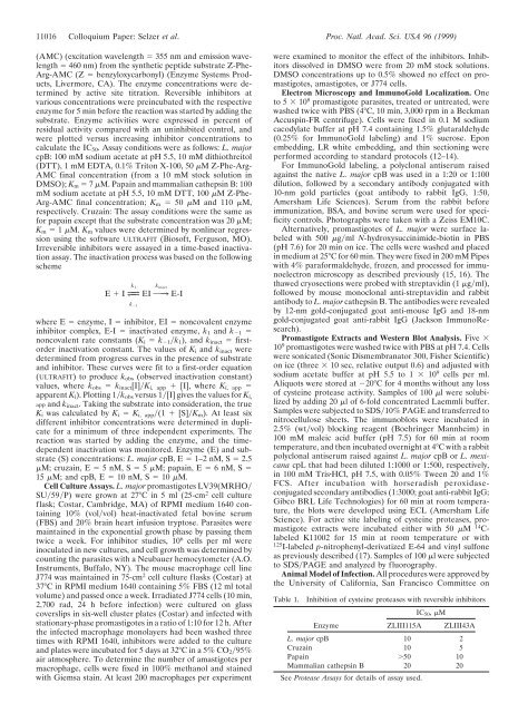
11016 Colloquium Paper: Selzer et al. Proc. Natl. Acad. Sci. USA 96 (1999)<br />
(AMC) (excitation wavelength � 355 nm and emission wavelength<br />
� 460 nm) from <strong>the</strong> syn<strong>the</strong>tic peptide substrate Z-Phe-<br />
Arg-AMC (Z � benzyloxycarbonyl) (Enzyme Systems Products,<br />
Livermore, CA). The enzyme concentrations were determined<br />
by active site titration. Reversible <strong>inhibitors</strong> at<br />
various concentrations were preincubated with <strong>the</strong> respective<br />
enzyme for 5 min before <strong>the</strong> reaction w<strong>as</strong> started by adding <strong>the</strong><br />
substrate. Enzyme activities were expressed in percent <strong>of</strong><br />
residual activity compared with an uninhibited control, and<br />
were plotted versus incre<strong>as</strong>ing inhibitor concentrations to<br />
calculate <strong>the</strong> IC50. Assay conditions were <strong>as</strong> follows: L. major<br />
cpB: 100 mM sodium acetate at pH 5.5, 10 mM dithiothreitol<br />
(DTT), 1 mM EDTA, 0.1% Triton X-100, 50 �M Z-Phe-Arg-<br />
AMC final concentration (from a 10 mM stock solution in<br />
DMSO); Km � 7 �M. Papain and mammalian ca<strong>the</strong>psin B: 100<br />
mM sodium acetate at pH 5.5, 10 mM DTT, 100 �M Z-Phe-<br />
Arg-AMC final concentration; Km � 50 �M and 110 �M,<br />
respectively. Cruzain: The <strong>as</strong>say conditions were <strong>the</strong> same <strong>as</strong><br />
for papain except that <strong>the</strong> substrate concentration w<strong>as</strong> 20 �M;<br />
Km � 1 �M. Km values were determined by nonlinear regression<br />
using <strong>the</strong> s<strong>of</strong>tware ULTRAFIT (Bios<strong>of</strong>t, Ferguson, MO).<br />
Irreversible <strong>inhibitors</strong> were <strong>as</strong>sayed in a time-b<strong>as</strong>ed inactivation<br />
<strong>as</strong>say. The inactivation process w<strong>as</strong> b<strong>as</strong>ed on <strong>the</strong> following<br />
scheme<br />
k1 E � I L|;<br />
k �1<br />
kinact EI O¡<br />
E-I<br />
where E � enzyme, I � inhibitor, EI � noncovalent enzyme<br />
inhibitor complex, E-I � inactivated enzyme, k1 and k�1 �<br />
noncovalent rate constants (Ki � k�1�k1), and kinact � firstorder<br />
inactivation constant. The values <strong>of</strong> Ki and kinact were<br />
determined from progress curves in <strong>the</strong> presence <strong>of</strong> substrate<br />
and inhibitor. These curves were fit to a first-order equation<br />
(ULTRAFIT) to produce kobs (observed inactivation constant)<br />
values, where kobs � kinact[I]�Ki, app � [I], where Ki, app �<br />
apparent Ki). Plotting 1�kobs versus 1�[I] gives <strong>the</strong> values for Ki,<br />
app and kinact. Taking <strong>the</strong> substrate into consideration, <strong>the</strong> true<br />
Ki w<strong>as</strong> calculated by Ki � Ki, app�(1 � [S]�Km). At le<strong>as</strong>t six<br />
different inhibitor concentrations were determined in duplicate<br />
for a minimum <strong>of</strong> three independent experiments. The<br />
reaction w<strong>as</strong> started by adding <strong>the</strong> enzyme, and <strong>the</strong> timedependent<br />
inactivation w<strong>as</strong> monitored. Enzyme (E) and substrate<br />
(S) concentrations: L. major cpB, E � 1–2 nM, S � 2.5<br />
�M; cruzain, E � 5 nM, S � 5 �M; papain, E � 6 nM, S �<br />
15 �M; and cpB, E � 10 nM, S � 10 �M.<br />
Cell Culture Assays. L. major prom<strong>as</strong>tigotes LV39(MRHO�<br />
SU�59�P) were grown at 27°C in 5 ml (25-cm 2 cell culture<br />
fl<strong>as</strong>k; Costar, Cambridge, MA) <strong>of</strong> RPMI medium 1640 containing<br />
10% (vol�vol) heat-inactivated fetal bovine serum<br />
(FBS) and 20% brain heart infusion tryptose. Par<strong>as</strong>ites were<br />
maintained in <strong>the</strong> exponential growth ph<strong>as</strong>e by p<strong>as</strong>sing <strong>the</strong>m<br />
twice a week. For inhibitor studies, 10 6 cells per ml were<br />
inoculated in new cultures, and cell growth w<strong>as</strong> determined by<br />
counting <strong>the</strong> par<strong>as</strong>ites with a Neubauer hemocytometer (A.O.<br />
Instruments, Buffalo, NY). The mouse macrophage cell line<br />
J774 w<strong>as</strong> maintained in 75-cm 2 cell culture fl<strong>as</strong>ks (Costar) at<br />
37°C in RPMI medium 1640 containing 5% FBS (12 ml total<br />
volume) and p<strong>as</strong>sed once a week. Irradiated J774 cells (10 min,<br />
2,700 rad, 24 h before infection) were cultured on gl<strong>as</strong>s<br />
coverslips in six-well cluster plates (Costar) and infected with<br />
stationary-ph<strong>as</strong>e prom<strong>as</strong>tigotes in a ratio <strong>of</strong> 1:10 for 12 h. After<br />
<strong>the</strong> infected macrophage monolayers had been w<strong>as</strong>hed three<br />
times with RPMI 1640, <strong>inhibitors</strong> were added to <strong>the</strong> culture<br />
and plates were incubated for 5 days at 32°C in a 5% CO2�95%<br />
air atmosphere. To determine <strong>the</strong> number <strong>of</strong> am<strong>as</strong>tigotes per<br />
macrophage, cells were fixed in 100% methanol and stained<br />
with Giemsa stain. At le<strong>as</strong>t 200 macrophages per experiment<br />
were examined to monitor <strong>the</strong> effect <strong>of</strong> <strong>the</strong> <strong>inhibitors</strong>. Inhibitors<br />
dissolved in DMSO were from 20 mM stock solutions.<br />
DMSO concentrations up to 0.5% showed no effect on prom<strong>as</strong>tigotes,<br />
am<strong>as</strong>tigotes, or J774 cells.<br />
Electron Microscopy and ImmunoGold Localization. One<br />
to 5 � 10 8 prom<strong>as</strong>tigote par<strong>as</strong>ites, treated or untreated, were<br />
w<strong>as</strong>hed twice with PBS (4°C, 10 min, 3,000 rpm in a Beckman<br />
Accuspin-FR centrifuge). Cells were fixed in 0.1 M sodium<br />
cacodylate buffer at pH 7.4 containing 1.5% glutaraldehyde<br />
(0.25% for ImmunoGold labeling) and 1% sucrose. Epon<br />
embedding, LR white embedding, and thin sectioning were<br />
performed according to standard protocols (12–14).<br />
For ImmunoGold labeling, a polyclonal antiserum raised<br />
against <strong>the</strong> native L. major cpB w<strong>as</strong> used in a 1:20 or 1:100<br />
dilution, followed by a secondary antibody conjugated with<br />
10-nm gold particles (goat antibody to rabbit IgG, 1:50,<br />
Amersham Life Sciences). Serum from <strong>the</strong> rabbit before<br />
immunization, BSA, and bovine serum were used for specificity<br />
controls. Photographs were taken with a Zeiss EM10C.<br />
Alternatively, prom<strong>as</strong>tigotes <strong>of</strong> L. major were surface labeled<br />
with 500 �g�ml N-hydroxysuccinimide-biotin in PBS<br />
(pH 7.6) for 20 min on ice. The cells were w<strong>as</strong>hed and placed<br />
in medium at 25°C for 60 min. They were fixed in 200 mM Pipes<br />
with 4% paraformaldehyde, frozen, and processed for immunoelectron<br />
microscopy <strong>as</strong> described previously (15, 16). The<br />
thawed cryosections were probed with streptavidin (1 �g�ml),<br />
followed by mouse monoclonal anti-streptavidin and rabbit<br />
antibody to L. major ca<strong>the</strong>psin B. The antibodies were revealed<br />
by 12-nm gold-conjugated goat anti-mouse IgG and 18-nm<br />
gold-conjugated goat anti-rabbit IgG (Jackson ImmunoResearch).<br />
Prom<strong>as</strong>tigote Extracts and Western Blot Analysis. Five �<br />
10 9 prom<strong>as</strong>tigotes were w<strong>as</strong>hed twice with PBS at pH 7.4. Cells<br />
were sonicated (Sonic Dismembranator 300, Fisher Scientific)<br />
on ice (three � 10 sec, relative output 0.6) and adjusted with<br />
sodium acetate buffer at pH 5.5 to 1 � 10 9 cells per ml.<br />
Aliquots were stored at �20°C for 4 months without any loss<br />
<strong>of</strong> cysteine <strong>prote<strong>as</strong>e</strong> activity. Samples <strong>of</strong> 100 �l were solubilized<br />
by adding 20 �l <strong>of</strong> 6-fold concentrated Laemmli buffer.<br />
Samples were subjected to SDS�10% PAGE and transferred to<br />
nitrocellulose sheets. The immunoblots were incubated in<br />
2.5% (wt�vol) blocking reagent (Boehringer Mannheim) in<br />
100 mM maleic acid buffer (pH 7.5) for 60 min at room<br />
temperature, and <strong>the</strong>n incubated overnight at 4°C with a rabbit<br />
polyclonal antiserum raised against L. major cpB or L. mexicana<br />
cpL that had been diluted 1:1000 or 1:500, respectively,<br />
in 100 mM Tris�HCl, pH 7.5, with 0.05% Tween 20 and 1%<br />
FCS. After incubation with horseradish peroxid<strong>as</strong>econjugated<br />
secondary antibodies (1:3000; goat anti-rabbit IgG;<br />
Gibco BRL Life Technologies) for 60 min at room temperature,<br />
<strong>the</strong> blots were developed using ECL (Amersham Life<br />
Science). For active site labeling <strong>of</strong> cysteine <strong>prote<strong>as</strong>e</strong>s, prom<strong>as</strong>tigote<br />
extracts were incubated ei<strong>the</strong>r with 50 �M 14 Clabeled<br />
K11002 for 15 min at room temperature or with<br />
125 I-labeled p-nitrophenyl-derivatized E-64 and vinyl sulfone<br />
<strong>as</strong> previously described (17). Samples <strong>of</strong> 100 �l were subjected<br />
to SDS�PAGE and analyzed by fluorography.<br />
Animal Model <strong>of</strong> Infection. All procedures were approved by<br />
<strong>the</strong> University <strong>of</strong> California, San Francisco Committee on<br />
Table 1. Inhibition <strong>of</strong> cysteine <strong>prote<strong>as</strong>e</strong>s with reversible <strong>inhibitors</strong><br />
IC50, �M<br />
Enzyme<br />
ZLIII115A ZLIII43A<br />
L. major cpB 10 2<br />
Cruzain 10 5<br />
Papain �50 10<br />
Mammalian ca<strong>the</strong>psin B 20 20<br />
See Prote<strong>as</strong>e Assays for details <strong>of</strong> <strong>as</strong>say used.