Cysteine protease inhibitors as chemotherapy - Proceedings of the ...
Cysteine protease inhibitors as chemotherapy - Proceedings of the ...
Cysteine protease inhibitors as chemotherapy - Proceedings of the ...

Create successful ePaper yourself
Turn your PDF publications into a flip-book with our unique Google optimized e-Paper software.
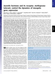
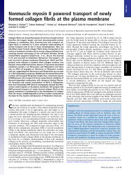
FIG. 3. Electron micrographs <strong>of</strong> Epon-embedded L. major prom<strong>as</strong>tigotes.<br />
Par<strong>as</strong>ites were untreated (A) or treated for 24 h with 50<br />
�M K11002 (B and C) or50�M ZLIII115A (D). Treatment <strong>of</strong> <strong>the</strong><br />
par<strong>as</strong>ite with ei<strong>the</strong>r inhibitor had very similar effects, resulting in <strong>the</strong><br />
appearance <strong>of</strong> diverse multivesicular and dense bodies (arrowheads),<br />
lipid inclusions (arrows), and myelin figures (<strong>as</strong>terisk). n, Nucleus; g,<br />
Golgi apparatus; f, flagellar pocket; m, mitochondrion; k, kinetopl<strong>as</strong>t.<br />
(Bars � 10 �m.)<br />
appeared within <strong>the</strong> abnormally dilated par<strong>as</strong>ite lysosomes and<br />
<strong>the</strong> flagellar pocket (Fig. 3), <strong>the</strong> site where endocytosis and<br />
exocytosis takes place (22). These abnormalities resemble<br />
alterations seen in lysosomal storage dise<strong>as</strong>es caused by <strong>the</strong><br />
deficiency or absence <strong>of</strong> specific lysosomal hydrol<strong>as</strong>es (23).<br />
The nucleus and <strong>the</strong> Golgi apparatus were not affected, but<br />
some cells showed dilated mitochondria. In <strong>the</strong>se latter cells<br />
<strong>the</strong> kinetopl<strong>as</strong>t DNA w<strong>as</strong> no longer condensed but appeared<br />
in diffuse patches. No effects on treated mammalian host cells<br />
at <strong>the</strong> light microscopic or ultr<strong>as</strong>tructural level were observed.<br />
C<br />
Colloquium Paper: Selzer et al. Proc. Natl. Acad. Sci. USA 96 (1999) 11019<br />
A B<br />
E F<br />
D<br />
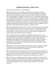
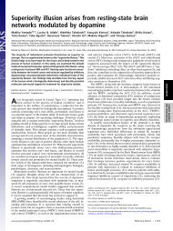
To localize <strong>the</strong> Leishmania cysteine <strong>prote<strong>as</strong>e</strong>s within <strong>the</strong><br />
par<strong>as</strong>ite cell, ImmunoGold electron microscopic analysis using<br />
a L. major cpB-specific antiserum w<strong>as</strong> performed. In untreated<br />
cells <strong>the</strong> gold label appeared only in lysosomes (Fig. 4 C and<br />
D). Treated cells were more heavily labeled in <strong>the</strong> dilated<br />
lysosome�endosome compartment and in <strong>the</strong> flagellar pocket<br />
(Fig. 4 E and F). Apparently empty flagellar pockets were also<br />
heavily labeled in treated par<strong>as</strong>ites, but not in untreated<br />
par<strong>as</strong>ites. To confirm target <strong>prote<strong>as</strong>e</strong> localization at <strong>the</strong> site <strong>of</strong><br />
inhibitor-induced abnormalities, untreated prom<strong>as</strong>tigotes<br />
were surface-labeled with N-hydroxysuccinimide-biotin and<br />
placed back in culture to facilitate internalization <strong>of</strong> labeled<br />
proteins. This method allows visualization <strong>of</strong> <strong>the</strong> endosomal�<br />
lysosomal network <strong>of</strong> <strong>the</strong> cells. ImmunoGold electron microscopy<br />
<strong>of</strong> <strong>the</strong>se par<strong>as</strong>ites revealed an abundance <strong>of</strong> ca<strong>the</strong>psin B<br />
in <strong>the</strong> flagellar pocket and in vesicles subtending that structure<br />
(Fig. 4 A and B). Some <strong>of</strong> <strong>the</strong>se vesicles contained biotinylated<br />
proteins, indicating that <strong>the</strong>y are endosomes or lysosomes,<br />
where<strong>as</strong> o<strong>the</strong>rs contained only ca<strong>the</strong>psin B, suggesting that<br />
<strong>the</strong>y may be secretory vesicles.<br />
Because <strong>of</strong> <strong>the</strong> selective arrest <strong>of</strong> par<strong>as</strong>ite versus host cell<br />
growth by <strong>inhibitors</strong> added to cultures, <strong>the</strong> efficacy <strong>of</strong> <strong>the</strong><br />
cysteine <strong>prote<strong>as</strong>e</strong> <strong>inhibitors</strong> in vivo w<strong>as</strong> evaluated in Leishmania-infected<br />
BALB�c mice. Twenty-four hours after infection,<br />
mice received intraperitoneal injections <strong>of</strong> K11002 or<br />
ZLIII115A dissolved in DMSO�H2O (70:30). By 2 weeks,<br />
control mice had already developed footpad lesions, which<br />
progressed in size and severity. In treated animals <strong>the</strong> lesion<br />
development w<strong>as</strong> significantly delayed, with no swelling <strong>of</strong> <strong>the</strong><br />
footpads until 3–4 weeks (Figs. 5 and 6). After 4 weeks <strong>of</strong><br />
treatment, inhibitor dosing w<strong>as</strong> stopped, and lesion development<br />
paralleled that seen in control mice. At <strong>the</strong> end <strong>of</strong> <strong>the</strong><br />
treatment period, whole footpad histology and limiting dilution<br />
<strong>as</strong>says <strong>of</strong> par<strong>as</strong>ites from extracted footpad tissues showed<br />
par<strong>as</strong>ite burden for treated animals w<strong>as</strong> at le<strong>as</strong>t two logs lower<br />
than that <strong>of</strong> <strong>the</strong> untreated animals (10 �7 versus 10 �5 ). This<br />
finding is consistent with results from previous studies that<br />
documented <strong>the</strong> correlation between footpad size and numbers<br />
<strong>of</strong> par<strong>as</strong>ites (18, 20). None <strong>of</strong> <strong>the</strong> compounds produced<br />
toxic effects in mice, <strong>as</strong> indicated by daily observation <strong>of</strong><br />
weight, activity, and appearance, <strong>as</strong> well <strong>as</strong> autopsy and<br />
histologic analysis. Also, <strong>the</strong>re w<strong>as</strong> no evidence <strong>of</strong> a switch<br />
from <strong>the</strong> usual TH2 cytokine response to Leishmania in<br />
FIG. 4. Immunoelectron micrographs <strong>of</strong> L. major<br />
prom<strong>as</strong>tigotes. (A and B) Electron micrographs <strong>of</strong> cryosections<br />
from L. major prom<strong>as</strong>tigotes that were surface<br />
biotinylated with N-hydroxysuccinimide-biotin and incubated<br />
in medium for 45 min prior to fixation. The<br />
sections were probed with streptavidin�mouse antistreptavidin<br />
mAb (12-nm gold particle conjugated to<br />
goat anti-mouse IgG) and rabbit anti-ca<strong>the</strong>psin B antibody<br />
(18-nm gold particle conjugated to goat anti-rabbit<br />
IgG). g, Golgi apparatus; e, endodome; n, nucleus; k,<br />
kinetopl<strong>as</strong>t; m, mitochondrion. (Bars � 0.25 �m.) (A)<br />
Ca<strong>the</strong>psin B label is observed in vesicles in <strong>the</strong> vicinity<br />
<strong>of</strong> <strong>the</strong> Golgi apparatus and in <strong>the</strong> flagellar pocket, which<br />
is strongly positive for biotinylated proteins. (B) Label<br />
is also observed in biotin-positive vesicles, or endosomes<br />
that subtend <strong>the</strong> flagellar pocket. The data indicated<br />
that ca<strong>the</strong>psin B is syn<strong>the</strong>sized and proceeds through <strong>the</strong><br />
Golgi apparatus into <strong>the</strong> secretory network, where it<br />
gains access to <strong>the</strong> flagellar pocket. (C–F) LR whiteembedded<br />
and ImmunoGold-labeled (anti-L. major cpB<br />
antiserum) prom<strong>as</strong>tigotes. Note <strong>the</strong> specific labeling in<br />
lysosomes <strong>of</strong> untreated cells (C and D) and in multivesicular<br />
bodies (arrowheads), <strong>as</strong> well <strong>as</strong> in <strong>the</strong> flagellar<br />
pocket <strong>of</strong> treated cells (E and F). (Bars � 0.5 �m.)