La Clasificacio´n IC3D de Las Distrofias Corneales - Cornea Society
La Clasificacio´n IC3D de Las Distrofias Corneales - Cornea Society
La Clasificacio´n IC3D de Las Distrofias Corneales - Cornea Society

Create successful ePaper yourself
Turn your PDF publications into a flip-book with our unique Google optimized e-Paper software.
Weiss et al Córnea Volumen 27, Supl. 2, Diciembre 2008<br />
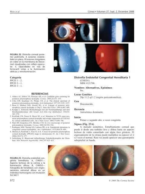
FIGURA 22. Distrofia corneal posterior<br />
polimorfa. A Lesiones endoteliales<br />
en placa. B Lesiones irregulares<br />
en cráter en la membrana <strong>de</strong> Descemet<br />
visualizadas con reflejo especular.<br />
C Opacida<strong>de</strong>s en vías <strong>de</strong><br />
ferrocarril vistas a la iluminación<br />
oblicua y retroiluminación.<br />
Categoría<br />
PPCD 1—2.<br />
PPCD 2—1.<br />
PPCD 3—1.<br />
REFERENCIAS<br />
1. Aldave AJ, Yellore VS, Principe AH, et al. Candidate gene screening for<br />
posterior polymorphous dystrophy. <strong>Cornea</strong>. 2005;24:151–155.<br />
2. Cibis GW, Krachmer JA, Phelps CD, et al. The clinical spectrum of<br />
posterior polymorphous dystrophy. Arch Ophthalmol. 1977;95:1529–1537.<br />
3. Heon E, Mathers WD, Alward WL, et al. Linkage of posterior polymorphous<br />
corneal dystrophy to 20q11. Hum Mol Genet. 1995;4:485–488.<br />
4. Koeppe L. Klinische Beobachtungen mit <strong>de</strong>r Nernstspaltlampe und <strong>de</strong>m<br />
Hornhautmikroskop. Albr Graefe Arch Klin Exp Ophthalmol. 1916;91:<br />
363–379.<br />
5. Krafchak CM, Pawar H, Moroi SE, et al. Mutations in TCF8 cause posterior<br />
polymorphous corneal dystrophy and ectopic expression of COL4A3<br />
by corneal endothelial cells. Am J Hum Genet. 2005;77:694–708.<br />
6. Patel DV, Grupcheva CN, McGhee CNJ. In vivo microscopy of posterior<br />
polymorphous dystrophy. <strong>Cornea</strong>. 2005;24:550–554.<br />
7. Rodrigues MM, Waring GO, <strong>La</strong>ibson PR, et al. Endothelial alterations in<br />
congenital corneal dystrophies. Am J Ophthalmol. 1975;80:678–689.<br />
8. Shimizu S, Krafchak C, Fuse N, et al. A locus for posterior polymorphous<br />
corneal dystrophy (PPCD3) maps to chromosome 10. Am J Med Genet.<br />
2004;130:372–377.<br />
9. Schlichting H. Blasen-und <strong>de</strong>llenförmige Endotheldystrophie <strong>de</strong>r Hornhaut.<br />
Klin Monatsbl Augenkeilkd. 1941;107:425–435.<br />
Distrofia Endotelial Congenital Hereditaria 1<br />
(CHED1).<br />
MIM #121700.<br />
Nombres Alternativos, Epónimos<br />
No.<br />
Locus Genético<br />
20p 11.2–q11.2 (región pericentromérica).<br />
Gen<br />
Desconocido.<br />
Herencia<br />
Autosómica dominante.<br />
Inicio<br />
Primer o segundo año, a veces congénita.<br />
Signos (Fig. 23A)<br />
A menudo asimétrico. Enturbiamiento corneal que<br />
pue<strong>de</strong> ir <strong>de</strong>s<strong>de</strong> una turbi<strong>de</strong>z leve y difusa hasta un aspecto<br />
lechoso <strong>de</strong> vidrio esmerilado con algún foco grisáceo. El<br />
engrosamiento <strong>de</strong> la córnea pue<strong>de</strong> multiplica su espesor x2 ó<br />
x3 veces el normal. Rara vez pue<strong>de</strong> aparecer una queratopatía<br />
subepitelial en banda.<br />
FIGURA 23. Distrofia endotelial congénita<br />
hereditaria. A, CHED1—<br />
Aspecto lechoso <strong>de</strong> la córnea a la<br />
iluminación difusa. B, CHED2—<br />
Fotografía <strong>de</strong> hendidura con engrosamiento<br />
estromal difuso en un<br />
individuo homocigoto con mutación<br />
SLC4A11<br />
S72<br />
q 2008 The <strong>Cornea</strong> <strong>Society</strong>