Occlusione degli pseudoaneurismi femorali ... - ConsultiMedici.it
Occlusione degli pseudoaneurismi femorali ... - ConsultiMedici.it
Occlusione degli pseudoaneurismi femorali ... - ConsultiMedici.it

You also want an ePaper? Increase the reach of your titles
YUMPU automatically turns print PDFs into web optimized ePapers that Google loves.
RADIOLOGIA VASCOLARE<br />
385<br />
La Radiologia Medica - Radiol Med 108: 385-393, 2004<br />
Edizioni Minerva Medica - Torino<br />
<strong>Occlusione</strong> <strong>degli</strong><br />
<strong>pseudoaneurismi</strong> <strong>femorali</strong> postcateterismo<br />
mediante iniezione percutanea<br />
di trombina sotto guida ecografica<br />
Rocco CORSO - Antonio RAMPOLDI - Franco RIOLO*<br />
Gianpaolo CARRAFIELLO** - Marco SOLCIA<br />
Marcello INTOTERO*** - Angelo VANZULLI<br />
Scopo. L’incidenza di <strong>pseudoaneurismi</strong> (PSA) iatrogeni dell’arteria femorale<br />
è compresa tra 1-7 % di tutti i cateterismi percutanei. I trattamenti tradizionali<br />
consistono nella compressione color-Doppler guidata o nella<br />
riparazione chirurgica. Riportiamo la nostra esperienza nell’occlusione<br />
<strong>degli</strong> PSA <strong>femorali</strong> postcateterismo mediante iniezione percutanea di<br />
trombina con guida eco-color-Doppler.<br />
Materiale e metodi. Dal giugno 2000 abbiamo trattato consecutivamente<br />
31 PSA in 30 pazienti, bilaterale in un caso, di età compresa tra<br />
45-81 anni, con diagnosi clinica di PSA femorale postcateterismo confermato<br />
in tutti i casi all’esame eco-color-Doppler. Abbiamo iniettato<br />
soluzione di trombina di origine bovina alla concentrazione di 1000<br />
U/mL, utilizzando aghi da 21-22 gauge, per via percutanea sotto guida<br />
eco-color-Doppler. Tutti i pazienti hanno esegu<strong>it</strong>o un controllo clinico<br />
ed eco-color-Doppler prima, durante ed a 24 ore dalla procedura ed un<br />
follow-up a 1 e 3 mesi.<br />
Risultati. Il tasso di successo primario è stato dell’83,8% ottenendo<br />
occlusione completa e persistente dello PSA in media in meno di 20<br />
secondi ed impiegando dosi medie di trombina di 880 U (0,8 mL di soluzione).<br />
In 5 casi (16,1%) si è osservata riperfusione dello PSA a distanza<br />
di 24 ore r<strong>it</strong>rattato iniettando ulteriori dosi di trombina. Il risultato<br />
finale è stato del 96,7% (30 su 31 casi) e non si è osservata alcuna complicanza<br />
di tipo tromboembolico. Immediatamente dopo l’iniezione il<br />
22,5% dei pazienti ha lamentato unicamente una fugace sensazione di<br />
calore in corrispondenza dell’arto trattato risoltasi spontaneamente e completamente<br />
nell’arco di qualche minuto.<br />
Conclusioni. Il trattamento <strong>degli</strong> PSA <strong>femorali</strong> postcateterismo con iniezione<br />
percutanea di trombina e guida eco-color-Doppler per la sua semplic<strong>it</strong>à<br />
d’impiego, sicurezza, efficacia e basso costo è da considerarsi terapia<br />
di elezione.<br />
PAROLE CHIAVE: Angiografia, complicanze - Pseudoaneurismi - Guida ecografica.<br />
Occlusion of postcatheterisation femoral<br />
pseudoaneurysms w<strong>it</strong>h percutaneous thrombin<br />
injection under ultrasound guidance<br />
Purpose. The incidence of iatrogenic femoral artery pseudoaneurysms<br />
is reported to occur in 1-7% by of all percutaneous<br />
catheterisations. These pseudoaneurysms are trad<strong>it</strong>ionally<br />
treated by ultrasound-guided compression or<br />
surgical repair. We report our experience in sealing<br />
postcatheterization femoral pseudoaneurysms w<strong>it</strong>h percutaneous<br />
thrombin injection under colour-Doppler ultrasound<br />
guidance.<br />
Materials and methods. Since June 2000 we have consecutively<br />
treated 31 pseudoaneurysms in 30 patients,<br />
(14 males and 16 females, age range 45 to 81 years); in one<br />
patient the pseudoaneurysm was bilateral. All patients<br />
had a clinical diagnosis of postcatheterization femoral<br />
pseudoaneurysm, later confirmed by colour-Doppler ultrasonography.<br />
We injected a bovine thrombin solution percutaneously<br />
at a concentration of 1000 U/mL using 21-22<br />
gauge needles under colour Doppler ultrasound guidance.<br />
All patients underwent clinical and colour-Doppler<br />
US examination before, during and 24 hours after the<br />
procedure and were followed up after 1 and 3 months.<br />
Results. The primary success rate was 83.8%. Complete<br />
and persistent occlusion of the pseudoaneurysm was<br />
achieved in less than 20 seconds by administering an<br />
average dose of 880 U of thrombin (0.8 mL of solution).<br />
In 5 cases (16.1%) reperfusion of the pseudoaneurysm<br />
was observed w<strong>it</strong>hin 24 hours. These patients underwent<br />
a repeat procedure. The final result was successful in<br />
96.7% of patients (30 of 31 cases). No thromboembolic<br />
complication was observed. Only 22.5% of patients reported<br />
a heat sensation in the treated limb, which resolved<br />
spontaneously w<strong>it</strong>hin minutes.<br />
Conclusions. The percutaneous injection of thrombin<br />
under ultrasound colour-Doppler guidance should be<br />
regarded as the first choice treatment for postcatheterization<br />
femoral pseudoaneurysms, owing to <strong>it</strong>s simplic<strong>it</strong>y,<br />
safety, effectiveness and inexpensiveness.<br />
KEY WORDS: Angiography, complications - Pseudoaneurysm -<br />
Ultrasound guidance.<br />
Introduzione<br />
Negli ultimi anni il successo sempre più evidente delle<br />
procedure endovascolari sia periferiche che coronariche<br />
rispetto alle alternative chirurgiche e le continue innovazioni<br />
tecnologiche hanno portato ad un allargamento delle indicazioni<br />
con realizzazione di procedure sempre più impegnative<br />
e complesse. Questo comporta maggiore utilizzo di<br />
sistemi introduttori e/o cateteri guida di ampio calibro, ele-<br />
Introduction<br />
Over the past few years, the growing success of peripheral<br />
and coronary endovascular procedures over surgical alternatives<br />
and the continuous technological innovations have<br />
extended the indications leading to the performance of<br />
increasingly challenging and complicated procedures. This<br />
involves an increased use of introducing systems and/or large<br />
guidance catheters, high doses of intravenous heparin (≥ 70<br />
Radiologia Diagnostica ed Interventistica - *Chirurgia Vascolare - Ospedale Niguarda Cà Granda - Milano - **Univers<strong>it</strong>à dell’Insubria - Varese -<br />
***Univers<strong>it</strong>à <strong>degli</strong> Studi - Milano.<br />
Pervenuto alla Redazione il 2.9.2003; revisionato il 4.9.2003; rest<strong>it</strong>u<strong>it</strong>o corretto il 3.12.2003; accettato per la pubblicazione l’1.3.2004.<br />
Indirizzo per la richiesta di estratti: Dott. R. Corso - Ospedale Niguarda Cà Granda - Piazza Ospedale Maggiore, 3 - 20162 Milano MI - Tel. 02/64442793<br />
- Fax 02/64442881. E-mail: roccocorso@jumpy.<strong>it</strong>
386 R. Corso et al: <strong>Occlusione</strong> <strong>degli</strong> <strong>pseudoaneurismi</strong> <strong>femorali</strong> postcateterismo<br />
vati dosaggi di eparina endovenosa (≥ 70 U/kg), trattamenti<br />
più aggressivi con farmaci fibrinol<strong>it</strong>ici o antiaggreganti<br />
piastrinici per via parenterale, con conseguente aumentato<br />
rischio di complicanze e danni iatrogeni, anche locali, legati<br />
al cateterismo arterioso tra cui gli <strong>pseudoaneurismi</strong> (PSA)<br />
dell’arteria femorale hanno un’incidenza stimabile tra l’1-7%,<br />
soprattutto se vengono ricercati sistematicamente con eco-<br />
Doppler [1]. Molti di questi PSA sono subclinici e si risolvono<br />
spontaneamente in particolare se di piccole dimensioni (diametro<br />
1,5 cm). Per i rimanenti, al fine di ev<strong>it</strong>are il rischio di<br />
una loro rottura, il trattamento tradizionale era di tipo chirurgico<br />
mentre in casi selezionati si impiegavano misure di<br />
tipo conservativo o endovascolare, quali embolizzazione con<br />
spirali metalliche o colle, esclusione con stent ricoperti o<br />
altre procedure più o meno complesse [2], a loro volta a<br />
rischio di ulteriori complicanze legate alle necessarie manovre<br />
di cateterismo.<br />
Dal 1991 dopo l’originale descrizione di Fellmeth [3] la<br />
compressione color-Doppler guidata si è imposta come trattamento<br />
di prima scelta negli PSA postcateterismo dell’arteria<br />
femorale per una netta riduzione dei rischi e dei costi associati<br />
alla riparazione chirurgica. Attualmente questa tecnica<br />
mininvasiva è impiegata nella maggior parte dei centri come<br />
prima linea nel trattamento <strong>degli</strong> PSA iatrogeni.<br />
Essa consiste in una prolungata compressione esterna sul<br />
colletto dello PSA con la sonda ecografica impiegata nella<br />
funzione color-Doppler per il mon<strong>it</strong>oraggio flussimetrico in<br />
real-time della procedura. Questa manovra comporta notevole<br />
rallentamento del flusso con stasi di sangue all’interno della<br />
camera dello PSA che es<strong>it</strong>a in trombosi della sacca.<br />
Impiegando questa tecnica vengono riportati tassi di successo<br />
variabili tra il 60-90% [4]. Tuttavia alcune importanti<br />
lim<strong>it</strong>azioni sono legate al disagio e spesso alla scarsa tollerabil<strong>it</strong>à<br />
del paziente ad una prolungata ed intensa compressione,<br />
all’affaticabil<strong>it</strong>à dell’operatore, allo scarso o nullo<br />
successo nei pazienti in concom<strong>it</strong>ante terapia anticoagulante<br />
[5]. Inoltre occasionalmente durante la manovra di compressione<br />
si possono verificare dolore intenso (che necess<strong>it</strong>a<br />
di sedazione profonda), trombosi dell’adiacente vena femorale,<br />
necrosi cutanea, infezioni o rottura dello PSA [6].<br />
Recentemente l’iniezione percutanea eco-guidata di trombina,<br />
sostanza nota per essere un potente attivatore della<br />
trombosi, è stata proposta come valida alternativa nel trattamento<br />
<strong>degli</strong> PSA <strong>femorali</strong> postcateterismo [7, 8].<br />
Scopo del nostro lavoro è stato valutare efficacia e sicurezza<br />
dell’iniezione percutanea di trombina nel trattamento <strong>degli</strong><br />
PSA iatrogeni <strong>femorali</strong>.<br />
Materiale e metodi<br />
Dal giugno 2000 abbiamo trattato consecutivamente 31<br />
PSA in 30 pazienti (1 caso bilaterale), 14 maschi e 16 femmine,<br />
di età compresa tra 45 e 81 anni, con diagnosi clinica<br />
e conferma eco-color-Doppler di PSA femorale iatrogeno, nella<br />
total<strong>it</strong>à dei casi come conseguenza di cateterismo dell’arteria<br />
femorale per esecuzione di procedure diagnostiche e/o<br />
interventistiche cardiologiche.<br />
In tutti i pazienti è stato ottenuto consenso informato dopo<br />
che sono state spiegate le modal<strong>it</strong>à tecniche della procedura<br />
e le alternative terapeutiche; nessun paziente aveva allergie<br />
U/kg), more aggressive treatments w<strong>it</strong>h parenterally delivered<br />
fibrinolytic or antiplatelet drugs, w<strong>it</strong>h a resulting higher risk<br />
of complications and iatrogenic damage, even local, linked<br />
to arterial catheterism. Among these, femoral artery pseudoaneurysms<br />
have an estimated incidence between 1% and<br />
7%, especially if they are systematically searched for by<br />
using Doppler ultrasound [1]. Many of these pseudoaneurysms,<br />
particularly if small (1.5 cm in diameter) are subclinical<br />
and resolve spontaneously. For the remaining ones<br />
requiring treatment to prevent rupture, trad<strong>it</strong>ional treatment<br />
was by surgery or in selected cases by conservative or endovascular<br />
procedures such as embolization w<strong>it</strong>h metal coils or<br />
glues, exclusion w<strong>it</strong>h coated stents or other more or less<br />
complex procedures [2], which in turn carry a risk of further<br />
complications resulting from catheterization manoeuvres.<br />
After Fellmeth’s original description in 1991 [3], colour-<br />
Doppler guided compression became established as the treatment<br />
of choice for femoral postcatheterisation pseudoaneurysms,<br />
because of the lower risks and costs involved compared<br />
to surgical repair. At present this minimally invasive technique<br />
is employed in most medical centres as the first-line treatment<br />
of iatrogenic pseudoaneurysms.<br />
The technique consists of a protracted external compression<br />
on the neck of the pseudoaneurysm w<strong>it</strong>h the ultrasound<br />
probe being used in colour-Doppler mode for real-time mon<strong>it</strong>oring<br />
of flow. The manoeuvre considerably slows the flow<br />
and creates blood stasis inside the pseudoaneurysm cav<strong>it</strong>y,<br />
thereby thrombosing the sac. The reported success rate ranges<br />
from 60% to 90% [4]. There are, however, important lim<strong>it</strong>ations,<br />
such as patient discomfort and often poor tolerance<br />
of the protracted intense compression, operator fatigue, poor<br />
success rates or failure of the procedure in patients undergoing<br />
anticoagulant therapy [5]. Furthermore, the compression<br />
manoeuvre may at times cause severe pain (requiring deep<br />
sedation), thrombosis of the adjacent femoral vein, skin<br />
necrosis, infections o rupture of the pseudoaneurysm [6].<br />
More recently, the US-guided percutaneous injection of<br />
thrombin, a substance known as a powerful promoter of<br />
thrombosis, has been proposed as an alternative in the<br />
treatment of postcatetherization femoral pseudoaneurysms<br />
[7, 8].<br />
The purpose of our study was to evaluate the efficacy and<br />
safety of the percutaneous injection of thrombin in the treatment<br />
of iatrogenic femoral pseudoaneurysms.<br />
Materials and methods<br />
Since June 2000 we have consecutively treated 31 pseudoaneurysms<br />
in 30 patients (bilateral in one case), 14 men and<br />
16 women, aged between 45 and 81 years, w<strong>it</strong>h a clinical<br />
diagnosis of iatrogenic femoral pseudoaneurysm confirmed<br />
by colour-Doppler US. All pseudoaneurysms were a consequence<br />
of femoral artery catheterization for diagnostic and/or<br />
interventional cardiological procedures.<br />
Informed consent was obtained from all patients after<br />
explaining the technique and possible alternatives. No patient<br />
had known allergies to bovine-derived products nor had<br />
undergone previous treatment w<strong>it</strong>h bovine-derived thrombin<br />
for other reasons.<br />
For each pseudoaneurysm we evaluated the origin, mor-
R. Corso et al: <strong>Occlusione</strong> <strong>degli</strong> <strong>pseudoaneurismi</strong> <strong>femorali</strong> postcateterismo 387<br />
note a prodotti di derivazione bovina ed in precedenza non era<br />
già stato trattato per altre patologie con trombina di origine<br />
bovina.<br />
Per ogni singolo PSA sono stati valutati: origine, morfologia,<br />
dimensioni, volume e profond<strong>it</strong>à; è stato identificato il<br />
colletto e la sua posizione rispetto all’arteria nativa; sono<br />
stati inoltre misurati il diametro e la lunghezza. Prima e sub<strong>it</strong>o<br />
dopo l’iniezione di trombina sono stati mon<strong>it</strong>orati i polsi<br />
periferici ed i segni e sintomi d’ischemia d’arto quali temperatura,<br />
colore e dolore al fine di rilevare immediatamente<br />
i potenziali rischi di embolizzazione periferica.<br />
Ai pazienti, posti in posizione supina, veniva preparata<br />
sterilmente la cute della regione inguinale da trattare. In nessun<br />
caso è stata utilizzata sedazione profonda o anestesia<br />
locale.<br />
Abbiamo impiegato trombina di origine bovina (D-Stat<br />
Flowable Hemostat, Vascular Solution - NGC Medical Italia)<br />
miscelando la trombina in polvere (5000 U) con 5 mL di<br />
diluente ottenendo una soluzione di trombina alla concentrazione<br />
di 1000 U/mL. Dal volume globale così formato (5<br />
mL) venivano, a seconda delle necess<strong>it</strong>à, prelevate 1000 U per<br />
volta impiegando per l’iniezione siringhe da 1 mL che consentivano<br />
un preciso e lento rilascio di trombina. Come guida<br />
ecografica sono state utilizzate due apparecchiature con<br />
funzioni color-Doppler: Acuson 128 XP (Acuson Computer<br />
Sonography, Mountain View, California, USA) e ATL HDI<br />
5000 (Advanced Technology Laboratories, Bothell, Washington,<br />
USA), impiegando sonde lineari da 7,5 Mhz, adattatore<br />
per biopsie e traccia elettronica (fig. 1). Sono stati utilizzati<br />
aghi sottili da 21-22 gauge eco-visibili introdotti per via<br />
percutanea sotto guida ecografia in funzione B-mode in quanto<br />
con questa modal<strong>it</strong>à la punta dell’ago risultava meglio<br />
visibile (fig. 2B). Si è sempre cercato di posizionare la punta<br />
dell’ago il più lontano possibile dal colletto: la conferma<br />
che l’ago fosse inser<strong>it</strong>o correttamente all’interno della camera<br />
dello PSA la si otteneva rimuovendone lo stiletto ed osservando<br />
la fuoriusc<strong>it</strong>a di sangue pulsante; quindi si attivava la<br />
funzione colore dell’ecografo per il mon<strong>it</strong>oraggio in realtime<br />
dell’infusione di trombina e della formazione del trombo.<br />
La soluzione veniva infusa molto lentamente interrompendo<br />
l’iniezione una volta osservata la formazione di trombo<br />
ecogeno che riempiva completamente la cav<strong>it</strong>à dello PSA<br />
in assenza di segnale colore (fig. 2C). Se veniva identificata<br />
una persistenza di flusso l’ago con stiletto veniva riposizionato<br />
ed iniettata ulteriore dose di trombina sino a completa<br />
trombosi dello PSA. Dopo la procedura il paziente rimaneva<br />
a letto per almeno 6 ore.<br />
Tutti i pazienti hanno esegu<strong>it</strong>o controllo clinico ed ecocolor-Doppler<br />
prima, durante e dopo 24 ore dalla procedura.<br />
Se a quest’ultimo controllo si osservava riperfusione dello<br />
PSA la procedura veniva ripetuta. In caso di successo il<br />
paziente poteva essere dimesso il giorno stesso ed eseguiva<br />
valutazione clinica ed eco-Doppler a distanza di 1 e 3 mesi.<br />
Risultati<br />
CUTE/SKIN<br />
COLLETTO<br />
NECK<br />
AFC<br />
SONDA<br />
TRANSDUCER<br />
PSA<br />
AGO/NEEDLE<br />
AFS<br />
AFP<br />
Fig. 1. — Disegno schematico raffigurante l’inserimento percutaneo dell’ago<br />
nella cav<strong>it</strong>à dello PSA con guida ecografica utilizzando un adattatore<br />
per biopsie: la punta dell’ago è posizionata lontana dal colletto e l’iniezione<br />
di trombina viene mon<strong>it</strong>orata in real-time con la sonda ecografica in funzione<br />
color-Doppler. PSA: pseudoaneurisma. AFC: arteria femorale comune.<br />
AFS: arteria femorale superficiale. AFP: arteria femorale profonda.<br />
Schematic drawing representing percutaneous insertion of the needle in<br />
the pseudoaneurysm under ultrasound guidance using an attachable biopsy-guide:<br />
the needle tip is placed far away from the neck and the thrombin<br />
injection is real-time mon<strong>it</strong>ored w<strong>it</strong>h color-Doppler sonography. PSA:<br />
pseudoaneurysm. AFC: common femoral artery. AFS: superficial femoral<br />
artery. AFP: profunda femoral artery.<br />
Le principali caratteristiche dei pazienti sono riassunte<br />
nella tabella I.<br />
Il diametro <strong>degli</strong> PSA era compreso tra 11 e 52 mm (media<br />
30 mm), la maggior parte di essi originava dall’arteria femophology,<br />
size, volume and depth; we identified the neck and<br />
<strong>it</strong>s pos<strong>it</strong>ion relative to the native artery, and measured the<br />
diameter and length. Before and immediately after the thrombin<br />
injection, we mon<strong>it</strong>ored the peripheral pulses and the<br />
signs and symptoms of limb ischaemia, such as temperature,<br />
colour and pain, in order to immediately detect the potential<br />
risks of peripheral embolization.<br />
The patients were placed supine, and the skin of the inguinal<br />
region was prepared. In no case was deep sedation or<br />
local anaesthesia used in a sterile fashion.<br />
We used bovine-derived thrombin (D-Stat Flowable<br />
Hemostat, Vascular Solution - NGC Medical Italy) mixing the<br />
thrombin powder (5000 U) w<strong>it</strong>h 5 ml of diluent to obtain a<br />
thrombin solution at a concentration of 1,000 U/mL. From this<br />
total volume (5 mL), we drew amounts of 1,000 U at a time<br />
—as needed—using 1 mL syringes for the injection so as to<br />
obtain an accurate and slow release of thrombin. Two US<br />
un<strong>it</strong>s w<strong>it</strong>h colour-Doppler functions were used for guidance:<br />
Acuson 128 XP (Acuson Computer Sonography, Mountain<br />
View, California, USA) and ATL HDI 5000 (Advanced<br />
Technology Laboratories, Bothell, Washington, USA), w<strong>it</strong>h 7.5<br />
Mhz linear-array probes, biopsy adapter and electronic tracing<br />
(fig. 1). Thin 21-22 gauge needles, visible at ultrasonography,<br />
were introduced percutaneously under ultrasound<br />
guidance, in B-mode function, since this mode made the tip
388 R. Corso et al: <strong>Occlusione</strong> <strong>degli</strong> <strong>pseudoaneurismi</strong> <strong>femorali</strong> postcateterismo<br />
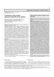
Fig. 2. — Pseudoaneurisma femorale iatrogeno. A) Eco-color-Doppler prima del trattamento mostra il tipico flusso turbolento<br />
all’interno della camera dello pseudoaneurisma, parzialmente trombizzato (frecce). B) Ecografia B-mode durante introduzione<br />
di ago da 21 gauge consente di meglio visualizzare la punta (freccia). C) Eco-color-Doppler esegu<strong>it</strong>o sub<strong>it</strong>o dopo infusione di<br />
600 U di trombina mostra assenza di flusso all’interno dello PSA: la cav<strong>it</strong>à è riemp<strong>it</strong>a da materiale ecogeno rappresentante il trombo<br />
fresco.<br />
Iatrogenic femoral pseudoaneurysm. A) Color-Doppler sonography performed before the treatment shows typical swirling flow<br />
into the pseudoaneurysm sac, partially thrombosed (arrows). B) Using of B-mode ultrasound during placement of the 21-gauge<br />
needle allows clearer visualization of the needle tip (arrows). C) Color-Doppler ultrasound obtained immediately after<br />
injection of 600 U of thrombin shows no flow w<strong>it</strong>hin the pseudoaneurysm; the sac is filled w<strong>it</strong>h echogenic material representing<br />
fresh thrombus.<br />
TABELLA I. — Caratteristiche di base dei pazienti sottoposti ad iniezione<br />
percutanea di trombina.<br />
Età, anni 62 (45-81)<br />
Maschi, N. (%) 14 (46,6)<br />
Cateterismo cardiaco, N. (%)<br />
— Diagnostico 17 (54,8)<br />
— Terapeutico (PTCA/stent) 14 (45,2)<br />
Calibro medio introduttore, French 7 (6-9)<br />
Origine pseudoaneurisma, N. (%)<br />
— Arteria femorale comune 18 (58,1)<br />
— Arteria femorale superficiale 10 (32,3)<br />
— Arteria femorale profonda 3 (9,6)<br />
Diametro medio cav<strong>it</strong>à, mm 30 (11-52)<br />
Sottoposti a terapia con anticoagulanti e/o antipiastri- 23 (76,6)<br />
nici, N. (%)<br />
TABLE I.—Baseline characteristics of patients undergoing percutaneous<br />
thrombin injection.<br />
Age, years 62 (45-81)<br />
Male gender, N. (%) 14 (46,6)<br />
Cardiac catheterization, N. (%)<br />
— Diagnostic 17 (54,8)<br />
— Therapeutic (PTCA/stent) 14 (45,2)<br />
Mean sheath size, French 7 (6-9)<br />
Origin of pseudoaneurysm, N. (%)<br />
— Common femoral artery 18 (58,1)<br />
— Superficial femoral artery 10 (32,3)<br />
— Profunda femoral artery 3 (9,6)<br />
Mean cav<strong>it</strong>y diameter, mm 30 (11-52)<br />
Underwent antiplatelet or anticoagulant therapies 23 (76,6)<br />
N. (%)<br />
rale comune ed era di tipo uniloculare. Soltanto in 4 casi<br />
(12,9%) era già presente spontaneamente all’interno della<br />
cav<strong>it</strong>à una sottile stratificazione trombotica; in 2 casi concom<strong>it</strong>ava<br />
fistola artero-venosa, anch’essa legata al cateterismo<br />
femorale ed in 1 caso lo PSA era presente bilateralmente<br />
come conseguenza di doppio cateterismo. La maggior<br />
parte dei pazienti (76,6%) assumeva terapia antiaggregante<br />
piastrinica o anticoagulante (tabella II).<br />
of the needle better visible (fig. 2B). In all cases we tried to<br />
place the tip of the needle as far from the neck as possible:<br />
confirmation of correct insertion of the needle inside the<br />
pseudoaneurysm cav<strong>it</strong>y was provided on removing the stylet<br />
and observing the outflow of pulsating blood; the colour<br />
function of ultrasound un<strong>it</strong> was then activated to mon<strong>it</strong>or<br />
the infusion of thrombin and production of the thrombus in<br />
real-time. The solution was infused very slowly and the injec-
R. Corso et al: <strong>Occlusione</strong> <strong>degli</strong> <strong>pseudoaneurismi</strong> <strong>femorali</strong> postcateterismo 389<br />
TABELLA II. — Terapia antiaggregante o anticoagulante assunta al<br />
momento dell’iniezione di trombina.<br />
N. pazienti<br />
Aspirina 6<br />
Aspirina + ticlopidina idrocloride 7<br />
Ticlopidina idrocloride 4<br />
Warfarin 3<br />
Eparina 3<br />
Totale 23<br />
TABLE II.—Antiplatelet or anticoagulation therapy used at the time<br />
of thrombin injection.<br />
No. patients<br />
Aspirin 6<br />
Aspirin + ticlopidine hydrochloride 7<br />
Ticlopidine hydrochloride 4<br />
Warfarin 3<br />
Heparin 3<br />
Total 23<br />
La trombina è stata iniettata in media a distanza di 5 giorni<br />
dal cateterismo femorale (range 3-29 giorni). In 26 casi si<br />
è ottenuta trombizzazione completa dello PSA in meno di 20<br />
secondi con un’unica iniezione di trombina e dose media di<br />
880 U (0,88 mL), con tasso di successo primario dell’83,8%.<br />
In 5 casi (16,1%) si è osservata riperfusione dello PSA a<br />
distanza di 24 ore r<strong>it</strong>rattato iniettando ulteriori dosi di trombina<br />
(in media
390 R. Corso et al: <strong>Occlusione</strong> <strong>degli</strong> <strong>pseudoaneurismi</strong> <strong>femorali</strong> postcateterismo<br />
Fig. 3. — Arteriografia selettiva. Pseudoaneurisma postcateterismo<br />
dell’arteria femorale superficiale destra: prima (A)<br />
e sub<strong>it</strong>o dopo (B) iniezione percutanea di 400 U di trombina.<br />
Completa occlusione della PSA con pervietà del lume dei<br />
vasi nativi.<br />
Selective arteriography. Right postcatheterization superficial<br />
femoral pseudoaneurysm: before (A) and immediately<br />
after (B) injection of 400 U of thrombin. Complete occlusion<br />
of the pseudoaneurysm while native vessels remained patent.<br />
come dolore inguinale, anemia, compressione sulle strutture<br />
vascolo-nervose adiacenti, tromboembolia distale fino<br />
anche la rottura del PSA. La causa principale della loro<br />
formazione risiede in una inadeguata compressione manuale<br />
dopo rimozione dell’introduttore vascolare. Fattori concom<strong>it</strong>anti<br />
sono legati ad un s<strong>it</strong>o di puntura troppo «basso»<br />
(spesso sulla biforcazione femorale o sull’arteria femorale<br />
superficiale o profonda), al calibro dell’introduttore<br />
impiegato (soprattutto se ≥7 French), all’alto dosaggio di<br />
farmaci anticoagulanti e/o antiaggreganti somministrati<br />
per via endovenosa durante o sub<strong>it</strong>o dopo la procedura<br />
endovascolare. La diagnosi è essenzialmente clinica (ematoma<br />
pulsante in sede inguinale) e strumentale con ecocolor-Doppler<br />
che evidenzia all’interno della sacca pseudoaneurismatica<br />
il classico pattern flussimetrico tipo «to<br />
and fro» legato ai fenomeni di turbolenza e di ricircolo<br />
del sangue (fig. 2A).<br />
La trombina è un potente attivatore della trombosi ed agisce<br />
nella parte terminale della cascata coagulativa. La sua<br />
principale azione consiste nel trasformare il fibrinogeno circolante<br />
in fibrina la quale polimerizza istantaneamente cost<strong>it</strong>uendo<br />
la matrice sulla quale si depos<strong>it</strong>eranno piastrine ed<br />
altri fattori coagulatori circolanti con formazione finale del<br />
trombo. Inoltre la trombina attiva il fattore XIII (che stabilizza<br />
la fibrina formandone una struttura estremamente resistente),<br />
stimola l’aggregazione piastrinica, attiva i fattori<br />
coagulatori VIII e V ed accelera la conversione della protrombina<br />
circolante in ulteriore trombina (feed-back pos<strong>it</strong>ivo).<br />
Essa non si lega ai fattori della cascata coagulatoria,<br />
che sono bersaglio delle terapie anticoagulanti ed è per questo<br />
che teoricamente il suo effetto terapeutico non viene<br />
influenzato dal concom<strong>it</strong>ante uso di questi farmaci che invece<br />
è causa di fallimento della compressione color-Dopplerguidata<br />
[5].<br />
La prima descrizione nella coagulazione di PSA perifein<br />
both procedures. It should be noted that in the 20 patients<br />
treated over the last year (66.6% of cases), thrombosis was<br />
obtained w<strong>it</strong>h a mean dose of less than 500 U (range 200-700<br />
U). No significant differences in the response to thrombin<br />
injection based on size, morphology and occlusion time, nor<br />
above all between patients taking or not taking anticoagulant<br />
or antiplatelet drugs which could in theory have interfered<br />
w<strong>it</strong>h the process thrombosis.<br />
In 7 cases (22.5%), during or immediately after the treatment,<br />
pain and/or a transient sensation of heat was reported<br />
distal to the treated limb, which resolved spontaneously<br />
w<strong>it</strong>hin seconds. After the thrombin injection no significant<br />
alterations or irregular<strong>it</strong>ies of the lumen of the native vessels<br />
were observed (fig. 3). We did not observe any allergic reactions<br />
or local complications at the injection s<strong>it</strong>e, or distal<br />
thromboembolic events (e<strong>it</strong>her immediate or late), or reperfusions<br />
of the pseudoaneurysm during the follow-up.<br />
Discussion<br />
The incidence of symptomatic femoral pseudoaneurysms<br />
after arterial catheterization is low, and may be estimated to<br />
be around 1% of all vascular procedures, both diagnostic<br />
and interventional [1]. Nonetheless, these pseudoaneurysms<br />
can cause the patient significant morbid<strong>it</strong>y, such as inguinal<br />
pain, anaemia, compression of the contiguous vascularnerve<br />
structures, distal thromboembolism, or even rupture of<br />
the pseudoaneurysm. The main cause of pseudoaneurysms is<br />
inadequate manual compression after removing the vascular<br />
sheat. Concurrent factors are too “low” a puncture s<strong>it</strong>e (often<br />
on the femoral bifurcation or on the superficial or deep femoral<br />
artery), inadequate caliber of the sheat (especially when<br />
7 French), and high dose of anticoagulation and/or antiplatelet<br />
drugs administered intravenously during or immediately<br />
after the endovascular procedure. The diagnosis is
R. Corso et al: <strong>Occlusione</strong> <strong>degli</strong> <strong>pseudoaneurismi</strong> <strong>femorali</strong> postcateterismo 391<br />
rici con trombina iniettata mediante puntura diretta è opera<br />
di Cope e Ze<strong>it</strong> [9] nel 1986. Successivamente venne applicata<br />
da Walker [10] nel 1987 per il trattamento di uno PSA<br />
dell’arteria femorale profonda mediante iniezione di trombina<br />
intravascolare attraverso un catetere angiografico.<br />
Tuttavia è solo nel 1997 ad opera di Liau [11] che vengono<br />
descr<strong>it</strong>ti i primi cinque casi di PSA <strong>femorali</strong> postcateterismo<br />
trattati con successo mediante soluzione di trombina bovina<br />
(1000 U/mL) iniettata attraverso un catetere per via transluminale.<br />
Tale tecnica venne successivamente modificata,<br />
perfezionata e soprattutto semplificata nel 1998 ad opera di<br />
Kang et al. [7] che per primi iniettarono soluzione di trombina<br />
direttamente nello PSA per via percutanea impiegando<br />
la guida ecografica. In questo modo hanno trattato consecutivamente<br />
20 su 21 PSA <strong>femorali</strong> postcateterismo riportando<br />
un tasso di successo globale del 95% in assenza di<br />
complicanze tromboemboliche e senza nessuna recidiva a<br />
distanza di tempo.<br />
Da allora in letteratura sono comparsi diversi articoli con<br />
risultati molto simili tra loro sia per quanto riguarda l’alto tasso<br />
di successo che la bassissima o nulla incidenza di complicanze<br />
[12]. Una recente revisione della letteratura ad opera<br />
di Friedman riporta che sul totale di 418 casi trattati in<br />
diversi Centri con iniezione percutanea di trombina si è ottenuta<br />
permanente trombizzazione dello PSA in 410 casi con<br />
tasso di successo di quasi il 99% [13].<br />
Questa nuova e semplice modal<strong>it</strong>à di trattamento <strong>degli</strong><br />
PSA <strong>femorali</strong> nella nostra serie ha comportato un’efficacia<br />
del 96,7% senza nessuna complicanza di tipo tromboembolico;<br />
in molti casi la trombosi è avvenuta istantaneamente<br />
ed in modo indipendente dalla contemporanea assunzione<br />
di farmaci antiaggreganti piastrinici o anticoagulanti<br />
orali come peraltro già evidenziato in precedenti studi<br />
[14].<br />
L’unico insuccesso si è osservato in una paziente con PSA<br />
biconcamerato (3×3,5 e 3,2×1,5 cm), associato ad un’ampia<br />
fistola artero-venosa tra arteria e vena femorale superficiale.<br />
Nonostante la trombizzazione raggiunta al termine di<br />
ogni seduta il controllo color-Doppler a distanza di 24 ore evidenziava<br />
per tre volte consecutive riperfusione parziale dello<br />
PSA sempre in prossim<strong>it</strong>à del colletto e pertanto si è deciso,<br />
data la sede e la presenza della fistola artero-venosa, di trattarlo,<br />
con successo, per via endovascolare posizionando uno<br />
stent ricoperto.<br />
In letteratura vengono riportate pochissime e spesso aneddotiche<br />
complicanze tromboemboliche causate dall’iniezione<br />
inavvert<strong>it</strong>a di trombina direttamente nel colletto dello<br />
PSA o nell’arteria nativa e dall’iniezione di eccessiva quant<strong>it</strong>à<br />
di trombina rispetto al volume dello PSA. La risoluzione<br />
della trombosi a volte è stata spontanea [15, 16] mentre in<br />
altri casi sono stati impiegati farmaci anticoagulanti [8] o<br />
fibrinol<strong>it</strong>ici [13, 17]. In alcuni casi si è ricorsi alla trombectomia<br />
per via chirurgica [18, 19].<br />
Al fine di ridurre al minimo possibile queste complicanze<br />
è di estrema importanza accertarsi che la punta dell’ago sia<br />
effettivamente all’interno dello PSA, lontana dal colletto e<br />
soprattutto che l’iniezione di trombina venga esegu<strong>it</strong>a molto<br />
lentamente, sotto continuo mon<strong>it</strong>oraggio color-Doppler,<br />
interrompendo l’iniezione non appena si osserva la formazione<br />
del trombo.<br />
Sette pazienti della nostra casistica, durante o sub<strong>it</strong>o dopo<br />
essentially clinical (pulsatile haematoma in the groin) and<br />
instrumental w<strong>it</strong>h colour-Doppler US showing the classical<br />
“to and fro” flow pattern inside the pseudoaneurysmatic<br />
sac related to turbulence and blood recirculation (fig. 2A).<br />
Thrombin is a powerful promoter of thrombosis and acts<br />
in the end phase of the coagulation cascade. Its main effect<br />
is to convert circulating fibrinogen into fibrin, which immediately<br />
polymerizes, cons<strong>it</strong>uting the matrix onto which platelets<br />
and other circulating coagulation factors will depos<strong>it</strong>,<br />
thereby forming the thrombus. Furthermore, thrombin activates<br />
factor XIII (which stabilizes the fibrin, giving rise to a<br />
very resistant structure), stimulates platelet aggregation,<br />
activates coagulation factors VIII and V and accelerates the<br />
conversion of circulating prothrombin into more thrombin<br />
(pos<strong>it</strong>ive feed-back). Not binding to factors of the coagulation<br />
cascade—the targets of anticoagulation therapies—<strong>it</strong>s<br />
therapeutic action is not in theory affected by the concurrent<br />
use of these drugs, which is instead responsible for the<br />
failure of colour-Doppler guided compression [5].<br />
Coagulation of peripheral pseudoaneurysms by directly<br />
injected thrombin was first reported by Cope and Ze<strong>it</strong> [9] in<br />
1986. The technique was later applied by Walker [10] in 1987<br />
to treat a pseudoaneurysm of the deep femoral artery by means<br />
of an intravascular thrombin injection and an angiographic<br />
catheter. Nonetheless, only in 1997 did Liau [11] describe the<br />
first five cases of postcatetherization femoral pseudoaneurysms<br />
successfully treated by bovine-derived thrombin solution<br />
(1,000 U/mL) injected via a transluminal catheter. This technique<br />
was subsequently modified, perfected and, more importantly,<br />
simplified in 1998 by Kang et al [7], the first to inject<br />
a thrombin solution percutaneously directly into the pseudoaneurysm<br />
by using ultrasound guidance. They consecutively<br />
treated 20 out of 21 postcatheterization femoral pseudoaneurysms,<br />
reporting an overall success rate of 95% w<strong>it</strong>h ne<strong>it</strong>her<br />
thromboembolic complications nor late recurrences.<br />
Since then, a number of papers have appeared in the l<strong>it</strong>erature<br />
w<strong>it</strong>h very similar results, in terms of both success rates<br />
and low, or null, incidence of complications [12]. A recent<br />
review of the l<strong>it</strong>erature by Friedman reports that in over 418<br />
cases treated at different centres, percutaneous thrombin<br />
injection induced permanent thrombosis of the pseudoaneurysm<br />
in 410 cases, w<strong>it</strong>h a success rate of almost 99% [13].<br />
This new and simple technique to treat femoral pseudoaneurysms<br />
in our series was effective in 96.7% of cases, w<strong>it</strong>h no<br />
thromboembolic complications; in many cases thrombosis<br />
was immediate, regardless of concurrent treatment w<strong>it</strong>h oral<br />
anticoagulant or antiplatelet drugs, as has already been<br />
shown by previous studies [14].<br />
The only failure was observed in a female patient w<strong>it</strong>h a<br />
lobulated pseudoaneurysm (3×3.5 and 3.2×1.5 cm), associated<br />
w<strong>it</strong>h a large arteriovenous fistula between the superficial<br />
femoral artery and vein. Desp<strong>it</strong>e the thrombosis obtained<br />
at the end of each session, the colour Doppler scans after 24<br />
hours showed partial reperfusion of the pseudoaneurysm<br />
three times in a row always near the neck. Given the s<strong>it</strong>e and<br />
the presence of the arteriovenous fistula, we therefore decided<br />
to treat <strong>it</strong> intravascularly by placing a coated stent, and<br />
obtained success.<br />
There are very few, and often anecdotal, reports in the l<strong>it</strong>erature<br />
on thromboembolic complications caused by the<br />
inadvertent injection of thrombin directly into the neck of
392 R. Corso et al: <strong>Occlusione</strong> <strong>degli</strong> <strong>pseudoaneurismi</strong> <strong>femorali</strong> postcateterismo<br />
l’iniezione di trombina, hanno avvert<strong>it</strong>o un lieve dolore e/o<br />
una fugace sensazione di calore distalmente all’arto trattato<br />
a risoluzione spontanea pressocchè immediata. Questi disturbi<br />
possono essere ricondotti al cosiddetto traboccamento o<br />
«spillage» causato dal possibile passaggio di piccole quant<strong>it</strong>à<br />
di trombina, attraverso il colletto dello PSA, all’interno<br />
dei vasi nativi. Questo tuttavia non comporta significative<br />
complicanze immediate o a distanza in quanto sia l’alto flusso<br />
del circolo sistemico e quindi l’immediata diluizione della<br />
trombina, sia le piccole quant<strong>it</strong>à di trombina impiegate, in<br />
presenza di fattori endogeni anticoagulanti di tipo endoteliale<br />
e circolante (trombomodulina, ant<strong>it</strong>rombina III), impediscono<br />
lo sviluppo di trombosi accidentale [15, 20, 21].<br />
Nell’ultimo anno, grazie all’esperienza maturata ed all’impiego<br />
di dosi sempre più basse di trombina, nessun paziente<br />
ha più avvert<strong>it</strong>o tali disturbi.<br />
La soluzione di trombina ad alta concentrazione (1000<br />
U/mL), iniettata direttamente in una camera contenente sangue<br />
a basso flusso (quale quello circolante all’interno dello<br />
PSA), comporta un’immediata conversione nella sua forma<br />
solida (trombo) che si arresta spontaneamente in prossim<strong>it</strong>à<br />
del colletto. A sua volta il colletto per le piccole dimensioni,<br />
tortuos<strong>it</strong>à ed alto flusso (legato alla circolazione sistemica),<br />
ostacola ulteriormente il reflusso di trombina all’interno del<br />
vaso nativo.<br />
La trombina di origine bovina essendo una sostanza estranea<br />
può causare reazioni di tipo allergico, in alcuni casi<br />
anche di tipo anafilattico. Sheldon [22] riporta un caso di reazione<br />
orticariode generalizzata, periodica e prolungata per<br />
circa 4 settimane, causata secondo gli autori dal possibile<br />
interm<strong>it</strong>tente e protratto rilascio di trombina intrappolata<br />
nello PSA man mano che il trombo veniva riassorb<strong>it</strong>o (anche<br />
se non era possibile escludere una reazione idiopatica da<br />
causa sconosciuta). Pope e Johnston [23] riportano il caso di<br />
un paziente in trattamento emodial<strong>it</strong>ico esposto più volte<br />
alla trombina in quanto utilizzata come emostatico dopo<br />
rimozione <strong>degli</strong> aghi da dialisi. Il paziente, dopo iniezione<br />
di 500 U di trombina per il trattamento di uno PSA femorale,<br />
ha sviluppato una reazione anafilattica grave che ha necess<strong>it</strong>ato,<br />
per la risoluzione, trattamento in terapia intensiva;<br />
successivamente l’allergia alla trombina è stata accertata<br />
dal prick-test. Da qui la necess<strong>it</strong>à di accertare per ogni<br />
paziente note allergie o precedenti esposizioni a prodotti di<br />
origine bovina.<br />
Studi ematologici hanno evidenziato che pazienti sottoposti<br />
a ripetute esposizioni di trombina di origine bovina per via<br />
topica, ad esempio per accelerare l’emostasi nel corso di<br />
sanguinamento da varici esofagee per via endoscopica o nel<br />
corso di interventi di cardiochirurgia o neurochirurgia, possono<br />
sviluppare anticorpi diretti contro proteine di origine<br />
bovina tra cui vi sono i fattori della coagulazione [24]. In<br />
alcuni di questi pazienti occasionalmente sono stati dimostrati<br />
anticorpi IgM e IgG diretti verso il fattore V bovino, contenuto<br />
nelle preparazioni di trombina, con cross-reattiv<strong>it</strong>à con<br />
quello di origine umana e conseguente defic<strong>it</strong> di fattore V<br />
con possibile sviluppo di coagulopatie più o meno gravi [25].<br />
Attualmente in letteratura non sono state segnalate reazioni<br />
di questo tipo in pazienti sottoposti a trombizzazione di PSA<br />
<strong>femorali</strong>.<br />
L’estrema rar<strong>it</strong>à di queste reazioni ha recentemente indotto<br />
alcuni gruppi di studio ad impiegare e testare soluzioni di<br />
the pseudoaneurysm or into the native artery and by the<br />
injection of an excessive amount of thrombin for the voloume<br />
of the pseudoaneurysm. In some cases the thrombosis resolved<br />
spontaneously [15, 16], but in others anticoagulation [8] or<br />
fibrinolytic [13, 17] drugs were used. In some cases surgical<br />
thrombectomy was necessary [18, 19].<br />
In order to minimize these complications, <strong>it</strong> is very important<br />
to make sure that the tip of the needle is actually inside<br />
the pseudoaneurysm and far from the neck and above all that<br />
the injection of thrombin is performed very slowly, under continuous<br />
colour Doppler mon<strong>it</strong>oring, and stopping the injection<br />
as soon as the thrombus can be seen to develop.<br />
Seven patients of our series, during or immediately after<br />
the thrombin injection, experienced mild pain and/or a transient<br />
heat sensation distal to the treated limb, which resolved<br />
spontaneously almost immediately. These disorders can be<br />
traced back to the spillage of small amounts of thrombin<br />
through the pseudoaneurysm neck into the native vessels.<br />
This however does not entail significant immediate or delayed<br />
complications since both the high flow of systemic circulation<br />
and the immediate dilution of thrombin, and the small<br />
amounts used, in the presence of endogenous endothelial<br />
and circulating anticoagulation factors (thrombomodulin,<br />
ant<strong>it</strong>hrombin III), prevent the development of incidental<br />
thrombosis [15, 20, 21]. Over the last year, thanks to the<br />
experience gained and the use of increasingly low doses of<br />
thrombin, no patient has experienced such disorders.<br />
High concentration thrombin solution (1,000 U/mL), injected<br />
directly into a cav<strong>it</strong>y containing low-flow blood (such as<br />
that circulating w<strong>it</strong>hin the pseudoaneurysm), involves immediate<br />
conversion into <strong>it</strong>s solid form (thrombus) which stops<br />
spontaneously at the neck. The neck <strong>it</strong>self, given <strong>it</strong>s small size,<br />
tortuosness and high flow (related to systemic circulation), further<br />
obstructs the reflux of thrombin inside the native vessel.<br />
Bovine-derived thrombin, being a foreign substance, may<br />
cause allergic and in some cases even anaphylactic reactions.<br />
Sheldon [22] reports on one case of generalized urticarial<br />
reaction, periodic and lasting around 4 weeks apparently<br />
caused by the possible interm<strong>it</strong>tent and protracted<br />
release of thrombin trapped in the pseudoaneurysm as the<br />
thrombus was being reabsorbed (although an idiopathic<br />
reaction of unknown origin could not be excluded). Pope<br />
and Johnston [23] report the case of a patient undergoing<br />
haemodialysis who was repeatedly exposed to thrombin, as<br />
<strong>it</strong> was used as a haemostatic agent after removing the dialysis<br />
needles. The patient, after receiving 500 U of thrombin<br />
to treat a femoral pseudoaneurysm, developed a severe anaphylactic<br />
reaction requiring intensive care; an allergy to thrombin<br />
was subsequently established by prick-test. Hence the<br />
need to establish any known allergies or previous exposure<br />
to bovine-derived products for each patient.<br />
Haematological studies have shown that patients repeatedly<br />
exposed locally to bovine-derived thrombin, for example<br />
to accelerate haemostasis during bleeding from esophageal<br />
varices treated endoscopically or during heart or brain<br />
surgery, may develop antibodies against bovine-derived proteins<br />
including coagulation factors [24]. Some of these<br />
patients occasionally showed IgM and IgG antibodies against<br />
bovine factor V, contained in thrombin preparations, w<strong>it</strong>h<br />
cross-reactiv<strong>it</strong>y w<strong>it</strong>h the human factor V, and resulting factor<br />
V defic<strong>it</strong>, w<strong>it</strong>h the possible development of more or less<br />
severe coagulopathies [25]. No such reactions in patients
R. Corso et al: <strong>Occlusione</strong> <strong>degli</strong> <strong>pseudoaneurismi</strong> <strong>femorali</strong> postcateterismo 393<br />
trombina a più bassa concentrazione (100-500 U/mL) [26] o<br />
trombina a differente origine quale quella di derivazione<br />
umana [27, 28] o autologa [29]. Tuttavia nel caso di soluzioni<br />
di trombina a bassa concentrazione, per infondere le stesse<br />
un<strong>it</strong>à terapeutiche, è necessario incrementare il volume globale<br />
di soluzione iniettata (esponendo il paziente a maggior<br />
rischio teorico di complicanze) mentre quelle di origine non<br />
bovina hanno costi più elevati in quanto la trombina umana<br />
è derivata da tecniche di ingegneria genetica mentre quella<br />
autologa necess<strong>it</strong>a del coinvolgimento di un laboratorio di<br />
ematologia per la separazione della trombina dagli altri emocomponenti.<br />
Conclusioni<br />
I nostri risultati evidenziano come negli <strong>pseudoaneurismi</strong><br />
postcateterismo dell’arteria femorale il trattamento innovativo<br />
mediante iniezione percutanea di trombina sotto guida<br />
eco-color-Doppler è da considerarsi terapia di prima scelta<br />
rispetto alle alternative terapeutiche per la sua ridottissima<br />
invasiv<strong>it</strong>à, rapid<strong>it</strong>à d’azione, sicurezza ed efficacia, con conseguente<br />
accettabil<strong>it</strong>à sia da parte del paziente che dell’operatore.<br />
undergoing thrombosis of femoral pseudoaneurysms have<br />
been reported in the l<strong>it</strong>erature.<br />
The extreme rar<strong>it</strong>y of these reactions recently prompted<br />
some research groups to use and test thrombin solutions w<strong>it</strong>h<br />
lower concentrations (100- 500 U/mL) [26] or human-derived<br />
[27, 28] or autologous thrombin [29]. However, in the case<br />
of low concentration thrombin solutions, to infuse the same<br />
therapeutic un<strong>it</strong>s, the overall volume of the injected solution<br />
has to be increased (exposing the patients to a theoretical<br />
higher risk of complications) while non-bovine thrombin is<br />
more expensive, since human thrombin is derived from genetical<br />
engineering while autologous thrombin requires a haematology<br />
laboratory to separate thrombin from the other<br />
blood components.<br />
Conclusions<br />
Our results show that in postcatheterization pseudoaneurysms<br />
of the femoral artery, the innovative treatment by percutaneously<br />
injecting thrombin under colour-Doppler US guidance<br />
should be considered the treatment of choice as <strong>it</strong> is minimally<br />
invasive, acts rapidly, <strong>it</strong> is safe and efficacious, and is consequently<br />
well accepted by both the patient and the operator.<br />
Bibliografia/References<br />
1) Katzenschlager R, Ugurluoglu A,<br />
Armadi A et al: Incidence of pseudoaneurysm<br />
after diagnostic and therapeutic<br />
angiography. Radiology 195: 463-466,<br />
1995.<br />
2) Marcello R, Cortese F, Mangialardi N<br />
et al: Una nuova metodica interventistica<br />
per il trattamento <strong>degli</strong> <strong>pseudoaneurismi</strong><br />
iatrogeni. Radiol Med 105: 63-68,<br />
2003.<br />
3) Fellmeth BD, Roberts AC, Bookstein<br />
JJ et al: Postangiographic femoral artery<br />
injuries: nonsurgical repair w<strong>it</strong>h US-guided<br />
compression. Radiology 178: 671-<br />
675, 1991.<br />
4) Anguissola R, Bramucci E, Campani<br />
R: Pseudoaneurisma dell’arteria femorale<br />
dopo procedure angiografiche. Trattamento<br />
non-chirurgico con compressione<br />
eco-guidata. Radiol Med 85: 280-282,<br />
1993.<br />
5) Lewis DR, Davies AH, Irvine CD et al:<br />
Compression ultrasonography for false<br />
femoral artery aneurysms: hypocoagulabil<strong>it</strong>y<br />
is a cause of failure. Eur J Vasc<br />
Endovasc Surg 16: 427-428, 1998.<br />
6) Hilborn M, Downey D: Deep venous<br />
thrombosis complicating sonographically<br />
guided compression repair of the common<br />
femoral artery. AJR 161: 1334-1335,<br />
1993.<br />
7)Kang SS, Labropoulos N, Mansour A<br />
et al: Percutaneous ultrasound guided<br />
thrombin injection: a new method for<br />
treating postcatheterisation femoral pseudoaneurysms.<br />
J Vasc Surgery 27: 1032-<br />
1038, 1998.<br />
8) La Perna DO, Olin JW, Goines D et<br />
al: Ultrasound-guided thrombin injection<br />
for the treatment of postcatheterisation<br />
pseudoaneurysms. Circulation 102:<br />
2391-2395, 2000.<br />
9) Cope C, Ze<strong>it</strong> R: Coagulation of aneurysms<br />
by direct percutaneous thrombin<br />
injection. AJR 147: 383-387, 1986.<br />
10) Walker TG, Geller SC, Brewster DC:<br />
Transcatheter occlusion of a profunda<br />
femoral pseudoaneurysm using thrombin.<br />
AJR 149: 185-186, 1987.<br />
11) Liau CS, Ho FM, Chen MF et al:<br />
Treatment of iatrogenic femoral pseudoaneurysm<br />
w<strong>it</strong>h percutaneous thrombin<br />
injection. J Vasc Surg 26: 18-23, 1997.<br />
12) Powell A, Benenati JF, Becker GJ et<br />
al: Percutaneous-guided thrombin injection<br />
for the treatment of pseudoaneurysms.<br />
J Am Coll Surg 194: S53-S57, 2002.<br />
13) Friedman SG, Peller<strong>it</strong>o JS, Scher L et<br />
al: Ultrasound-guided injection is the<br />
treatment of choice for femoral pseudoaneurysms.<br />
Arch Surg 137: 462-464,<br />
2002.<br />
14) Lennox AF, Delis KT, Szendro G et<br />
al: Duplex-guided thrombin injection for<br />
iatrogenic femoral artery pseudoaneurysm<br />
is effective even in anticoagulated<br />
patients. Br J Surg 87: 796-801, 2000.<br />
15) Pezzullo JA, Dupuy DE, Cronan JJ:<br />
Percutaneous injection of thrombin for<br />
the treatment of pseudoaneurysms after<br />
catheterisation: an alternative to sonographically<br />
guided compression. AJR<br />
175: 1035-1040, 2000.<br />
16) Paulson EK, Nelson RC, Mayes CE<br />
et al: Sonographically guided thrombin<br />
injection of iatrogenic femoral pseudoaneurysms:<br />
further experience of a single<br />
inst<strong>it</strong>ution. AJR 177: 309-316, 2001.<br />
17) Sadiq S, Ibrahim W: Thromboembolism<br />
complicating thrombin injection of<br />
femoral artery pseudoaneurysm: management<br />
w<strong>it</strong>h intrarterial thrombolysis. J<br />
Vasc Intervent Radiol 12: 633-636, 2001.<br />
18) Sackett WR, Taylor SM, Coffey CB<br />
et al: Ultrasound-guided thrombin injection<br />
of iatrogenic femoral pseudoaneurysms:<br />
a prospective analysis. Am<br />
Surg 66: 937-940, 2000.<br />
19) Calton WC, Franklin DP, Elmore JR<br />
et al: Ultrasound-guided thrombin injection<br />
is a safe and durable treatment for<br />
femoral pseudoaneurysms. Vasc Surg 35:<br />
379-383, 2001.<br />
20) Morrison SL, Obrand DA, Steinmetz<br />
OK et al: Treatment of femoral artery<br />
pseudoaneurysms w<strong>it</strong>h percutaneous<br />
thrombin injection. Ann Vasc Surg 14:<br />
634-639, 2000.<br />
21) Krüger K, Zähringer M, Söhngen F et<br />
al: Femoral pseudoaneurysms: management<br />
w<strong>it</strong>h percutaneous thrombin injection<br />
- success rates and effect on systemic<br />
coagulation. Radiology 226: 452-458,<br />
2003.<br />
22) Sheldon PJ, Oglevie SB, Kaplan LA:<br />
Prolonged generalized urticarial reaction<br />
after percutaneous thrombin injection for<br />
treatment of a femoral artery pseudoaneurysm.<br />
J Vasc Interv Radiol 11: 759-<br />
761, 2000.<br />
23) Pope M, Johnston KW: Anaphylaxis<br />
after thrombin injection of a femoral<br />
pseudoaneurysm: recommendations for<br />
prevention. J Vasc Surg 32: 190-191,<br />
2000.<br />
24) Ortel TL, Charles LA, Keller FG et al:<br />
Topical thrombin and acquired coagulation<br />
factor inhib<strong>it</strong>ors: clinical spectrum<br />
and laboratory diagnosis. Am J Hematol<br />
45: 128-135, 1994.<br />
25) Chonhan VD, De la Cadena RA,<br />
Nagaswamic C: Simultaneous occurrence<br />
of human antibodies directed against<br />
fibrinogen, thrombin and factor V following<br />
the exposure to bovine thrombin:<br />
effects on blood coagulation, platelet C<br />
activation and platelet function. Thromb<br />
Haemost 77: 343-349, 1997.<br />
26) Reeder SB, Widlus DM, Lazinger M:<br />
Low-dose thrombin injection to treat<br />
iatrogenic femoral artery pseudoaneurysms.<br />
AJR 177: 595-598, 2001.<br />
27) Elford J, Burrell C, Freeman S,<br />
Roobottom C: Human thrombin injection<br />
for the percutaneous treatment of<br />
iatrogenic pseudoaneurysms. Cardiovasc<br />
Intervent Radiol 25: 115-118, 2002.<br />
28) Maleux G, Hendrickx S, Vaninbroukx<br />
et al: Percutaneous injection of human<br />
thrombin to treat iatrogenic femoral pseudoaneurysms:<br />
short- and midterm ultrasound<br />
follow-up. Eur Radiol 13: 209-<br />
212, 2003.<br />
29) Quarmby JW, Engelke C, Ch<strong>it</strong>olie A<br />
et al: Autologous thrombin for treatment<br />
of pseudoaneurysms. Lancet 359: 946-<br />
947, 2002.<br />
Dott. R. Corso<br />
Ospedale Niguarda Cà Granda<br />
Piazza Ospedale Maggiore 3<br />
20162 Milano MI<br />
Tel. 02/64442793<br />
Fax 02/64442881