Indications for the use of the Amplatzer vascular ... - ConsultiMedici.it
Indications for the use of the Amplatzer vascular ... - ConsultiMedici.it
Indications for the use of the Amplatzer vascular ... - ConsultiMedici.it

You also want an ePaper? Increase the reach of your titles
YUMPU automatically turns print PDFs into web optimized ePapers that Google loves.
Radiol med (2008) 113:707–718<br />
DOI 10.1007/s11547-008-0306-1<br />
VASCULAR AND INTERVENTIONAL RADIOLOGY<br />
RADIOLOGIA VASCOLARE ED INTERVENTISTICA<br />
<strong>Indications</strong> <strong>for</strong> <strong>the</strong> <strong>use</strong> <strong>of</strong> <strong>the</strong> <strong>Amplatzer</strong> <strong>vascular</strong> plug in interventional<br />
radiology<br />
Indicazioni in radiologia interventistica all’utilizzo del sistema Vascular<br />
Plug <strong>Amplatzer</strong> (VPA)<br />
D. Laganà • G. Carrafiello • M. Mangini • D. Lumia • F. Fontana • A. Ianniello • C. Fugazzola<br />
Vascular and Interventional Radiology, Department <strong>of</strong> Radiology, Univers<strong>it</strong>y <strong>of</strong> Insubria, Viale Borri 57, 21100 Varese, Italy<br />
Correspondence to: D. Laganà, Tel.: +39-0332-278763, Fax: +39-0332-278656, e-mail: donlaga@gmail.com<br />
Received: 22 October 2006 / Accepted: 14 December 2006 / Published online: 1 July 2008<br />
© Springer-Verlag 2008<br />
Abstract<br />
Purpose. This study was undertaken to assess <strong>the</strong><br />
indications and effectiveness <strong>of</strong> <strong>the</strong> <strong>Amplatzer</strong> <strong>vascular</strong><br />
plug (AVP) system in interventional radiology.<br />
Materials and methods. Over <strong>the</strong> past year, we selected 12<br />
patients (seven men and five women; mean age 65.8 years,<br />
range 45–82) <strong>for</strong> <strong>the</strong> occlusion <strong>of</strong> five internal iliac arteries<br />
(in three aortoiliac aneurysms, one internal iliac aneurysm<br />
and one isolated common iliac artery aneurysm), two<br />
common iliac arteries (in two ruptured abdominal aortic<br />
aneurysms), two subclavian arteries (in aortic arch<br />
aneurysms) and three splenic artery aneurysms. We <strong>use</strong>d<br />
15 AVPs (splenic artery aneurysms were excluded, w<strong>it</strong>h<br />
one AVP in <strong>the</strong> feeding vessel and one in <strong>the</strong> draining<br />
vessel).<br />
Results. We achieved immediate technical success in<br />
12/12 cases. No rupture or dissection <strong>of</strong> <strong>the</strong> treated arteries<br />
occurred. During <strong>the</strong> follow-up (mean 4.6 months, range<br />
3–6) computed tomography (CT) angiography and/or<br />
contrast-enhanced ultrasound demonstrated complete<br />
artery occlusion and aneurysm exclusion.<br />
Conclusions. Ease and speed <strong>of</strong> <strong>use</strong> combined w<strong>it</strong>h<br />
precise, controlled delivery justify <strong>the</strong> growing <strong>use</strong> <strong>of</strong> <strong>the</strong><br />
AVP in interventional radiology. No doubt, <strong>the</strong> system’s<br />
versatil<strong>it</strong>y will extend <strong>it</strong>s indications, and larger studies<br />
w<strong>it</strong>h longer follow-up periods will validate <strong>the</strong> results<br />
achieved so far.<br />
Keywords Interventional radiology · Vascular plug ·<br />
Embolization<br />
Riassunto<br />
Obiettivo. Valutare le possibili indicazioni e l’efficacia del<br />
sistema Vascular Plug <strong>Amplatzer</strong> (VPA) in radiologia<br />
interventistica.<br />
Materiali e metodi. Nell’ultimo anno abbiamo selezionato<br />
12 pazienti (7 maschi e 5 femmine), (età media 65,8 anni,<br />
range 45–82) per l’occlusione di: 5 arterie ipogastriche (in<br />
3 aneurismi aorto-iliaci, 1 aneurisma dell’arteria<br />
ipogastrica e 1 aneurisma isolato dell’arteria iliaca<br />
comune), 2 arterie iliache comuni (in aneurismi dell’aorta<br />
addominale rotti), 2 arterie succlavie (in aneurismi<br />
dell’arco aortico) e di 3 aneurismi dell’arteria splenica.<br />
Sono stati utilizzati 15 VPA (gli aneurismi splenici sono stati<br />
esclusi mediante 2 VPA a monte e a valle).<br />
Risultati. È stato ottenuto successo tecnico immediato 12/12<br />
casi. Non si sono verificate complicanze maggiori quali<br />
rottura, per<strong>for</strong>azione o dissezione del vaso trattato. Durante<br />
il follow-up (medio 4,6 mesi, range 3–6) il controllo con<br />
angio-TC e/o ecografia con MdC ha dimostrato la completa<br />
occlusione dei vasi trattati e l’esclusione degli aneurismi.<br />
Conclusioni. La rapid<strong>it</strong>à e semplic<strong>it</strong>à di utilizzo e il preciso<br />
e controllato rilascio dell’AVP, con immediato successo<br />
tecnico, ne giustificano la divulgazione in radiologia<br />
interventistica. La versatil<strong>it</strong>à del dispos<strong>it</strong>ivo ne amplierà<br />
sicuramente le indicazioni con risultati avvalorati da<br />
pubblicazioni ulteriori e studi numericamente più ampi e<br />
con follow-up più protratto.<br />
Parole chiave Radiologia interventistica · Vascular Plug ·<br />
Embolizzazione
708 Radiol med (2008) 113:707–718<br />
Introduction<br />
The <strong>Amplatzer</strong> <strong>vascular</strong> plug (AVP) consists <strong>of</strong> a self-expandable<br />
n<strong>it</strong>inol cylinder secured to a stainless-steel delivery<br />
cable by a microscrew. Anticlockwise rotation <strong>of</strong> <strong>the</strong> cable<br />
unscrews <strong>it</strong> from <strong>the</strong> cylinder, allowing controlled release.<br />
The device grew out <strong>of</strong> two precursors designed exclusively<br />
<strong>for</strong> cardiological purposes, <strong>the</strong> <strong>Amplatzer</strong> septal<br />
occluder and <strong>the</strong> <strong>Amplatzer</strong> duct occluder, <strong>use</strong>d <strong>for</strong> <strong>the</strong> occlusion<br />
<strong>of</strong> interatrial defects and patent ductus arteriosus,<br />
respectively [1, 2]. The system’s plug-like shape and availabil<strong>it</strong>y<br />
in several sizes have progressively expanded <strong>it</strong>s applications<br />
in interventional radiology, where <strong>it</strong> <strong>of</strong>fers an alternative<br />
to permanent embolising material such as metallic<br />
coils or acrylic glue, in <strong>the</strong> occlusion <strong>of</strong> small- to mediumsize<br />
feeding and draining vessels in <strong>the</strong> treatment <strong>of</strong><br />
aneurysms or arteriovenous mal<strong>for</strong>mations (AVM) [3–14].<br />
The aim <strong>of</strong> this study was to review our experience to<br />
confirm <strong>the</strong> reported indications <strong>for</strong> <strong>the</strong> AVP, identify possible<br />
fur<strong>the</strong>r indications in interventional radiology and<br />
evaluate <strong>the</strong> immediate and long-term technical success <strong>of</strong><br />
<strong>the</strong> AVP.<br />
Materials and methods<br />
Over <strong>the</strong> past year, we selected 12 patients (seven men and<br />
five women; mean age 65.8 years; age range 45–82) <strong>for</strong> <strong>the</strong><br />
occlusion <strong>of</strong> five internal iliac arteries close to <strong>the</strong> origin (in<br />
three aortoiliac aneurysms, one internal iliac aneurysm and<br />
one isolated common iliac aneurysm), two common iliac arteries<br />
(in ruptured abdominal aortic aneurysms treated on<br />
an emergency basis), two subclavian arteries (in aortic arch<br />
aneurysms) and three aneurysms <strong>of</strong> <strong>the</strong> mid splenic artery<br />
(two AVPs were <strong>use</strong>d, one proximal and one distal to <strong>the</strong><br />
aneurysm). Preprocedural planning was done w<strong>it</strong>h computed<br />
tomography (CT) angiography to assess <strong>the</strong> procedure’s<br />
feasibil<strong>it</strong>y in relation to <strong>the</strong> tortuos<strong>it</strong>y and diameter <strong>of</strong> <strong>the</strong><br />
target vessel (Figs. 1a,b and 2a). We <strong>use</strong>d 15 AVPs (AGA<br />
Medical Corporation, Golden Valley, MN, USA) w<strong>it</strong>h a diameter<br />
30%–50% larger than that <strong>of</strong> <strong>the</strong> target vessel: 14<br />
mm or 16 mm in diameter delivered through an 8-Fr guiding<br />
ca<strong>the</strong>ter (Envoy, Cordis, Miami, Fl, USA) <strong>for</strong> <strong>the</strong> iliac arteries;<br />
10 mm or 12 mm in diameter delivered through 6- to 7-<br />
Fr guiding ca<strong>the</strong>ters (Envoy) <strong>for</strong> splenic, subclavian and internal<br />
iliac arteries, respectively. A guiding ca<strong>the</strong>ter is preferred<br />
to a long introducer sheath beca<strong>use</strong> <strong>the</strong> AVP device<br />
and <strong>it</strong>s loader cannot cross <strong>the</strong> haemostasis valve <strong>of</strong> <strong>the</strong><br />
sheath.<br />
Oversizing <strong>of</strong> <strong>the</strong> AVP by 30%–50% w<strong>it</strong>h respect to <strong>the</strong><br />
target vessel is justified by <strong>the</strong> elastic<strong>it</strong>y <strong>of</strong> n<strong>it</strong>inol, which allows<br />
<strong>the</strong> cylinder to expand to f<strong>it</strong> <strong>the</strong> diameter <strong>of</strong> <strong>the</strong> vessel.<br />
After expanding, <strong>the</strong> device becomes elongated and assumes<br />
Introduzione<br />
Il sistema VPA è un device cost<strong>it</strong>u<strong>it</strong>o da un cilindro autoespandibile<br />
in N<strong>it</strong>inol fissato mediante una microv<strong>it</strong>e ad un<br />
cavo di introduzione in acciaio inossidabile; il cavo, con un<br />
movimento antiorario, viene sv<strong>it</strong>ato dal cilindro consentendone<br />
un rilascio controllato. Il dispos<strong>it</strong>ivo è nato da due<br />
precursori per utilizzo esclusivo in amb<strong>it</strong>o cardiologico,<br />
l’Amplater Septal Occluder e l’<strong>Amplatzer</strong> Duct Occluder,<br />
per l’occlusione rispettivamente dei difetti interatriali e dei<br />
dotti arteriosi pervi [1, 2]. Le caratteristiche morfologiche<br />
“a tappo” e la disponibil<strong>it</strong>à in più diametri ne hanno progressivamente<br />
ampliato le indicazioni specie in radiologia<br />
interventistica, in alternativa all’utilizzo di materiale embolizzante<br />
di tipo defin<strong>it</strong>ivo, quali spirali metalliche o la<br />
colla acrilica, nell’occlusione di afferenze o efferenze di arterie<br />
e vene di piccolo e medio calibro per il trattamento<br />
della patologia aneurismatica o delle mal<strong>for</strong>mazioni arterovenose<br />
(MAV) [3–14].<br />
Scopo del nostro lavoro è presentare la nostra esperienza<br />
nel confermare le indicazioni all’utilizzo del VPA apparse<br />
in letteratura e nell’individuare possibili ulteriori indicazioni<br />
in radiologia interventistica e valutarne il successo<br />
tecnico immediato e a distanza.<br />
Materiali e metodi<br />
Nell’ultimo anno abbiamo selezionato 12 pazienti (7 maschi<br />
e 5 femmine, età media 65,8 anni, range 45–82) per l’occlusione<br />
mediante 1 VPA di: 5 arterie ipogastriche all’origine<br />
(in 3 aneurismi aorto-iliaci, 1 aneurisma dell’arteria ipogastrica<br />
e 1 aneurisma isolato dell’arteria iliaca comune), 2<br />
arterie iliache comuni (in aneurismi dell’aorta addominale<br />
rotti trattati in urgenza), 2 arterie succlavie (in aneurismi<br />
dell’arco aortico) e di 3 aneurismi dell’arteria splenica del<br />
tratto intermedio, mediante 2 VPA a monte e a valle<br />
dell’aneurisma. Il “planning” pre-procedura è stato effettuato<br />
mediante angio-TC per valutarne la fattibil<strong>it</strong>à in relazione<br />
alla tortuos<strong>it</strong>à vascolare e il calibro del vaso da occludere<br />
(Figg. 1a,b e 2a). Sono stati utilizzati complessivamente<br />
15 VPA (Vascular Plug <strong>Amplatzer</strong>, AGA Medical Corporation,<br />
Golden Valley, MN, USA) del diametro del 30%–50%<br />
superiore rispetto al calibro del vaso da occludere: da 14 o<br />
16 mm di diametro attraverso cateteri guida da 8 F (Envoy,<br />
Cordis, Miami, Fl, USA) per le arterie iliache; del diametro<br />
da 10 o 12 mm mediante cateteri guida da 6–7 F (Envoy,<br />
Cordis, Miami, Fl, USA) rispettivamente per l’arteria splenica,<br />
succlavia e ipogastrica. Il catetere guida è da preferirsi<br />
all’introduttore lungo in quanto il dispos<strong>it</strong>ivo VPA con il suo<br />
caricatore non supera la valvola emostatica di quest’ultimo.<br />
La sovradimensione del calibro del VAP dal 30% al 50%<br />
superiore al calibro del vaso da occludere è giustificata
Radiol med (2008) 113:707–718 709<br />
a<br />
b<br />
c<br />
d<br />
e<br />
f<br />
g<br />
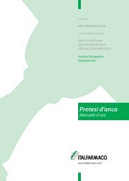
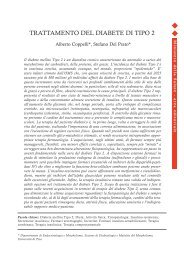
Fig. 1a-g Splenic artery aneurysm. a-c Preprocedural computed tomography (CT) angiography<br />
(a volume-rendered reconstruction; b axial image) and c angiography: a large saccular<br />
aneurysm <strong>of</strong> <strong>the</strong> middle third <strong>of</strong> <strong>the</strong> splenic artery. d Endo<strong>vascular</strong> ligation w<strong>it</strong>h two <strong>Amplatzer</strong><br />
<strong>vascular</strong> plugs (AVPs) (asterisk) in <strong>the</strong> feeding and draining vessels; a contrast-agent<br />
collection is well evident inside <strong>the</strong> sac. e Postprocedural angiography: complete aneurysm<br />
exclusion; inferior pole <strong>of</strong> <strong>the</strong> spleen is supplied by collateral vessels (wh<strong>it</strong>e arrow). f,g CT<br />
angiography per<strong>for</strong>med 1 month after <strong>the</strong> procedure (contiguous axial images): AVP (asterisk)<br />
w<strong>it</strong>hout metallic artefacts, aneurysm exclusion (black arrow) and large splenic ischemia<br />
(arrowhead).<br />
Fig. 1a-g Aneurisma dell’arteria splenica. a-c Angio-TC (a ricostruzione VR, b immagine<br />
assiale e c angiografia pre-procedura: voluminoso aneurisma sacci<strong>for</strong>me del terzo medio<br />
dell’arteria splenica. d “Legatura endovascolare” mediante VPA (asterisco) a monte e a<br />
valle, nei rami afferente ed efferente; si osserva ristagno di mezzo di contrasto nella sacca<br />
aneurismatica esclusa. e Angiografia post-procedura: completa esclusione dell’aneurisma;<br />
circoli collaterali rivascolarizzano il polo inferiore della milza (freccia bianca). f,g Angio-<br />
TC espletata 1 mese dopo la procedura (scansioni assiali contigue): ben riconoscibile il VPA<br />
(asterisco) senza artefatti da indurimento del fascio che consente la valutazione della completa<br />
esclusione dell’aneurisma (freccia nera); ampio infarto splenico (testa di freccia).
710 Radiol med (2008) 113:707–718<br />
a<br />
b<br />
c<br />
Fig. 2a-c Aortoiliac aneurysm. a Preprocedural computed tomography (CT) angiography (volume-rendered<br />
reconstruction): aortoiliac aneurysm involving <strong>the</strong> origin <strong>of</strong> both internal iliac<br />
arteries and small aneurysm <strong>of</strong> <strong>the</strong> left internal iliac artery. b Angiography per<strong>for</strong>med after deployment<br />
<strong>of</strong> a customised bifurcated stent-graft w<strong>it</strong>h a branch preserving <strong>the</strong> patency <strong>of</strong> <strong>the</strong><br />
right internal iliac artery (black arrow); aneurysm <strong>of</strong> <strong>the</strong> left internal iliac artery is excluded<br />
w<strong>it</strong>h endo<strong>vascular</strong> ligation w<strong>it</strong>h <strong>Amplatzer</strong> <strong>vascular</strong> plug (AVP) in <strong>the</strong> feeding artery (black<br />
asterisk) and coils in <strong>the</strong> draining tract (wh<strong>it</strong>e arrow). c CT angiography per<strong>for</strong>med 6 months<br />
after <strong>the</strong> procedure (maximum intens<strong>it</strong>y projection reconstruction): complete exclusion <strong>of</strong> <strong>the</strong><br />
aneurysm, patency <strong>of</strong> <strong>the</strong> stent-graft branch <strong>of</strong> <strong>the</strong> right internal iliac artery (black arrow) and<br />
exclusion <strong>of</strong> <strong>the</strong> left internal iliac artery aneurysm w<strong>it</strong>h AVP (asterisk) and coils (wh<strong>it</strong>e arrow).<br />
Fig. 2a-c Aneurisma aorto-iliaco. a Angio-TC pre-procedura (ricostruzione VR): aneurisma<br />
aorto-iliaco con coinvolgimento dell’emergenza delle arterie ipogastriche e piccolo aneurisma<br />
dell’arteria ipogastrica sinistra. b Angiografia espletata dopo posizionamento di endoprotesi<br />
bi<strong>for</strong>cata “custom-made” con braccietto protesico che preserva la pervietà dell’arteria ipogastrica<br />
destra (freccia nera); l’aneurisma dell’arteria ipogastrica sinistra è stato escluso<br />
mediante “legatura endovascolare” con VPA a monte (asterisco nero) e spirali metalliche a<br />
valle (freccia bianca). c Follow-up con angio-TC esegu<strong>it</strong>a 6 mesi dopo la procedura (ricostruzione<br />
MIP): completa esclusione dell’aneurisma con pervietà dell’arteria ipogastrica destra<br />
dove è riconoscibile braccietto protesico (freccia nera); esclusione anche dell’aneurisma<br />
dell’arteria ipogastrica sinistra con VPA (asterisco) e spirali metaliche (freccia bianca).<br />
<strong>it</strong>s typical plug shape. The AVP is 7 mm or 8 mm long, and<br />
<strong>it</strong>s elongation is inversely proportional to <strong>the</strong> vessel diameter.<br />
In vessels w<strong>it</strong>h a diameter less than 50% that <strong>of</strong> <strong>the</strong> cylinder,<br />
<strong>the</strong> device can reach more than 1 cm in length.<br />
In 10/12 cases, we <strong>use</strong>d percutaneous transfemoral access,<br />
whereas in 2/12 cases, requiring occlusion <strong>of</strong> <strong>the</strong> subclavian<br />
artery, percutaneous access was transhumeral. The<br />
target vessel was first ca<strong>the</strong>terised selectively w<strong>it</strong>h an appropriately<br />
shaped 5-Fr ca<strong>the</strong>ter (Cordis) and 0.035”<br />
guidewire (Glidewire, Terumo, Tokyo, Japan) (Fig. 3a-c).<br />
Then, w<strong>it</strong>h <strong>the</strong> aid <strong>of</strong> a 0.035” stiff guidewire (Amplatz,<br />
Boston Scientific, Ratingen, Germany), we advanced <strong>the</strong><br />
guiding ca<strong>the</strong>ter (Envoy, Cordis, Miami, FI, USA). In two<br />
cases <strong>of</strong> aortoiliac aneurysm, <strong>the</strong> internal iliac artery was<br />
ca<strong>the</strong>terised w<strong>it</strong>h <strong>the</strong> crossover technique, whereas in <strong>the</strong> remaining<br />
cases, <strong>the</strong> ostium <strong>of</strong> <strong>the</strong> internal iliac artery was<br />
ca<strong>the</strong>terised through an ipsilateral access owing to more<br />
favourable anatomy. Once <strong>the</strong> guiding ca<strong>the</strong>ter was close to<br />
dalla elastic<strong>it</strong>à del N<strong>it</strong>inol che permette l’espansione sino al<br />
raggiungimento del calibro vaso; successivamente il dispos<strong>it</strong>ivo<br />
si allunga conferendo la caratteristica morfologia “a<br />
tappo”. Il VAP è lungo 7 o 8 mm e l’allungamento è inversamente<br />
proporzionale al calibro del vaso; con vasi di calibro<br />
inferiore al 50% si può allungare più di 1 cm.<br />
In 10/12 casi l’approccio percutaneo è stato effettuato<br />
per via trans-femorale; in 2/12 casi, per l’occlusione<br />
dell’arteria succlavia, è stato utilizzato l’approccio transomerale.<br />
Il vaso da occludere è stato dapprima cateterizzato<br />
selettivamente mediante catetere da 5 F (Cordis, Miami,<br />
USA) di morfologia appropriata e guida 0,035” (Glidewire,<br />
Terumo, Tokyo, Giappone) (Fig. 3a-c); successivamente<br />
con l’ausilio di guida rigida 0,035” (Amplatz, Boston Scientific,<br />
Ratingen, Germania) viene avanzato il catetere guida<br />
(Envoy, Cordis, Miami, Fl, USA); in 2 casi di aneurisma<br />
aorto-iliaco, l’arteria ipogastrica è stata cateterizzata con<br />
tecnica “cross-over”, mentre nei rimanenti casi l’ostio
Radiol med (2008) 113:707–718 711<br />
a b c<br />
*<br />
d e f<br />
Fig. 3a-f Isolated common iliac artery aneurysm. a Preprocedural angiography: isolated common iliac artery aneurysm involving <strong>the</strong> origin <strong>of</strong> internal iliac<br />
artery; complete exclusion requires occlusion <strong>of</strong> <strong>the</strong> orifice <strong>of</strong> <strong>the</strong> internal iliac artery. b Selective ca<strong>the</strong>terisation <strong>of</strong> <strong>the</strong> internal iliac artery w<strong>it</strong>h<br />
crossover technique (wh<strong>it</strong>e arrow). c Angiography per<strong>for</strong>med after <strong>Amplatzer</strong> <strong>vascular</strong> plug (AVP) deployment (asterisk): occlusion <strong>of</strong> <strong>the</strong> internal iliac<br />
artery. d Angiography per<strong>for</strong>med after pos<strong>it</strong>ioning <strong>of</strong> a covered stent-graft: complete aneurysm exclusion. e,f Computed tomography (CT) angiography per<strong>for</strong>med<br />
3 months after <strong>the</strong> procedure (e oblique multiplanar reconstruction w<strong>it</strong>h maximum intens<strong>it</strong>y algor<strong>it</strong>hm; f volume-rendered reconstruction w<strong>it</strong>h bone<br />
segmentation): complete aneurysm exclusion, w<strong>it</strong>h calcified shell (black arrow) and iliac axis patency; AVP at <strong>the</strong> origin <strong>of</strong> <strong>the</strong> internal iliac artery (asterisk);<br />
branches <strong>of</strong> <strong>the</strong> internal iliac artery are recanalised by collateral vessels (wh<strong>it</strong>e arrow).<br />
Fig. 3a-f Aneurisma isolato dell’arteria iliaca comune destra. a Angiografia pre-procedura: aneurisma isolato dell’arteria iliaca comune che coinvolge<br />
l’emergenza dell’arteria ipogastrica; l’esclusione completa necess<strong>it</strong>a dell’occlusione all’origine dell’arteria ipogastrica. b Cateterismo selettivo dell’arteria<br />
ipogastrica con tecnica “cross-over”(freccia bianca). c Angiografia espletata dopo posizionamento di VPA (asterisco): occlusione all’origine dell’arteria<br />
ipogastrica. d Angiografia espletata dopo posizionamento di stent ricoperto: completa esclusione dell’aneurisma. e,f Follow-up con angio-TC esegu<strong>it</strong>a<br />
3 mesi dopo la procedura (ricostruzione MPR obliqua con algor<strong>it</strong>mo MIP: e ricostruzione VR con segmentazione dell’osso; f completa esclusione<br />
dell’aneurisma, con guscio calcifico ben evidente in e (freccia nera) e regolare pervietà dell’asse iliaco; all’origine dell’arteria ipogastrica è presente <strong>vascular</strong><br />
plug (asterisco); i rami di suddivisione dell’arteria ipogastrica sono ricanalizzati da circoli collaterali (frecce bianche).<br />
<strong>the</strong> target s<strong>it</strong>e, <strong>the</strong> AVP was inserted into <strong>it</strong> and advanced to<br />
<strong>the</strong> s<strong>it</strong>e by pushing <strong>the</strong> 135-mm steel cable. Correct pos<strong>it</strong>ioning<br />
<strong>of</strong> <strong>the</strong> AVP was <strong>the</strong>n verified, and <strong>the</strong> guiding<br />
ca<strong>the</strong>ter was slightly w<strong>it</strong>hdrawn. At this point, <strong>the</strong> cylinder<br />
expands until <strong>it</strong> adheres firmly to <strong>the</strong> vessel, thanks to <strong>the</strong><br />
outward radial <strong>for</strong>ce <strong>of</strong> n<strong>it</strong>inol. Once expanded, <strong>the</strong> device<br />
can no longer be moved. Then, <strong>the</strong> delivery cable was rotated<br />
anticlockwise to detach <strong>it</strong> and allow defin<strong>it</strong>ive deployment<br />
<strong>of</strong> <strong>the</strong> device. The AVP can also be released by pushdell’arteria<br />
ipogastrica, in relazione alla favorevole anatomia,<br />
è stato cateterizzato per via omolaterale. Posizionato il<br />
catetere guida in prossim<strong>it</strong>à del s<strong>it</strong>o del rilascio, il VPA è<br />
stato inser<strong>it</strong>o con appos<strong>it</strong>o caricatore nel catetere guida e<br />
quindi avanzato mediante il cavo in acciaio lungo 135 cm<br />
che permette di spingerlo nel s<strong>it</strong>o del rilascio; verificata la<br />
giusta posizione del VPA il catetere guida è stato per breve<br />
tratto retratto; il cilindro quindi si espande sino ad aderire<br />
saldamente al vaso grazie alla <strong>for</strong>za radiale del N<strong>it</strong>inol; a
712 Radiol med (2008) 113:707–718<br />
ing <strong>the</strong> delivery cable out <strong>of</strong> <strong>the</strong> guiding ca<strong>the</strong>ter until <strong>the</strong><br />
distal end expands and <strong>the</strong>n w<strong>it</strong>hdrawing <strong>the</strong> guiding<br />
ca<strong>the</strong>ter while firmly holding <strong>the</strong> delivery cable in pos<strong>it</strong>ion<br />
until <strong>the</strong> device has expanded completely.<br />
Treatment <strong>of</strong> splenic and internal iliac aneurysms required,<br />
where possible, <strong>the</strong> placement <strong>of</strong> two AVPs, one<br />
proximal and one distal to <strong>the</strong> aneurysm, to create an endo<strong>vascular</strong><br />
ligation (per<strong>for</strong>med in <strong>the</strong> three splenic<br />
aneurysms) (Fig. 3c-e). Where deployment is precluded by<br />
<strong>the</strong> small diameter <strong>of</strong> <strong>the</strong> outflow vessel or <strong>vascular</strong> tortuos<strong>it</strong>y<br />
hampering advancement <strong>of</strong> <strong>the</strong> guiding ca<strong>the</strong>ter, <strong>the</strong> outflow<br />
vessel can be ca<strong>the</strong>terised w<strong>it</strong>h a microca<strong>the</strong>ter and occluded<br />
w<strong>it</strong>h metallic coils. The proximal feeding vessel can<br />
<strong>the</strong>n be occluded w<strong>it</strong>h <strong>the</strong> AVP (this procedure was done in<br />
<strong>the</strong> internal iliac aneurysm) (Fig. 2b). Follow-up was carried<br />
out w<strong>it</strong>h CT angiography and/or contrast-enhanced ultrasound<br />
(US) at 3 and 6 months (Figs. 1f,g, 2c and 3e,f). Subsequent<br />
follow-up was carried out according to <strong>the</strong> cond<strong>it</strong>ion<br />
being treated.<br />
Results<br />
Immediate technical success was achieved in 12/12 cases<br />
and documented by postprocedural angiography, which<br />
showed firm anchoring <strong>of</strong> <strong>the</strong> AVP by outward radial <strong>for</strong>ce<br />
w<strong>it</strong>h artery occlusion and aneurysm exclusion (Figs. 1e, 2b,<br />
3d, and 4b,c). No major complication such as rupture, per<strong>for</strong>ation<br />
or dissection was observed.<br />
During <strong>the</strong> follow-up period (mean 4.6 months, range<br />
3–6) CT angiography and/or contrast-enhanced US demonstrated<br />
occlusion <strong>of</strong> <strong>the</strong> artery and aneurysm. No cases <strong>of</strong><br />
device migration occurred. Reduced metallic artefacts, compared<br />
w<strong>it</strong>h metallic coils, allowed <strong>for</strong> precise depiction <strong>of</strong><br />
occlusion <strong>of</strong> vessels and aneurysms at follow-up CT angiography<br />
(Figs. 1f,g, 2c and 3e,f).<br />
Discussion<br />
The AVP is a self-expanding cylindrical device made <strong>of</strong> a<br />
n<strong>it</strong>inol wire mesh and secured on both ends w<strong>it</strong>h platinum<br />
marker bands. A stainless-steel microscrew is welded to <strong>the</strong><br />
proximal marker band to allow attachment <strong>of</strong> <strong>the</strong> delivery<br />
cable (Fig. 5). The AVP is available in diameters from 4<br />
mm to 16 mm, w<strong>it</strong>h 2-mm increments, and is 7 mm or 8 mm<br />
long. The AVP grew out <strong>of</strong> two cardiac devices <strong>use</strong>d <strong>for</strong> <strong>the</strong><br />
closure <strong>of</strong> interatrial defects or patent ductus arteriosus (<strong>the</strong><br />
<strong>Amplatzer</strong> septal occluder and <strong>the</strong> <strong>Amplatzer</strong> duct occluder,<br />
respectively) [1, 2]. The AVP in now indicated <strong>for</strong> <strong>the</strong> occlusion<br />
<strong>of</strong> medium- to small-size arteries or veins in interventional<br />
radiology [3–14].<br />
The main advantage <strong>of</strong> <strong>the</strong> AVP, compared w<strong>it</strong>h o<strong>the</strong>r<br />
questo punto non è più possibile muovere il dispos<strong>it</strong>ivo. Successivamente<br />
mediante un movimento antiorario il cavo in<br />
acciaio è stato sv<strong>it</strong>ato dalla microv<strong>it</strong>e s<strong>it</strong>a nella porzione<br />
prossimale con rilascio defin<strong>it</strong>ivo del dispos<strong>it</strong>ivo. Il VPA<br />
può essere anche posizionato spingendolo mediante il cavo<br />
al di fuori del catetere guida sino all’espansione della parte<br />
distale quindi retrarre il catetere guida tenendo in tensione<br />
il cavo in acciaio sino alla completa espansione.<br />
Il trattamento degli aneurismi splenici e dell’arteria ipogastrica<br />
ha previsto, dove possibile, il posizionamento di 2<br />
VPA a monte e a valle dell’aneurisma al fine di realizzare<br />
una “legatura endovascolare” (effettuata nei 3 aneurismi<br />
splenici) (Fig. 3c-e). Qualora il ridotto calibro dell’arteria<br />
efferente o la difficoltà di portare in sede un catetere guida<br />
per tortuos<strong>it</strong>à vascolare, non consentono il posizionamento,<br />
è possibile cateterizzare le efferenze con microcatetere e occluderle<br />
con spirali metalliche e successivamente occludere<br />
l’afferenza prossimale mediante il VPA (tale procedura è<br />
stata effettuata nell’aneurisma dell’arteria ipogastrica)<br />
(Fig. 2b). Il follow-up è stato espletato mediante angio-TC<br />
e/o ecografia con MdC a 3, 6 mesi (Figg. 1f,g, 2c e 3e,f).<br />
Successivamente i pazienti hanno esegu<strong>it</strong>o il follow-up in<br />
relazione alla patologia trattata.<br />
Risultati<br />
È stato ottenuto successo tecnico immediato 12/12 casi, documentato<br />
all’angiografia post-procedura che ha dimostrato<br />
l’ancoraggio per <strong>for</strong>za radiale al vaso e la successiva occlusione<br />
dell’arteria e l’esclusione degli aneurismi trattati<br />
(Figg. 1e, 2b, 3d e 4b,c). Non si sono verificate complicanze<br />
maggiori quali rottura, per<strong>for</strong>azione o dissezione del vaso<br />
trattato.<br />
Durante il follow-up (medio 4,6 mesi, range 3–6) il controllo<br />
con angio-TC e/o ecografia con MdC ha dimostrato<br />
l’occlusione dell’arteria e quindi dell’aneurisma trattato.<br />
Non si sono verificate migrazioni dei dispos<strong>it</strong>ivi impiantati.<br />
In relazione ai ridotti artefatti da indurimento del fascio<br />
causati del dispos<strong>it</strong>ivo, rispetto alle spirali metalliche, ai<br />
controlli angio-TC, è stato possibile documentare con precisione<br />
l’occlusione del vaso e degli aneurismi trattati<br />
(Figg. 1f,g, 2c e 3e,f).<br />
Discussione<br />
Il VPA è un dispos<strong>it</strong>ivo cilindrico autoespandibile realizzato<br />
in maglia metallica di N<strong>it</strong>inol fissato alle due estrem<strong>it</strong>à<br />
con fasce di marker in platino; a quella prossimale è saldata<br />
una microv<strong>it</strong>e in acciaio che consente il fissaggio al cavo<br />
di introduzione (Fig. 5). È disponibile in calibri da 4 a<br />
16 mm, con incremento di 2 mm, lungo 7 o 8 mm. È nato da
Radiol med (2008) 113:707–718 713<br />
embolising devices such as metallic coils lies in <strong>the</strong> opportun<strong>it</strong>y<br />
to occlude <strong>the</strong> feeding and/or draining vessels <strong>of</strong><br />
aneurysms or AVMs w<strong>it</strong>h a single device and thus w<strong>it</strong>h<br />
faster immediate results. Ano<strong>the</strong>r advantage is <strong>the</strong> increased<br />
precision and control during deployment af<strong>for</strong>ded by <strong>the</strong> delivery<br />
system [5, 7, 9, 13]. The elastic<strong>it</strong>y <strong>of</strong> n<strong>it</strong>inol allows<br />
<strong>the</strong> device to become firmly anchored to <strong>the</strong> vessel wall by<br />
outward radial <strong>for</strong>ce, which prevents migration and allows<br />
leng<strong>the</strong>ning to be predicted. As a result, selection <strong>of</strong> <strong>the</strong> diameter<br />
<strong>of</strong> <strong>the</strong> device can be less precise compared w<strong>it</strong>h selection<br />
<strong>of</strong> <strong>the</strong> diameter, length and type <strong>of</strong> coil. Use <strong>of</strong> <strong>the</strong><br />
AVP <strong>the</strong>re<strong>for</strong>e <strong>of</strong>fers an alternative to occlusion w<strong>it</strong>h metallic<br />
coil embolisation, which requires <strong>the</strong> placement <strong>of</strong> several<br />
coils <strong>of</strong> different sizes to occlude a single vessel w<strong>it</strong>h<br />
4- or 5-Fr ca<strong>the</strong>ters or <strong>the</strong> more expensive microca<strong>the</strong>ters.<br />
Moreover, <strong>the</strong> inabil<strong>it</strong>y to control coil release w<strong>it</strong>h <strong>the</strong><br />
metallic coils creates a risk <strong>of</strong> malpos<strong>it</strong>ioning or displacement.<br />
Although this lim<strong>it</strong>ation can be overcome by controlled-release<br />
coils, <strong>the</strong>ir <strong>use</strong> increases overall procedure<br />
cost and time. Ano<strong>the</strong>r advantage <strong>of</strong> <strong>the</strong> AVP is that, due to<br />
<strong>it</strong>s morphological and structural characteristics, <strong>it</strong> ca<strong>use</strong>s<br />
less metallic artefacts on CT angiography compared w<strong>it</strong>h<br />
platinum coils (Figs. 1f,g, 2c, and 3e,f). This allows <strong>for</strong> a<br />
more precise assessment <strong>of</strong> aneurysm exclusion and possible<br />
endoleak feeding vessels.<br />
All <strong>of</strong> <strong>the</strong>se features have allowed <strong>the</strong> AVP to be proposed<br />
<strong>for</strong> <strong>use</strong> in interventional radiology and <strong>it</strong>s indications<br />
to be progressively extended. Over <strong>the</strong> past year, a number<br />
<strong>of</strong> early reports have been published in <strong>the</strong> l<strong>it</strong>erature (Table<br />
1). These are mostly case reports regarding extracardiac applications<br />
<strong>of</strong> <strong>the</strong> <strong>Amplatzer</strong> systems <strong>for</strong> treating pulmonary<br />
AVM [4, 7, 9, 13], subclavian [8], internal iliac [6, 14], pulmonary<br />
[12] and cerebral aneurysms [5], <strong>for</strong> <strong>the</strong> occlusion<br />
<strong>of</strong> a retroper<strong>it</strong>oneal shunt in a patient w<strong>it</strong>h portal hypertension<br />
during transjugular intrahepatic portosystemic shunt<br />
(TIPS) creation [3] and <strong>for</strong> <strong>the</strong> occlusion <strong>of</strong> <strong>the</strong> internal iliac<br />
artery in patients w<strong>it</strong>h aortoiliac or iliac aneurysm [11]. One<br />
<strong>of</strong> <strong>the</strong> earliest reports described <strong>the</strong> <strong>use</strong> <strong>of</strong> <strong>the</strong> AVP to occlude<br />
<strong>the</strong> origin <strong>of</strong> <strong>the</strong> internal iliac artery in <strong>the</strong> endo<strong>vascular</strong><br />
treatment <strong>of</strong> aortoiliac and iliac aneurysms to prevent<br />
type II endoleaks [11].<br />
The major<strong>it</strong>y <strong>of</strong> published case reports deals w<strong>it</strong>h pulmonary<br />
AVM. In particular, pulmonary AVM w<strong>it</strong>h large<br />
feeding arteries, and thus at a greater risk <strong>of</strong> device migration,<br />
were successfully treated w<strong>it</strong>h <strong>the</strong> AVP, which showed<br />
excellent anchoring to <strong>the</strong> vessel and immediate occlusion<br />
[4, 7, 9, 13]. The only alternative treatment <strong>for</strong> pulmonary<br />
AVM is <strong>the</strong> <strong>use</strong> <strong>of</strong> metallic coils [15, 16], as indications <strong>for</strong><br />
occlusion w<strong>it</strong>h a detachable latex balloon have become obsolete.<br />
Among later studies, a sporadic report <strong>of</strong> a ruptured<br />
isolated aneurysm <strong>of</strong> <strong>the</strong> internal iliac artery [14] described<br />
embolisation <strong>of</strong> <strong>the</strong> outflow branches and <strong>the</strong> origin <strong>of</strong> <strong>the</strong><br />
internal iliac artery w<strong>it</strong>h coils and an AVP using <strong>the</strong> endue<br />
precursori per utilizzo esclusivo in amb<strong>it</strong>o cardiologico<br />
per la chiusura dei difetti interatriali o dei dotti arteriosi<br />
pervii (Amplater Septal Occluder e l’<strong>Amplatzer</strong> Duct Occluder<br />
rispettivamente) [1, 2]. Il VPA trova oggi indicazione<br />
in radiologia interventistica nell’occlusione di arterie o<br />
vene di medio e piccolo calibro [3–14].<br />
Il vantaggio principale, rispetto ad altri “devices” embolizzanti<br />
come le spirali metalliche, è la possibil<strong>it</strong>à di utilizzare<br />
un unico dispos<strong>it</strong>ivo per l’occlusione di vasi afferenti e/o<br />
efferenti a patologia aneurismatica o mal<strong>for</strong>mazioni arterovenose<br />
(MAV), quindi con un più veloce risultato immediato;<br />
inoltre le caratteristiche del sistema di rilascio permettono<br />
un più preciso e controllato posizionamento [5, 7, 9, 13].<br />
L’elastic<strong>it</strong>à del N<strong>it</strong>inol consente un saldo ancoraggio al vaso<br />
per <strong>for</strong>za radiale ev<strong>it</strong>ando le migrazioni con un allungamento<br />
prevedibile; è quindi possibile una scelta del calibro del<br />
dispos<strong>it</strong>ivo sicuramente meno precisa rispetto alla scelta del<br />
calibro, della lunghezza e del tipo di spirale. Si pone quindi,<br />
attualmente, quale alternativa all’occlusione mediante embolizzazione<br />
con spirali metalliche; quest’ultima prevede il<br />
posizionamento di più spirali di calibro diverso per occlusioni<br />
di un unico vaso con cateteri da 4 o 5 F o l’utilizzo più<br />
costoso di microcateteri; inoltre l’impossibil<strong>it</strong>à di controllarne<br />
il rilascio comporta il rischio di malposizionamento o<br />
dislocazione delle stesse. Questo lim<strong>it</strong>e può essere superato<br />
dall’uso di spirali metalliche a distacco o a rilascio controllato<br />
che però accresce ulteriormente i costi complessivi e<br />
prolunga i tempi della procedura. Un altro vantaggio è cost<strong>it</strong>u<strong>it</strong>o<br />
dal fatto che il VPA, per le sue caratteristiche morfologiche<br />
e strutturali, comporta minori artefatti da indurimento<br />
del fascio all’indagine angio-TC rispetto alle spirali<br />
metalliche in platino (Figg. 1f,g, 2c e 3e,f); questo permette<br />
una più precisa valutazione dell’esclusione degli aneurismi e<br />
l’eventuale ri<strong>for</strong>nimento degli endoleak.<br />
Le caratteristiche peculiari descr<strong>it</strong>te hanno consent<strong>it</strong>o di<br />
proporne l’utilizzo in radiologia interventistica e di ampliarne<br />
progressivamente le indicazioni. Durante l’ultimo anno<br />
sono apparsi in letteratura i primi lavori (Tabella 1), la<br />
maggior parte “case report”, riguardanti l’utilizzo extracardiologico<br />
dei sistemi Ampltazer (Tabella 1) per il trattamento<br />
di MAV polmonari [4, 7, 9, 13], aneurismi dell’arteria<br />
succlavia [8], ipogastrica [6, 14], polmonare [12], cerebrali<br />
[5], per l’occlusione di shunt retroper<strong>it</strong>oneale in paziente<br />
con ipertensione portale durante TIPS [3] e per l’occlusione<br />
dell’arteria ipogastrica in pazienti con aneurisma aorto-iliaco<br />
o iliaco [11]. Uno dei primi studi riporta l’utilizzo del<br />
VPA nell’occlusione dell’origine dell’arteria ipogastrica nel<br />
trattamento endovascolare degli aneurismi aorto-iliaci e<br />
iliaci per la prevenzione dell’endoleak di tipo II [11].<br />
La maggior parte dei casi pubblicati riguarda le MAV<br />
polmonari per le quali è stato riportato il trattamento con<br />
successo di MAV con arteria afferente di grosso calibro nelle<br />
quali è maggiore il rischio di migrazione del dispos<strong>it</strong>ivo
714 Radiol med (2008) 113:707–718<br />
Table 1 L<strong>it</strong>erature about <strong>the</strong> extracardiac <strong>use</strong> <strong>of</strong> <strong>Amplatzer</strong> <strong>vascular</strong> plug (AVP) systems<br />
Author; publication year Disease treated Number <strong>of</strong> cases device Result<br />
Ha et al., 2005 [11] Internal arteries in aortoiliac and iliac aneurysms 5 AVP Success<br />
Pate et al., 2005 [12] Pulmonary artery aneurysm 1 <strong>Amplatzer</strong> septal occluder Success<br />
Ferro et al., 2006 [13] Pulmonary AVM 1 AVP Success<br />
de Medici et al., 2006 [14] Isolated aneurysm internal iliac artery 1 AVP+coils Success<br />
Ho<strong>it</strong> et al., 2006 [5] Cerebral aneurysms 2 AVP+coils Success<br />
Dorenberg et al., 2006 [6] Ruptured isolated aneurysm internal iliac artery 1 AVP Recanalisation<br />
at 1 month<br />
Rossi et al., 2006 [7] Pulmonary AVM 2 AVP Success<br />
Hoppe et al., 2006 [8] Aneurysm <strong>of</strong> aberrant right subclavian artery 1 <strong>Amplatzer</strong> septal occluder Success<br />
Beck et al., 2006 [9] Pulmonary AVM 14: 12 <strong>Amplatzer</strong> duct Success<br />
occluders, 2 AVPs<br />
Mylonas et al., 2006 [10] Anastomotic aneurysm <strong>of</strong> aortocoronary bypass graft 1 AVP Success<br />
Rabenstein et al., 2006 [20] Broncho-oesophageal fistula 1 <strong>Amplatzer</strong> septal occluder Success<br />
Kessler et al., 2006 [3] Retroper<strong>it</strong>oneal shunt during TIPS 1 AVP+coils Success<br />
Cil et al., 2006 [4] Bilateral pulmonary AVM 2 AVP Success<br />
AVM, arteriovenous mal<strong>for</strong>mations; TIPS, transjugular intrahepatic portosystemic shunt<br />
Tabella 1 Letteratura riguardante l’utilizzo extra-cardiaco dei sistemi <strong>Amplatzer</strong><br />
Autore; anno di pubblicazione Patologia trattata Numero casi (device) Risultato<br />
Ha et al., 2005 [11] Arterie ipogastriche in aneurismi aorto-iliaci e iliaci 5 (VPA) Successo<br />
Pate et al., 2005 [12] Aneurisma arteria polmonare 1 (<strong>Amplatzer</strong> Septal Occluder) Successo<br />
Ferro et al., 2007 [13] MAV polmonare 1 (VPA) Successo<br />
de Medici et al., 2006 [14] Aneurisma isolato arteria ipogastrica 1 (VPA+spirali) Successo<br />
Ho<strong>it</strong> et al., 2006 [5] Aneurismi cerebrali 2 (VPA+spirali) Successo<br />
Dorenberg et al., 2006 [6] Aneurisma isolato arteria ipogastrica rotto 1 (VPA) Ricanalizzazione<br />
a 1 mese<br />
Rossi et al., 2006 [7] MAV polmonari 2 (VPA) Successo<br />
Hoppe et al., 2006 [8] Aneurisma arteria lusoria 1 (<strong>Amplatzer</strong> Septal Occluder) Successo<br />
Beck et al., 2006 [9] MAV polmonari 14: 12 (<strong>Amplatzer</strong> Duct Successo<br />
Occluder) 2 (VPA)<br />
Mylonas et al., 2006 [10] Aneurisma anastomotico in by-pass aorto-coronarico 1 (VPA) Successo<br />
Rabenstein et al., 2006 [20] Fistola bronco-es<strong>of</strong>agea 1 (<strong>Amplatzer</strong> Septal Occluder) Successo<br />
Kessler et al., 2006 [3] Shunt retroper<strong>it</strong>oneale durante TIPS 1 (VPA+spirali) Successo<br />
Cil et al., 2006 [4] MAV polmonari bilaterali 2 (VPA) Successo<br />
MAV, mal<strong>for</strong>mazioni arterovenose; TIPS, shunt portosistemico intraepatico transgiugulare<br />
do<strong>vascular</strong> ligation technique. This technique has been successfully<br />
applied to <strong>the</strong> treatment <strong>of</strong> visceral aneurysms [17]<br />
and, given that <strong>the</strong> 10-mm AVP can be <strong>use</strong>d w<strong>it</strong>h 6-Fr guiding<br />
ca<strong>the</strong>ters, we recognised <strong>the</strong> possibil<strong>it</strong>y <strong>of</strong> excluding<br />
aneurysms <strong>of</strong> <strong>the</strong> mid splenic artery. As an alternative, when<br />
vessel tortuos<strong>it</strong>y and <strong>the</strong> diameter <strong>of</strong> <strong>the</strong> outflow vessels<br />
preclude advancement <strong>of</strong> <strong>the</strong> device, <strong>the</strong> distal outflow vessels<br />
can be embolised w<strong>it</strong>h metallic coils and <strong>the</strong> proximal<br />
feeding vessel w<strong>it</strong>h <strong>the</strong> AVP, as done by us in <strong>the</strong> case <strong>of</strong><br />
<strong>the</strong> internal iliac aneurysm.<br />
In <strong>the</strong> treatment <strong>of</strong> peripheral aneurysms, where maintenance<br />
<strong>of</strong> <strong>vascular</strong> continu<strong>it</strong>y is not a prior<strong>it</strong>y, possible<br />
options include endo<strong>vascular</strong> ligation or a hybrid approach.<br />
The hybrid approach has been reported in <strong>the</strong> treatment <strong>of</strong><br />
embolizzante; in questi casi si è osservato l’ottimo ancoraggio<br />
del VPA con immediato effetto occlusivo [4, 7, 9, 13]. In<br />
questi casi l’alternativa è l’utilizzo di spirali metalliche [15,<br />
16]; l’indicazione all’occlusione mediante palloncino staccabile<br />
in lattice è ormai per le MAV polmonari obsoleta.<br />
Tra i lavori pubblicati successivamente, un caso sporadico<br />
di aneurisma isolato dell’arteria ipogastrica in rottura [14]<br />
descrive l’embolizzazione mediante spirali metalliche dei<br />
rami efferenti di suddivisione e dell’origine dell’arteria ipogastrica<br />
mediante VPA con la tecnica della “ legatura endovascolare”.<br />
Quest’ultima tecnica è stata adottata con<br />
successo nel trattamento degli aneurismi viscerali [17] e<br />
poiché il VPA del calibro da 10 mm scorre su cateteri guida<br />
da 6 F abbiamo individuato la possibil<strong>it</strong>à di escludere gli
Radiol med (2008) 113:707–718 715<br />
an aneurysm <strong>of</strong> an aberrant right subclavian artery. The origin<br />
<strong>of</strong> <strong>the</strong> subclavian artery was occluded w<strong>it</strong>h an AVP,<br />
followed by ligation <strong>of</strong> <strong>the</strong> subclavian artery above <strong>the</strong><br />
origin <strong>of</strong> <strong>the</strong> vertebral artery and carotid-subclavian bypass,<br />
resulting in less invasiveness and intraoperative mortal<strong>it</strong>y<br />
[8].<br />
In agreement w<strong>it</strong>h Ha et al. [11], we successfully explored<br />
<strong>the</strong> possibil<strong>it</strong>y <strong>of</strong> treating isolated iliac aneurysms by<br />
pos<strong>it</strong>ioning an AVP to exclude <strong>the</strong> internal iliac ostium,<br />
when involved, and prevent a type II endoleak be<strong>for</strong>e endograft<br />
placement, as an alternative to occlusion w<strong>it</strong>h metallic<br />
coils. Moreover, in endo<strong>vascular</strong> repair <strong>of</strong> aortoiliac<br />
aneurysms, <strong>the</strong> AVP can be <strong>use</strong>d to exclude <strong>the</strong> internal iliac<br />
ostium provided that <strong>the</strong> aneurysm involves a single axis.<br />
Where both ostia <strong>of</strong> <strong>the</strong> internal iliac arteries are involved,<br />
customised stent-grafts w<strong>it</strong>h small pros<strong>the</strong>tic branches to be<br />
inserted at <strong>the</strong> origin <strong>of</strong> one <strong>of</strong> <strong>the</strong> arteries can seal <strong>the</strong><br />
aneurysm while guaranteeing <strong>the</strong> patency <strong>of</strong> one <strong>of</strong> <strong>the</strong> internal<br />
iliac arteries [18] and allowing exclusion <strong>of</strong> <strong>the</strong> contralateral<br />
artery w<strong>it</strong>h an AVP (Fig. 2).<br />
In <strong>the</strong> endo<strong>vascular</strong> treatment <strong>of</strong> aortic-arch aneurysms,<br />
coverage <strong>of</strong> <strong>the</strong> origin <strong>of</strong> <strong>the</strong> subclavian artery by <strong>the</strong> stentgraft<br />
may give rise to a type II endoleak due to retrograde<br />
flow. These endoleaks are treated w<strong>it</strong>h coil embolisation <strong>of</strong><br />
<strong>the</strong> subclavian artery through a transhumeral access. Rapid<br />
placement and availabil<strong>it</strong>y <strong>of</strong> several sizes, all <strong>of</strong> which can<br />
be <strong>use</strong>d w<strong>it</strong>h 6- or 7-Fr guiding ca<strong>the</strong>ters and transhumeral<br />
approach, makes <strong>the</strong> AVP a valuable alternative to coil embolisation.<br />
In add<strong>it</strong>ion, <strong>the</strong> procedure can be proposed in all<br />
aortic-arch aneurysms to prevent <strong>for</strong>mation <strong>of</strong> type II endoleaks,<br />
given that in <strong>the</strong>se cases, <strong>the</strong> left transhumeral access<br />
is already <strong>use</strong>d <strong>for</strong> <strong>the</strong> sentinel ca<strong>the</strong>ter to be <strong>use</strong>d during<br />
<strong>the</strong> various phases <strong>of</strong> <strong>the</strong> procedure.<br />
In our experience, rapid placement also allowed <strong>for</strong> successful<br />
treatment <strong>of</strong> abdominal aorta aneurysms on an<br />
emergency basis by means <strong>of</strong> aorto-uni-iliac endograft and<br />
an AVP in <strong>the</strong> contralateral common iliac artery (Fig. 4).<br />
This preserved patency <strong>of</strong> <strong>the</strong> external and internal iliac<br />
artery and avoided surgical ligation, w<strong>it</strong>h faster exclusion<br />
<strong>of</strong> ruptured aneurysms. This approach can, however, also<br />
be adopted in elective treatment. The alternative is a stentgraft<br />
occluder available only <strong>for</strong> some uni-iliac stent-grafts<br />
(Zen<strong>it</strong>, Cook and Talent, Medtronic) or coil embolisation<br />
[19]. The advantage <strong>of</strong> <strong>the</strong> AVP over <strong>the</strong>se o<strong>the</strong>r occluding<br />
devices is <strong>it</strong>s more precise pos<strong>it</strong>ioning close to <strong>the</strong> iliac<br />
bifurcation, in that a single AVP device can accommodate<br />
a range <strong>of</strong> diameters, perm<strong>it</strong>ting better preservation <strong>of</strong><br />
patency <strong>of</strong> <strong>the</strong> external and internal iliac arteries. Add<strong>it</strong>ionally,<br />
<strong>the</strong> AVP is advanced through smaller arterial introducers.<br />
The only case <strong>of</strong> AVP failure was reported in a ruptured<br />
aneurysm <strong>of</strong> <strong>the</strong> internal iliac artery in which AVP recanalisation<br />
occurred at 4 weeks after treatment [6].<br />
aneurismi dell’arteria splenica del tratto intermedio. In alternativa,<br />
qualora la tortuos<strong>it</strong>à del vaso e il calibro dei rami<br />
efferenti all’aneurisma non consentano l’avanzamento<br />
del “device” è possibile embolizzare le efferenze distali mediante<br />
spirali metalliche e l’afferenza prossimale mediante<br />
il VPA come effettuato nella nostra esperienza nel trattamento<br />
di un aneurisma dell’arteria ipogastrica.<br />
Nel trattamento degli aneurismi periferici qualora non<br />
sia necessario garantire una continu<strong>it</strong>à vascolare è possibile<br />
una “legatura endovascolare” o un approccio ibrido come<br />
apparso in letteratura di trattamento di un aneurisma<br />
dell’arteria lusoria con occlusione dell’origine della succlavia<br />
mediante VPA e successiva legatura dell’arteria succlavia<br />
a monte dell’origine dell’arteria vertebrale e successivo<br />
bypass carotido-succlavio di minore invasiv<strong>it</strong>à e mortal<strong>it</strong>à<br />
perioperatoria [8].<br />
Come riportato da Ha et al. [11] abbiamo valutato con<br />
successo la possibil<strong>it</strong>à di trattare gli aneurismi isolati<br />
dell’asse iliaco con esclusione dell’origine dell’arteria ipogastrica<br />
mediante VPA qualora coinvolta, prima del posizionamento<br />
dello stent-graft, al fine di escludere il ri<strong>for</strong>nimento<br />
retrogrado, in alternativa all’occlusione mediante<br />
spirali metalliche. Inoltre, nel trattamento endovascolare<br />
degli aneurismi aorto-iliaci è possibile escludere l’origine<br />
di una arteria ipogastrica mediante VPA qualora l’aneurisma<br />
interessi un unico asse; mentre qualora interessi ambedue<br />
le origini delle arterie ipogastriche, le protesi “custom<br />
made” provviste di piccoli braccetti protesici da inserire<br />
all’origine di una delle arterie ipogastriche che sigillano<br />
perfettamente l’aneurisma garantiscono la pervietà di una<br />
delle arterie ipogastriche [18] e permettono di escludere la<br />
controlaterale con VPA (Fig. 2).<br />
Nel trattamento endovascolare degli aneurismi dell’arco<br />
dell’aorta, la copertura da parte dell’endoprotesi dell’origine<br />
dell’arteria succlavia può realizzare, mediante inversione<br />
del flusso, un endoleak di tipo II; per tali endoleaks è<br />
prevista l’embolizzazione con spirali metalliche con approccio<br />
trans-omerale dell’origine dell’arteria succlavia<br />
stessa. Attualmente il VPA, per la rapid<strong>it</strong>à di posizionamento<br />
e per la disponibil<strong>it</strong>à di diametri idonei al calibro del vaso<br />
da occludere avanzabili su cateteri guida da 6 o 7 F con<br />
approccio trans-omerale, è una valida alternativa all’embolizzazione<br />
con spirali; inoltre la procedura può essere<br />
proposta in tutti gli aneurismi dell’arco nella prevenzione<br />
dell’endoleak di tipo II, in quanto in questi casi è di già<br />
quasi sempre previsto un approccio transomerale di sinistra<br />
per posizionare un catetere “sentinella” da utilizzare nelle<br />
varie fasi della procedura.<br />
Nella nostra esperienza, la rapid<strong>it</strong>à di posizionamento ha<br />
consent<strong>it</strong>o di trattare con successo anche aneurismi<br />
dell’aorta addominale in urgenza mediante endoprotesi aorto-uniliaca<br />
e VPA posizionato in arteria iliaca comune controlaterale<br />
(Fig. 4) preservando la pervietà delle arterie ilia-
716 Radiol med (2008) 113:707–718<br />
a b c<br />
Fig. 4a-c Aneurysm <strong>of</strong> <strong>the</strong> infrarenal abdominal aorta. a Preprocedural angiography: aneurysm <strong>of</strong> <strong>the</strong> infrarenal abdominal aorta w<strong>it</strong>hout involvement <strong>of</strong> <strong>the</strong><br />
iliac arteries; <strong>the</strong> small size <strong>of</strong> <strong>the</strong> aortic bifurcation does not allow pos<strong>it</strong>ioning <strong>of</strong> a bifurcated stent-graft. b,c Angiography per<strong>for</strong>med after deployment <strong>of</strong><br />
an aorto-uni-iliac stent-graft and <strong>Amplatzer</strong> <strong>vascular</strong> plug (AVP) at <strong>the</strong> origin <strong>of</strong> <strong>the</strong> right common iliac artery: complete aneurysm exclusion and internal<br />
and external iliac artery patency.<br />
Fig. 4a-c Aneurisma dell’aorta addominale sottorenale. a Angiografia pre-procedura: aneurisma dell’aorta addominale sottorenale che non coinvolge gli<br />
assi iliaci. Le dimensioni del carrefour aortico non consentono il posizionamento di un’endoprotesi bi<strong>for</strong>cata. b,c Angiografia espletata per via anterograda<br />
(b) e retrograda (c) dopo posizionamento di endoprotesi aorto-uniliaca e AVP in corrispondenza dell’arteria iliaca comune destra: completa esclusione<br />
dell’aneurisma e regolare pervietà delle arterie ipogastrica e iliaca esterna destra.<br />
Fig. 5 <strong>Amplatzer</strong> <strong>vascular</strong> plug (AVP): A self-expandable, cylindrical device<br />
made from a n<strong>it</strong>inol wire mesh, secured on both ends w<strong>it</strong>h platinum<br />
marker bands. A stainless-steel microscrew welded to one <strong>of</strong> <strong>the</strong> platinum<br />
marker bands allows attachment to <strong>the</strong> long delivery cable. When delivered,<br />
<strong>the</strong> AVP assumes a plug shape.<br />
Fig. 5 Vascular Plug <strong>Amplatzer</strong>. Dispos<strong>it</strong>ivo cilindrico autoespandibile<br />
realizzato in maglia metallica di N<strong>it</strong>inol fissato alle due estrem<strong>it</strong>à con fasce<br />
di marker in platino; a quella prossimale è saldata una microv<strong>it</strong>e in acciaio<br />
che consente il fissaggio al cavo di introduzione. Il dispos<strong>it</strong>ivo, una<br />
volta rilasciato nel vaso assume una caratteristica morfologia “a tappo”.<br />
ca esterna e ipogastrica ed ev<strong>it</strong>ando la legatura chirurgica<br />
con una più rapida esclusione degli aneurismi rotti. Questo<br />
tipo di approccio è comunque fattibile anche nel trattamento<br />
in elezione; l’alternativa è lo stent-graft occlusore disponibile<br />
solo per alcune endoprotesi uniliache (Zen<strong>it</strong>, Cook e Talent,<br />
Medtronic) o l’embolizzazione con spirali [19]; il vantaggio<br />
rispetto a questi dispos<strong>it</strong>ivi occlusori è un più preciso<br />
posizionamento a ridosso della bi<strong>for</strong>cazione iliaca, in quanto<br />
un unico VPA abbraccia più calibri, con una conseguente<br />
più sicura preservazione della pervietà delle arterie iliaca<br />
esterna e ipogastrica; inoltre il VPA è avanzato attraverso<br />
introduttori arteriosi di minor calibro.<br />
L’unico caso di insuccesso del VPA è stato riportato nel<br />
trattamento di un aneurisma rotto dell’arteria ipogastrica<br />
nel quale si è verificata una ricanalizzazione del VPA a 4<br />
settimane dal trattamento [6].<br />
Conclusioni<br />
In conclusione, la semplic<strong>it</strong>à della scelta e dell’utilizzo del
Radiol med (2008) 113:707–718 717<br />
Conclusions<br />
In conclusion, easy selection and <strong>use</strong> <strong>of</strong> <strong>the</strong> device combined<br />
w<strong>it</strong>h precise and controlled release and immediate<br />
technical success justifies <strong>the</strong> widespread <strong>use</strong> <strong>of</strong> <strong>the</strong> AVP in<br />
interventional radiology. The AVP can – and in some cases<br />
has already – replaced metallic coils <strong>for</strong> endo<strong>vascular</strong> repair<br />
<strong>of</strong> aortic aneurysms, <strong>for</strong> <strong>the</strong> prevention <strong>of</strong> type II endoleaks<br />
from <strong>the</strong> internal iliac and subclavian arteries, and in endo<strong>vascular</strong><br />
ligation <strong>of</strong> visceral aneurysms where <strong>the</strong> device<br />
can be placed distal and proximal to <strong>the</strong> aneurysms. The<br />
AVP may also replace <strong>the</strong> occluder device <strong>of</strong> <strong>the</strong> aortic<br />
stent-graft in <strong>the</strong> aorto-uni-iliac k<strong>it</strong>, especially in emergencies<br />
due to ruptured aneurysm. The versatil<strong>it</strong>y <strong>of</strong> <strong>the</strong> device<br />
and fur<strong>the</strong>r <strong>use</strong> by operators addressing a variety <strong>of</strong> <strong>the</strong>rapeutic<br />
problems will extend <strong>the</strong> indications, no doubt also in<br />
extra<strong>vascular</strong> contexts [20].<br />
dispos<strong>it</strong>ivo, nonché il preciso e controllato rilascio, con<br />
immediato successo tecnico, ne giustificano, secondo la<br />
nostra esperienza, la divulgazione in radiologia interventistica.<br />
Il VPA può e in alcuni casi ha di già sost<strong>it</strong>u<strong>it</strong>o l’utilizzo<br />
delle spirali metalliche nel trattamento endovascolare<br />
degli aneurismi dell’aorta, per la prevenzione degli<br />
endoleak di tipo II da arteria ipogastrica e da arteria<br />
succlavia, e nel trattamento mediante “legatura endovascolare”<br />
degli aneurismi viscerali qualora sia possibile<br />
il posizionamento a monte e a valle. È possibile che sost<strong>it</strong>uisca<br />
il dispos<strong>it</strong>ivo stent-graft occlusore delle endop<br />
rotesi aortiche incluso nel k<strong>it</strong> aorto-uniliaco specie in<br />
urgenza per aneurisma rotto. La versatil<strong>it</strong>à del dispos<strong>it</strong>ivo<br />
e l’utilizzo ulteriore da parte più operatori che affrontano<br />
problematiche terapeutiche diverse ne amplieranno le<br />
indicazioni, sicuramente anche in amb<strong>it</strong>o extravascolare<br />
[20].<br />
References/Bibliografia<br />
1. Wang JK, Tsai SK, Wu MH et al<br />
(2004) Short- and intermediate-term<br />
results <strong>of</strong> transca<strong>the</strong>ter closure <strong>of</strong> atrial<br />
septal defect w<strong>it</strong>h <strong>the</strong> <strong>Amplatzer</strong> Septal<br />
Occluder. Am Heart J 148:511–517<br />
2. Santoro G, Bigazzi MC, Palladino MT<br />
et al (2004) Comparison <strong>of</strong><br />
percutaneous closure <strong>of</strong> large patent<br />
ductus arteriosus by multiple coils<br />
versus <strong>the</strong> <strong>Amplatzer</strong> duct occluder<br />
device. Am J Cardiol 94:252–255<br />
3. Kessler J, Trerotola SO (2006). Use <strong>of</strong><br />
<strong>the</strong> <strong>Amplatzer</strong> Vascular Plug fo<br />
embolization <strong>of</strong> a Retroper<strong>it</strong>oneal<br />
Shunt during Transjugular intrahepatic<br />
portosystemic shunt creation <strong>for</strong> gastric<br />
variceal bleeding. JVIR 17:135–140<br />
4. Cil B, Canyig<strong>it</strong> M, Ozkan OS et al<br />
(2006). Bilateral multiple pulmonary<br />
arteriovenous mal<strong>for</strong>mations:<br />
endo<strong>vascular</strong> treatment w<strong>it</strong>h <strong>the</strong><br />
<strong>Amplatzer</strong> <strong>vascular</strong> plug. JVIR<br />
17:141–145<br />
5. Ho<strong>it</strong> DA, Schirmer CM, Malek AM<br />
(2006) Use <strong>of</strong> <strong>the</strong> <strong>Amplatzer</strong> <strong>vascular</strong><br />
plug as an anchoring scaffold <strong>for</strong> coilmediated<br />
parent vessel occlusion:<br />
technical case report. Neurosurgery<br />
59:171–172<br />
6. Dorenberg EJ, Hafsahl G, Andersen R,<br />
Krohg-Sorensen K (2006) Recurrent<br />
rupture <strong>of</strong> a hypogastric aneurysm<br />
ca<strong>use</strong>d by spontaneous recanalization<br />
<strong>of</strong> an <strong>Amplatzer</strong> <strong>vascular</strong> plug. J Vasc<br />
Interv Radiol 17:1037–1041<br />
7. Rossi M, Rebonato A, Greco L et al<br />
(2006) A new device <strong>for</strong> <strong>vascular</strong><br />
embolization: report on case <strong>of</strong> two<br />
pulmonary arteriovenous fistulas<br />
embolization using <strong>the</strong> amplatzer<br />
<strong>vascular</strong> plug. Cardiovasc Intervent<br />
Radiol 29:902–906<br />
8. Hoppe H, Hohenwalter EJ, Kaufman<br />
JA, Petersen B (2006) Percutaneous<br />
treatment <strong>of</strong> aberrant right subclavian<br />
artery aneurysm w<strong>it</strong>h <strong>use</strong> <strong>of</strong> <strong>the</strong><br />
<strong>Amplatzer</strong> septal occluder. J Vasc<br />
Interv Radiol 17:889–894<br />
9. Beck A, Dagan T, Mat<strong>it</strong>iau A,<br />
Bruckheimer E (2006) Transca<strong>the</strong>ter<br />
closure <strong>of</strong> pulmonary arteriovenous<br />
mal<strong>for</strong>mations w<strong>it</strong>h amplatzer devices.<br />
Ca<strong>the</strong>ter Cardiovasc Interv 67:932–937<br />
10. Mylonas I, Sakata Y, Salinger MH,<br />
Feldman T (2006) Successful closure <strong>of</strong><br />
a giant true saphenous vein graft<br />
aneurysm using <strong>the</strong> <strong>Amplatzer</strong> <strong>vascular</strong><br />
plug. Ca<strong>the</strong>ter Cardiovasc Interv<br />
67:611–616<br />
11. Ha CD, Calcagno D (2005) <strong>Amplatzer</strong><br />
Vascular Plug to occlude <strong>the</strong> internal<br />
iliac arteries in patients undergoing<br />
aortoiliac aneurysm repair. J Vasc Surg<br />
42:1058–1062<br />
12. Pate GE, Carere RG (2005)<br />
Percutaneous occlusion <strong>of</strong> a pulmonary<br />
aneurysm causing hemoptysis in a<br />
patient w<strong>it</strong>h pulmonary atresia and<br />
aortopulmonary collaterals. Ca<strong>the</strong>ter<br />
Cardiovasc Interv 65:310–312<br />
13. Ferro C, Rossi UG, Bovio G et al<br />
(2007) Percutaneous transca<strong>the</strong>ter<br />
embolization <strong>of</strong> a large pulmonary<br />
arteriovenous fistula w<strong>it</strong>h an <strong>Amplatzer</strong><br />
Vascular Plug. Cardiovasc Intervent<br />
Radiol 30:328–331<br />
14. de Medici L, Bucci F, Nesi F, Rab<strong>it</strong>ti G<br />
(2006) Embolization <strong>of</strong> isolated<br />
hypogastric artery aneurysm: a case<br />
report and a review <strong>of</strong> <strong>the</strong> l<strong>it</strong>erature.<br />
Cardiovasc Intervent Radiol<br />
29:893–896<br />
15. Prasad V, Chan RP, Faughnan ME<br />
(2004) Embolo<strong>the</strong>rapy <strong>of</strong> pulmonary<br />
arteriovenous mal<strong>for</strong>mations: efficacy<br />
<strong>of</strong> platinum versus stainless steel coils.<br />
J Vasc Interv Radiol 15:153–160<br />
16. Dinkel HP, Triller J (2002) Pulmonary<br />
arteriovenous mal<strong>for</strong>mations:<br />
embolo<strong>the</strong>rapy w<strong>it</strong>h superselective<br />
coaxial ca<strong>the</strong>ter placement and filling<br />
<strong>of</strong> venous sac w<strong>it</strong>h Guglielmi<br />
detachable coils. Radiology<br />
223:709–714<br />
17. Lagana D, Carrafiello G, Mangini M et<br />
al (2006) Multimodal approach to<br />
endo<strong>vascular</strong> treatment <strong>of</strong> visceral<br />
artery aneurysms and<br />
pseudoaneurysms. Eur J Radiol<br />
59:104–111
718 Radiol med (2008) 113:707–718<br />
18. Muhs BE, Verhoeven EL, Zeebregts CJ<br />
et al (2006) Mid-term results <strong>of</strong><br />
endo<strong>vascular</strong> aneurysm repair w<strong>it</strong>h<br />
branched and fenestrated endografts.<br />
J Vasc Surg 44:9–15<br />
19. Lagana D, Carrafiello G, Mangini M et<br />
al (2006) Emergency endo<strong>vascular</strong><br />
treatment <strong>of</strong> abdominal aortic<br />
aneurysms: feasibil<strong>it</strong>y and results.<br />
Cardiovasc Intervent Radiol<br />
29:241–248<br />
20. Rabenstein T, Boosfeld C, Henrich R,<br />
Ell C (2006) First <strong>use</strong> <strong>of</strong> ventricular<br />
septal defect occlusion device <strong>for</strong><br />
endoscopic closure <strong>of</strong> an<br />
esophagorespiratory fistula using<br />
bronchoscopy and esophagoscopy.<br />
Chest 130:906–909