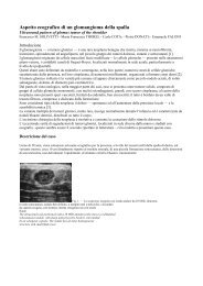
Indications for the use of the Amplatzer vascular ... - ConsultiMedici.it
Indications for the use of the Amplatzer vascular ... - ConsultiMedici.it
Indications for the use of the Amplatzer vascular ... - ConsultiMedici.it

Create successful ePaper yourself
Turn your PDF publications into a flip-book with our unique Google optimized e-Paper software.
708 Radiol med (2008) 113:707–718<br />
Introduction<br />
The <strong>Amplatzer</strong> <strong>vascular</strong> plug (AVP) consists <strong>of</strong> a self-expandable<br />
n<strong>it</strong>inol cylinder secured to a stainless-steel delivery<br />
cable by a microscrew. Anticlockwise rotation <strong>of</strong> <strong>the</strong> cable<br />
unscrews <strong>it</strong> from <strong>the</strong> cylinder, allowing controlled release.<br />
The device grew out <strong>of</strong> two precursors designed exclusively<br />
<strong>for</strong> cardiological purposes, <strong>the</strong> <strong>Amplatzer</strong> septal<br />
occluder and <strong>the</strong> <strong>Amplatzer</strong> duct occluder, <strong>use</strong>d <strong>for</strong> <strong>the</strong> occlusion<br />
<strong>of</strong> interatrial defects and patent ductus arteriosus,<br />
respectively [1, 2]. The system’s plug-like shape and availabil<strong>it</strong>y<br />
in several sizes have progressively expanded <strong>it</strong>s applications<br />
in interventional radiology, where <strong>it</strong> <strong>of</strong>fers an alternative<br />
to permanent embolising material such as metallic<br />
coils or acrylic glue, in <strong>the</strong> occlusion <strong>of</strong> small- to mediumsize<br />
feeding and draining vessels in <strong>the</strong> treatment <strong>of</strong><br />
aneurysms or arteriovenous mal<strong>for</strong>mations (AVM) [3–14].<br />
The aim <strong>of</strong> this study was to review our experience to<br />
confirm <strong>the</strong> reported indications <strong>for</strong> <strong>the</strong> AVP, identify possible<br />
fur<strong>the</strong>r indications in interventional radiology and<br />
evaluate <strong>the</strong> immediate and long-term technical success <strong>of</strong><br />
<strong>the</strong> AVP.<br />
Materials and methods<br />
Over <strong>the</strong> past year, we selected 12 patients (seven men and<br />
five women; mean age 65.8 years; age range 45–82) <strong>for</strong> <strong>the</strong><br />
occlusion <strong>of</strong> five internal iliac arteries close to <strong>the</strong> origin (in<br />
three aortoiliac aneurysms, one internal iliac aneurysm and<br />
one isolated common iliac aneurysm), two common iliac arteries<br />
(in ruptured abdominal aortic aneurysms treated on<br />
an emergency basis), two subclavian arteries (in aortic arch<br />
aneurysms) and three aneurysms <strong>of</strong> <strong>the</strong> mid splenic artery<br />
(two AVPs were <strong>use</strong>d, one proximal and one distal to <strong>the</strong><br />
aneurysm). Preprocedural planning was done w<strong>it</strong>h computed<br />
tomography (CT) angiography to assess <strong>the</strong> procedure’s<br />
feasibil<strong>it</strong>y in relation to <strong>the</strong> tortuos<strong>it</strong>y and diameter <strong>of</strong> <strong>the</strong><br />
target vessel (Figs. 1a,b and 2a). We <strong>use</strong>d 15 AVPs (AGA<br />
Medical Corporation, Golden Valley, MN, USA) w<strong>it</strong>h a diameter<br />
30%–50% larger than that <strong>of</strong> <strong>the</strong> target vessel: 14<br />
mm or 16 mm in diameter delivered through an 8-Fr guiding<br />
ca<strong>the</strong>ter (Envoy, Cordis, Miami, Fl, USA) <strong>for</strong> <strong>the</strong> iliac arteries;<br />
10 mm or 12 mm in diameter delivered through 6- to 7-<br />
Fr guiding ca<strong>the</strong>ters (Envoy) <strong>for</strong> splenic, subclavian and internal<br />
iliac arteries, respectively. A guiding ca<strong>the</strong>ter is preferred<br />
to a long introducer sheath beca<strong>use</strong> <strong>the</strong> AVP device<br />
and <strong>it</strong>s loader cannot cross <strong>the</strong> haemostasis valve <strong>of</strong> <strong>the</strong><br />
sheath.<br />
Oversizing <strong>of</strong> <strong>the</strong> AVP by 30%–50% w<strong>it</strong>h respect to <strong>the</strong><br />
target vessel is justified by <strong>the</strong> elastic<strong>it</strong>y <strong>of</strong> n<strong>it</strong>inol, which allows<br />
<strong>the</strong> cylinder to expand to f<strong>it</strong> <strong>the</strong> diameter <strong>of</strong> <strong>the</strong> vessel.<br />
After expanding, <strong>the</strong> device becomes elongated and assumes<br />
Introduzione<br />
Il sistema VPA è un device cost<strong>it</strong>u<strong>it</strong>o da un cilindro autoespandibile<br />
in N<strong>it</strong>inol fissato mediante una microv<strong>it</strong>e ad un<br />
cavo di introduzione in acciaio inossidabile; il cavo, con un<br />
movimento antiorario, viene sv<strong>it</strong>ato dal cilindro consentendone<br />
un rilascio controllato. Il dispos<strong>it</strong>ivo è nato da due<br />
precursori per utilizzo esclusivo in amb<strong>it</strong>o cardiologico,<br />
l’Amplater Septal Occluder e l’<strong>Amplatzer</strong> Duct Occluder,<br />
per l’occlusione rispettivamente dei difetti interatriali e dei<br />
dotti arteriosi pervi [1, 2]. Le caratteristiche morfologiche<br />
“a tappo” e la disponibil<strong>it</strong>à in più diametri ne hanno progressivamente<br />
ampliato le indicazioni specie in radiologia<br />
interventistica, in alternativa all’utilizzo di materiale embolizzante<br />
di tipo defin<strong>it</strong>ivo, quali spirali metalliche o la<br />
colla acrilica, nell’occlusione di afferenze o efferenze di arterie<br />
e vene di piccolo e medio calibro per il trattamento<br />
della patologia aneurismatica o delle mal<strong>for</strong>mazioni arterovenose<br />
(MAV) [3–14].<br />
Scopo del nostro lavoro è presentare la nostra esperienza<br />
nel confermare le indicazioni all’utilizzo del VPA apparse<br />
in letteratura e nell’individuare possibili ulteriori indicazioni<br />
in radiologia interventistica e valutarne il successo<br />
tecnico immediato e a distanza.<br />
Materiali e metodi<br />
Nell’ultimo anno abbiamo selezionato 12 pazienti (7 maschi<br />
e 5 femmine, età media 65,8 anni, range 45–82) per l’occlusione<br />
mediante 1 VPA di: 5 arterie ipogastriche all’origine<br />
(in 3 aneurismi aorto-iliaci, 1 aneurisma dell’arteria ipogastrica<br />
e 1 aneurisma isolato dell’arteria iliaca comune), 2<br />
arterie iliache comuni (in aneurismi dell’aorta addominale<br />
rotti trattati in urgenza), 2 arterie succlavie (in aneurismi<br />
dell’arco aortico) e di 3 aneurismi dell’arteria splenica del<br />
tratto intermedio, mediante 2 VPA a monte e a valle<br />
dell’aneurisma. Il “planning” pre-procedura è stato effettuato<br />
mediante angio-TC per valutarne la fattibil<strong>it</strong>à in relazione<br />
alla tortuos<strong>it</strong>à vascolare e il calibro del vaso da occludere<br />
(Figg. 1a,b e 2a). Sono stati utilizzati complessivamente<br />
15 VPA (Vascular Plug <strong>Amplatzer</strong>, AGA Medical Corporation,<br />
Golden Valley, MN, USA) del diametro del 30%–50%<br />
superiore rispetto al calibro del vaso da occludere: da 14 o<br />
16 mm di diametro attraverso cateteri guida da 8 F (Envoy,<br />
Cordis, Miami, Fl, USA) per le arterie iliache; del diametro<br />
da 10 o 12 mm mediante cateteri guida da 6–7 F (Envoy,<br />
Cordis, Miami, Fl, USA) rispettivamente per l’arteria splenica,<br />
succlavia e ipogastrica. Il catetere guida è da preferirsi<br />
all’introduttore lungo in quanto il dispos<strong>it</strong>ivo VPA con il suo<br />
caricatore non supera la valvola emostatica di quest’ultimo.<br />
La sovradimensione del calibro del VAP dal 30% al 50%<br />
superiore al calibro del vaso da occludere è giustificata