Clinical practice Terminology SPECT FDG-PET - NVALT
Clinical practice Terminology SPECT FDG-PET - NVALT
Clinical practice Terminology SPECT FDG-PET - NVALT

Create successful ePaper yourself
Turn your PDF publications into a flip-book with our unique Google optimized e-Paper software.
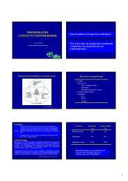
Nuclear medicine in pulmonology<br />
• NSCLC<br />
– SCLC<br />
• Pulmonary carcinoid (NET)<br />
• Sarcoidosis<br />
• Pulmonary embolism<br />
MPM Stokkel<br />
Nuclear Medicine<br />
NKI-AVL<br />
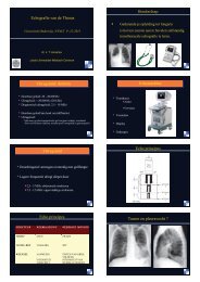
<strong>Clinical</strong> <strong>practice</strong><br />
<strong>SPECT</strong><br />
<strong>FDG</strong>-<strong>PET</strong><br />
Resolution = 1 cm<br />
3 <strong>SPECT</strong> vs 1 <strong>PET</strong><br />
Cost (- effectiveness)<br />
Cardiac perfusion = € 100,00<br />
Lungperfusion = € 25,00<br />
Bonescanning = € 15,00<br />
Ejectionfractions = € 12,50<br />
Brainpathology = € 75,00<br />
Leerdoelen<br />
• Inzicht in verschillende nucleair geneeskundige technieken bij<br />
pulmonale pathologie<br />
• Inzicht in de plaats van de botscan versus F-18 DG <strong>PET</strong> bij<br />
bronchusca<br />
• F-18 DG <strong>PET</strong> bij sarcoidose versus Ga-67 scintigrafie<br />
• Inzicht in de plaats van het perfusie/ventilatiescintigram<br />
– Relatie tot andere diagnostiek<br />
– Antelope, Christopher studie<br />
• <strong>SPECT</strong><br />
• S<strong>PET</strong><br />
• <strong>SPECT</strong>/CT<br />
• <strong>PET</strong><br />
• D<strong>PET</strong><br />
• <strong>PET</strong>/CT<br />
<strong>Terminology</strong><br />
Single photon emission<br />
tomography<br />
Positron emission<br />
tomography = Dual photon<br />
emission tomography<br />
<strong>PET</strong><br />
<strong>PET</strong>/CT
F-MISO = hypoxia<br />
FGD FMT<br />
Somatostatin receptor<br />
• 5 Subtypes<br />
<strong>FDG</strong><br />
FLT<br />
• NET: variable expression<br />
SSTR subtype
F-18 DG <strong>PET</strong> versus Ga-67<br />
scintigrafie in Sarcoidosis<br />
Gouden Standaard?<br />
• Sensitiviteit en specificiteit van Ga-67 scan en F-18 DG<br />
<strong>PET</strong> is moeilijk te meten door ontbreken gouden<br />
standaard<br />
• De clinicus vaart nu op: Klachten pat., ACE, IL2 rec, BAL,<br />
X-thorax en CT<br />
• Ga-67 scintigrafie sinds enkele decaden afh. van instituut<br />
(o.a. AMC, AZ Nieuwegein)<br />
• Rol F-18 DG <strong>PET</strong>?<br />
Sarcoidosis activity<br />
• Serological markers: Angiotensine Converting<br />
Enzyme (ACE) and soluble Interleukin-2<br />
Receptor (sIL2-R)<br />
• Sensitivity ACE 50-77%; genotyping improves<br />
sensitivity up to 86%<br />
Bons et. al. Repsir Med, 2007<br />
Studdy et. al. J Clin Pathol, 1983<br />
Klech et. al. Chest, 1982<br />
Sensitivity and specificity of Ga-67 scan<br />
and F-18-DG <strong>PET</strong><br />
Results<br />
• Sensitivity to detect active sarcoidosis was 88%<br />
for Ga-67 imaging and 97% for F-18 DG <strong>PET</strong><br />
• In Ga-67 imaging, extra pulmonary lesions were<br />
found in 24% of the patients while this was 65%<br />
in F-18 DG <strong>PET</strong><br />
Sarcoidosis<br />
• Sensitivity sIL2-R: 63-82%<br />
Bons et. al. Respir Med, 2007<br />
Rothkranz-Kos et. al. Clin Chem, 2003<br />
• F-18 DG <strong>PET</strong> is sensitive in detecting active<br />
sarcoidosis<br />
Nishiyama et. al. JNM, 2006<br />
Braun et. al. EJNMMI, 2008<br />
Aim: To correlate F-18 DG <strong>PET</strong>, ACE and sIL2-R
Results<br />
• F-18 DG <strong>PET</strong> positive in 34 patients (94%)<br />
• ACE increased in 13 patients (36%)<br />
• sIL2-R increased in 17 patients (47%)<br />
• No correlation was found between SUV max or<br />
avg and ACE or sIL-2R<br />
• Technetium-99m<br />
gelabeld difosfonaat<br />
• hydroxy-apatiet<br />
• functie osteoblasten<br />
Botscan principe<br />
• Benigne aandoeningen:<br />
– Fracturen<br />
– Osteomyelitis/arthritis/discitis<br />
– M. Paget<br />
– Verhoogde bot turnover<br />
– Orthopaedische/post-traumatische<br />
aandoeningen<br />
– Benigne bottumoren, enz<br />
• Maligne aandoeningen:<br />
– skeletmetastasen<br />
– primaire bottumoren<br />
Botscan indicaties<br />
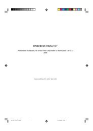
Normale botscan<br />
M, 70 jr, Prostaatca, hormonaal<br />
ontsnapt. Pijn in benen<br />
F-18 DG <strong>PET</strong>, ACE and sIL-2R<br />
Skeletscintigrafie/Botscan<br />
Tc-99m HPD<br />
• 1e fase = perfusiefase<br />
• 2e fase = diffusiefase<br />
• 3e fase = statische fase whole<br />
body of details<br />
• Osteoblastisch:<br />
– Prostaat<br />
– Mammaca<br />
• Osteoclastisch:<br />
– M. Kahler<br />
• Mixed:<br />
– bronchusca,<br />
– Grawitzca,<br />
– blaasca, cervixca.<br />
Botscan techniek<br />
Single Photon<br />
Emissie Computed<br />
Tomografie (<strong>SPECT</strong>)<br />
3-D bewerking<br />
V, 60 jr, microcalcificaties longen en<br />
maagwand bij hyperparathyreoidie<br />
Botscan skeletmetastasen<br />
M, 71 jr, AF 3984 U/L, PSA 3,2<br />
µg/L. St. na prostaatca, non-<br />
Hodgkin lymfoom en niercelca
Paraneoplastisch:<br />
hypertrofische pulmonale<br />
osteoarthropathie<br />
• Prednisongebruik;<br />
pijn linkerheup<br />
Planaire<br />
BS<br />
+F-18 DG<br />
<strong>PET</strong><br />
Botscan V 42 jr<br />
Botscan versus F-18 DG <strong>PET</strong><br />
<br />
<br />
Botscan + F-18 DG <strong>PET</strong> in NSCLC<br />
sens spec PPV NPV<br />
81,2% 74,3% 35% 96%<br />
87,5% 97,3% 90% 98%<br />
<br />
• Lytische laesies<br />
• Specificiteit is laag<br />
• Stralingsdosis<br />
Botscan beperkingen<br />
Botscan M 66 jr<br />
• Prednisongebruik; pijn<br />
in rug<br />
<br />
<br />
<br />
M, 75 jr, verdenking levermeta’s.<br />
Botscan: osteolytische meta Th10/<br />
osteoblastisch Th7-9<br />
Botscan versus F-18 DG <strong>PET</strong><br />
Botscan NaF-18 <strong>PET</strong><br />
V 62 jr, st na<br />
mammaca 2006 T1c<br />
pN2a Mx. Rugpijn<br />
lumbaal en<br />
thoracaal.<br />
Skeletmeta’s?
Long P/V scan indicaties<br />
• Diagnostiek naar longembolieën<br />
• Chronische dyspnoe e.c.i., CTEPH?<br />
• Preoperatief<br />
– TAAA<br />
– Resectie longca<br />
– Bullectomie/LVRC<br />
– Longtransplantatie<br />
– CTEPH/ Endarteriëctomie<br />
Long P/V scan methode<br />
Long segmentenkaart<br />
“Ideale” V/Qpatiënt<br />
• normale X-thorax<br />
• geen COPD<br />
• jong<br />
• mobiel<br />
• De inter-observer kappa<br />
stijgt van 0,62 naar 0,86<br />
• De intra-observer kappa<br />
stijgt van 0,8 naar 1,0<br />
• E.J.R. van Beek 1994<br />
• kan goed met mondkapje op ademen<br />
Long P/V scan methode<br />
• Longperfusie: Tc-99m-MAA<br />
macroaggregaten,<br />
Ø 20-90 micrometer<br />
– i.v. in rugligging<br />
• Ventilatie: Kr-81m gas<br />
T½= 13 sec.<br />
– of radio-aerosolen, Tc-99m<br />
DTPA, Technegas,<br />
Pertechnegas<br />
– Xe-133 gas<br />
Long perfusie/ventilatiescan<br />
Vascular occlusive disease<br />
Obstructive disease<br />
Normaal<br />
Perfusie/ventilatie patronen<br />
Consolidative disease<br />
Restrictive disease<br />
Criteria voor long embolieën<br />
• Pioped: High probability (≥ 2 segmenten),<br />
intermediate<br />
low<br />
normal<br />
• Hull: high (≥ 1 segment)<br />
intermediate (non-diagnostic)<br />
normal<br />
• Gestalt: Niet beoordelen volgens het rigide systeem<br />
van diagnostische criteria, maar volgens ‘intellectuele<br />
synthese van bekende algoritmen, ervaring en<br />
additionele klinische bevindingen’
Diagnostische criteria<br />
Specifieke patronen die de beoordeling<br />
beinvloeden<br />
Defecten door anatomische structuren en artefacten: hart,<br />
diafragma hoogstand, pacemaker etc.<br />
Specifieke patronen die de beoordeling<br />
beïnvloeden<br />
• Fissure sign door interlobair vocht<br />
• Pleuravocht: negatief voorspellende waarde 96.3%<br />
(Bedont et al, 1985)<br />
• Ventilatie/perfusie omgekeerde mismatch<br />
komt alleen op non-embolische basis voor<br />
Belangrijk:<br />
• Opeenvolgende longscans/controlescans<br />
WOMEN OF REPRODUCTIVE AGE<br />
Stralingsdosis op mammae:<br />
• CTPA/4slice: 20-60 mSv<br />
• Mammografie 3 mSv<br />
• V/Q: 0,28 mSv<br />
• D-dimeer positief? Echo<br />
benen<br />
• V/Q wordt geadviseerd<br />
• CT angiogram acceptabel<br />
alternatief<br />
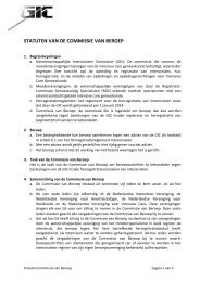
V. 34 jr<br />
High probability voor PE<br />
Specifieke patronen die de beoordeling<br />
beïnvloeden<br />
• Redistributie van flow naar de bovenvelden bij<br />
decompensatio cordis<br />
• ‘The stripe sign’, negatieve voorspellende waarde voor<br />
PE is 93% (Sostman, 1992)<br />
V, 54 jr. COPD en consolidatie<br />
Non-high probability voor PE<br />
Pregnant women<br />
• De stralingsdosis voor de<br />
foetus is bij CTPA en V/Q<br />
ws. ongeveer gelijk.<br />
• Mathematische modellen<br />
en fantoom-experimenten<br />
spreken elkaar tegen
Stralenbelasting<br />
V/Q pre-operatief<br />
Indicaties:<br />
– TAAA<br />
– Resectie longca<br />
– Bullectomie/LVRC<br />
– Longtransplantatie<br />
– CTEPH/ Endarteriëctomie<br />
EINDE<br />
V/Q pre-operatief<br />
CBO 2004:<br />
• Hoofdstuk 2. Criteria voor medische (in)operabiliteit<br />
• 2.3 Longfunctie<br />
• Wanneer de geforceerde-expiratievolume in 1 seconde (FEV1)<br />
en de diffusiecapaciteit (TLCO) beide > 80% zijn en er geen<br />
onverwachte dyspnoe d’effort is, is het operatierisico als niet<br />
verhoogd te beschouwen.<br />
• Geadviseerd wordt, indien niet aan deze criteria wordt voldaan,<br />
door middel van de calculatiemethode (perfusiescan met linksrechtsverhouding<br />
en bij lobectomie aangevuld met de<br />
segmentenmethode) de voorspelde postoperatieve longfunctie<br />
te berekenen.<br />
V 69 jr pre-TAAA<br />
Li:re kwantificatie