Akut pulmoner embolizm ile parankimal ve plevral anormalliklerin ...
Akut pulmoner embolizm ile parankimal ve plevral anormalliklerin ...
Akut pulmoner embolizm ile parankimal ve plevral anormalliklerin ...
Create successful ePaper yourself
Turn your PDF publications into a flip-book with our unique Google optimized e-Paper software.
Kaynaklar<br />
1. Weiss CR, Scatarige JC, Diette GB, Haponik<br />
EF, Merriman B, Fishman EK. CT pulmonary<br />
angiography is the first-line imaging<br />
test for acute pulmonary embolism: a sur<strong>ve</strong>y<br />
of US clinicians. Acad Radiol 2006;<br />
13:434–446.<br />
2. Ghaye B, Ghuysen A, Bruyere PJ, D’Orio<br />
V, Dondelinger RF. Can CT pulmonary<br />
angiography allow assessment of se<strong>ve</strong>rity<br />
and prognosis in patients presenting with<br />
pulmonary embolism? What the radiologist<br />
needs to know. Radiographics 2006; 26:23–<br />
39.<br />
3. Stein PD, Woodard PK, Weg JG, et al.<br />
Diagnostic pathways in acute pulmonary<br />
embolism: recommendations of the<br />
PIOPED II in<strong>ve</strong>stigators. Radiology 2007;<br />
242:15–21.<br />
4. Ghaye B, Remy J, Remy-Jardin M. Nontraumatic<br />
thoracic emergencies: CT diagnosis<br />
of acute pulmonary embolism—the first<br />
10 years. Eur Radiol 2002; 12:1886–1905.<br />
5. Schoepf UJ, Costello P. CT angiography for<br />
diagnosis of pulmonary embolism: state of<br />
the art. Radiology 2004; 230:329–337.<br />
6. Hayashino Y, Goto M, Noguchi Y, Fukui<br />
T. Ventilation-perfusion scanning and helical<br />
CT in suspected pulmonary embolism:<br />
meta-analysis of diagnostic performance.<br />
Radiology 2005; 234:740–748.<br />
7. Stein PD, Fowler SE, Goodman LR, et al.<br />
Multidetector computed tomography for<br />
acute pulmonary embolism. N Engl J Med<br />
2006; 354:2317–2327.<br />
8. Coche EE, Muller NL, Kim KI, Wiggs<br />
BR, Mayo JR. Acute pulmonary embolism:<br />
ancillary findings at spiral CT. Radiology<br />
1998; 207:753–758.<br />
9. Shah AA, Davis SD, Gamsu G, Intriere<br />
L. Parenchymal and pleural findings in<br />
patients with and patients without acute<br />
pulmonary embolism detected at spiral CT.<br />
Radiology 1999; 211:147–153.<br />
10. Reissig A, Heyne JP, Kroegel C. Ancillary<br />
lung parenchymal findings at spiral CT scanning<br />
in pulmonary embolism. Relationship<br />
to chest sonography. Eur J Radiol 2004;<br />
49:250–257.<br />
11. Johnson PT, Wechsler RJ, Salazar AM,<br />
Fisher AM, Nazarian LN, Steiner RM.<br />
Spiral CT of acute pulmonary thromboembolism:<br />
evaluation of pleuroparenchymal<br />
abnormalities. J Comput Assist Tomogr<br />
1999; 23:369–373.<br />
12. Qanadli SD, El Hajjam M, Vieillard-Baron<br />
A, et al. New CT index to quantify arterial<br />
obstruction in pulmonary embolism:<br />
comparison with angiographic index and<br />
echocardiography. AJR Am J Roentgenol<br />
2001; 176:1415–1420.<br />
13. van der Meer RW, Pattynama PM, van<br />
Strijen MJ, et al. Right <strong>ve</strong>ntricular dysfunction<br />
and pulmonary obstruction index at<br />
helical CT: prediction of clinical outcome<br />
during 3-month follow-up in patients with<br />
acute pulmonary embolism. Radiology<br />
2005; 235:798–803.<br />
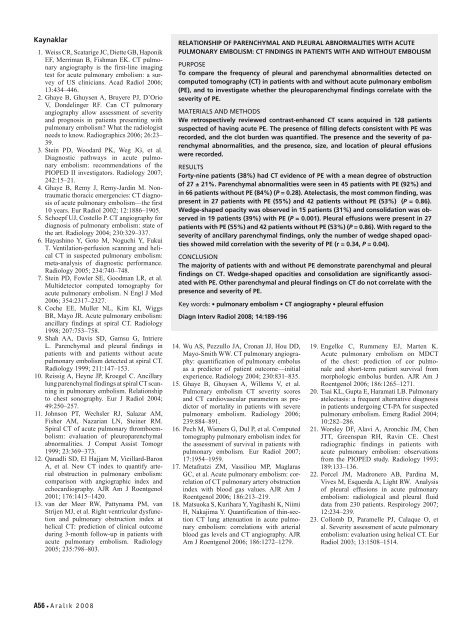
RELATIONSHIP OF PARENCHYMAL AND PLEURAL ABNORMALITIES WITH ACUTE<br />
PULMONARY EMBOLISM: CT FINDINGS IN PATIENTS WITH AND WITHOUT EMBOLISM<br />
PURPOSE<br />
To compare the frequency of pleural and parenchymal abnormalities detected on<br />
computed tomography (CT) in patients with and without acute pulmonary embolism<br />
(PE), and to in<strong>ve</strong>stigate whether the pleuroparenchymal findings correlate with the<br />
se<strong>ve</strong>rity of PE.<br />
MATERIALS AND METHODS<br />
We retrospecti<strong>ve</strong>ly reviewed contrast-enhanced CT scans acquired in 128 patients<br />
suspected of having acute PE. The presence of filling defects consistent with PE was<br />
recorded, and the clot burden was quantified. The presence and the se<strong>ve</strong>rity of parenchymal<br />
abnormalities, and the presence, size, and location of pleural effusions<br />
were recorded.<br />
RESULTS<br />
Forty-nine patients (38%) had CT evidence of PE with a mean degree of obstruction<br />
of 27 ± 21%. Parenchymal abnormalities were seen in 45 patients with PE (92%) and<br />
in 66 patients without PE (84%) (P = 0.28). Atelectasis, the most common finding, was<br />
present in 27 patients with PE (55%) and 42 patients without PE (53%) (P = 0.86).<br />
Wedge-shaped opacity was obser<strong>ve</strong>d in 15 patients (31%) and consolidation was obser<strong>ve</strong>d<br />
in 19 patients (39%) with PE (P = 0.001). Pleural effusions were present in 27<br />
patients with PE (55%) and 42 patients without PE (53%) (P = 0.86). With regard to the<br />
se<strong>ve</strong>rity of ancillary parenchymal findings, only the number of wedge shaped opacities<br />
showed mild correlation with the se<strong>ve</strong>rity of PE (r = 0.34, P = 0.04).<br />
CONCLUSION<br />
The majority of patients with and without PE demonstrate parenchymal and pleural<br />
findings on CT. Wedge-shaped opacities and consolidation are significantly associated<br />
with PE. Other parenchymal and pleural findings on CT do not correlate with the<br />
presence and se<strong>ve</strong>rity of PE.<br />
Key words: • pulmonary embolism • CT angiography • pleural effusion<br />
Diagn Interv Radiol 2008; 14:189-196<br />
14. Wu AS, Pezzullo JA, Cronan JJ, Hou DD,<br />
Mayo-Smith WW. CT pulmonary angiography:<br />
quantification of pulmonary embolus<br />
as a predictor of patient outcome—initial<br />
experience. Radiology 2004; 230:831–835.<br />
15. Ghaye B, Ghuysen A, Willems V, et al.<br />
Pulmonary embolism CT se<strong>ve</strong>rity scores<br />
and CT cardiovascular parameters as predictor<br />
of mortality in patients with se<strong>ve</strong>re<br />
pulmonary embolism. Radiology 2006;<br />
239:884–891.<br />
16. Pech M, Wieners G, Dul P, et al. Computed<br />
tomography pulmonary embolism index for<br />
the assessment of survival in patients with<br />
pulmonary embolism. Eur Radiol 2007;<br />
17:1954–1959.<br />
17. Metafratzi ZM, Vassiliou MP, Maglaras<br />
GC, et al. Acute pulmonary embolism: correlation<br />
of CT pulmonary artery obstruction<br />
index with blood gas values. AJR Am J<br />
Roentgenol 2006; 186:213–219.<br />
18. Matsuoka S, Kurihara Y, Yagihashi K, Niimi<br />
H, Nakajima Y. Quantification of thin-section<br />
CT lung attenuation in acute pulmonary<br />
embolism: correlations with arterial<br />
blood gas le<strong>ve</strong>ls and CT angiography. AJR<br />
Am J Roentgenol 2006; 186:1272–1279.<br />
19. Engelke C, Rummeny EJ, Marten K.<br />
Acute pulmonary embolism on MDCT<br />
of the chest: prediction of cor pulmonale<br />
and short-term patient survival from<br />
morphologic embolus burden. AJR Am J<br />
Roentgenol 2006; 186:1265–1271.<br />
20. Tsai KL, Gupta E, Haramati LB. Pulmonary<br />
atelectasis: a frequent alternati<strong>ve</strong> diagnosis<br />
in patients undergoing CT-PA for suspected<br />
pulmonary embolism. Emerg Radiol 2004;<br />
10:282–286.<br />
21. Worsley DF, Alavi A, Aronchic JM, Chen<br />
JTT, Greenspan RH, Ravin CE. Chest<br />
radiographic findings in patients with<br />
acute pulmonary embolism: observations<br />
from the PIOPED study. Radiology 1993;<br />
189:133–136.<br />
22. Porcel JM, Madronero AB, Pardina M,<br />
Vi<strong>ve</strong>s M, Esquerda A, Light RW. Analysis<br />
of pleural effusions in acute pulmonary<br />
embolism: radiological and pleural fluid<br />
data from 230 patients. Respirology 2007;<br />
12:234–239.<br />
23. Collomb D, Paramelle PJ, Calaque O, et<br />
al. Se<strong>ve</strong>rity assessment of acute pulmonary<br />
embolism: evaluation using helical CT. Eur<br />
Radiol 2003; 13:1508–1514.<br />
A56 Aralı k 2008


