Molekulare Physiologie und Pathobiochemie ausgewählter Magen ...
Molekulare Physiologie und Pathobiochemie ausgewählter Magen ...
Molekulare Physiologie und Pathobiochemie ausgewählter Magen ...

You also want an ePaper? Increase the reach of your titles
YUMPU automatically turns print PDFs into web optimized ePapers that Google loves.
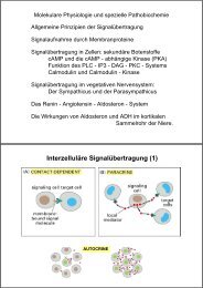
ZIEL<br />
<strong>Molekulare</strong> <strong>Physiologie</strong> - <strong>Pathobiochemie</strong><br />
Depiction of the intestinal mucosa with emphasis on the factors that take part in the development and control of coeliac disease. a | The parts of gluten that are<br />
resistant to processing p gby y luminal and brush-border enzymes y will survive digestion, g , and can be transported p across the mucosal epithelium p as polypeptides.<br />
p yp p<br />
Gluten peptides are deamidated by tissue transglutaminase (TG2), which, in the intestinal mucosa, is located mainly extracellularly in the subepithelial region,<br />
but is also fo<strong>und</strong> in the brush border. CD4+ T cells in the lamina propria recognize deamidated gluten peptides predominantly, presented by HLA-DQ2 or -DQ8<br />
molecules on the cell surface of antigen-presenting cells (APCs). b | Immunofluorescence staining of TG2 (pink), HLA-DQ (green) and T cells (CD3; purple) in<br />
the small-intestine mucosa of an untreated coeliac-disease patient. Note that there is a close spatial relationship between TG2, APCs that express HLA-DQ and<br />
T cells just beneath the epithelium.