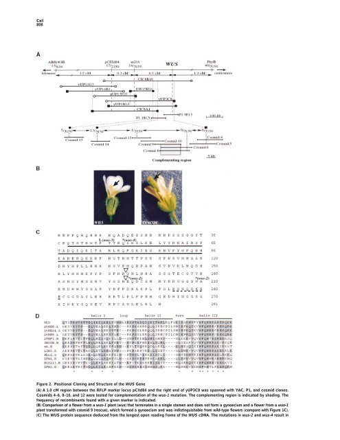
<strong>Cell</strong> 808 Figure 2. Positional Clon<strong>in</strong>g and Structure <strong>of</strong> <strong>the</strong> WUS Gene (A) A 1.0 cM region between <strong>the</strong> RFLP marker locus pCitd84 and <strong>the</strong> right end <strong>of</strong> yUP3C8 was spanned with YAC, P1, and cosmid clones. Cosmids 4–6, 8–10, and 12 were tested for complementation <strong>of</strong> <strong>the</strong> wus-1 mutation. The complement<strong>in</strong>g region is <strong>in</strong>dicated by shad<strong>in</strong>g. The frequency <strong>of</strong> recomb<strong>in</strong>ants found with a given marker is <strong>in</strong>dicated. (B) Comparison <strong>of</strong> a flower from a wus-1 plant (wus) that term<strong>in</strong>ates <strong>in</strong> a s<strong>in</strong>gle stamen and does not form a gynoecium and a flower from a wus-1 plant transformed with cosmid 9 (rescue), which formed a gynoecium and was <strong>in</strong>dist<strong>in</strong>guishable from wild-type flowers (compare with Figure 1C). (C) The WUS prote<strong>in</strong> sequence deduced from <strong>the</strong> longest open read<strong>in</strong>g frame <strong>of</strong> <strong>the</strong> WUS cDNA. The mutations <strong>in</strong> wus-2 and wus-4 result <strong>in</strong>
<strong>Role</strong> <strong>of</strong> <strong>WUSCHEL</strong> <strong>in</strong> <strong>Shoot</strong> and Floral <strong>Meristem</strong>s 809 expressed throughout <strong>the</strong> shoot meristem <strong>in</strong> late-stage wild-type embryos (Figure 4L). In wus <strong>the</strong>re are a few slightly enlarged cells <strong>in</strong> place <strong>of</strong> <strong>the</strong> shoot meristem at this stage (Laux et al., 1996). At least some <strong>of</strong> <strong>the</strong>se cells expressed STM (Figure 4M). However, we did not detect STM expression <strong>in</strong> term<strong>in</strong>ated apices <strong>of</strong> wus seedl<strong>in</strong>gs (data not shown). Therefore, expression <strong>of</strong> both WUS and STM dur<strong>in</strong>g embryo development appears to be <strong>in</strong>itiated <strong>in</strong>dependently <strong>of</strong> <strong>the</strong> respective o<strong>the</strong>r gene, but it was no longer found <strong>in</strong> apparently differentiated apices <strong>of</strong> <strong>the</strong> correspond<strong>in</strong>g mutant seedl<strong>in</strong>gs. Figure 3. Nuclear Localization <strong>of</strong> <strong>the</strong> WUS-GUS Prote<strong>in</strong> WUS Expression <strong>in</strong> Postembryonic Development (A) WUS-GUS fusion prote<strong>in</strong> is localized <strong>in</strong> <strong>the</strong> nucleus <strong>of</strong> onion The wus phenotype suggested that dur<strong>in</strong>g postembryepidermis cells. (B) Negative control: GUS prote<strong>in</strong> is evenly distributed throughout <strong>the</strong> cell. onic development, WUS is required <strong>in</strong> central cells <strong>of</strong> shoot and floral meristems (Laux et al., 1996). To test this hypo<strong>the</strong>sis, we studied WUS expression <strong>in</strong> postembryonic shoot meristems and dur<strong>in</strong>g flower development. In both <strong>the</strong> vegetative (Figure 5A) and <strong>the</strong> <strong>in</strong>flores- Dur<strong>in</strong>g subsequent development, <strong>the</strong> embryonic shoot meristem <strong>in</strong>creases <strong>in</strong> size and <strong>in</strong>itiates <strong>the</strong> first two leaf primordia. In late-stage embryos, WUS mRNA was detected <strong>in</strong> a small group <strong>of</strong> central cells <strong>in</strong> and underneath <strong>the</strong> L3 <strong>of</strong> <strong>the</strong> shoot meristem (Figure 4H). By contrast, WUS mRNA was absent from <strong>the</strong> two outer cell layers. In summary, WUS expression was <strong>in</strong>itiated <strong>in</strong> <strong>the</strong> 16-cell embryo and was gradually conf<strong>in</strong>ed to <strong>the</strong> center <strong>of</strong> <strong>the</strong> develop<strong>in</strong>g embryonic shoot meristem, but it was absent from all o<strong>the</strong>r parts <strong>of</strong> <strong>the</strong> embryo. cence shoot meristem (Figure 5B), WUS mRNA was conf<strong>in</strong>ed to a small group <strong>of</strong> cells <strong>in</strong> <strong>the</strong> center <strong>of</strong> <strong>the</strong> shoot meristem underneath <strong>the</strong> three outermost cell layers. Floral meristems emerge as small bulges (stage 1 <strong>in</strong> Figure 5B; stages accord<strong>in</strong>g to Bowman, 1994) at <strong>the</strong> periphery <strong>of</strong> <strong>the</strong> <strong>in</strong>florescence meristem and develop <strong>in</strong>to a dome <strong>of</strong> cells (stage 2, Figure 5D). Subsequently, sepals, petals, and stamens are successively <strong>in</strong>itiated at <strong>the</strong> periphery <strong>of</strong> <strong>the</strong> floral meristem (Figures 5B and 5E). The floral meristem term<strong>in</strong>ates when its central cells WUS and STM Gene Expression Are Initiated Independently <strong>of</strong> Each O<strong>the</strong>r In stm mutants <strong>the</strong> shoot meristem term<strong>in</strong>ates prema- turely <strong>in</strong> fused organs. Genetic analysis showed that mutations <strong>in</strong> <strong>the</strong> WUS gene did not lead to morphological changes <strong>in</strong> seedl<strong>in</strong>gs <strong>of</strong> strong stm alleles (Endrizzi et al., 1996). However, WUS expression is <strong>in</strong>itiated <strong>in</strong> shoot meristem precursor cells several embryo stages before STM is expressed. To exam<strong>in</strong>e <strong>the</strong> relation between WUS and STM at <strong>the</strong> transcriptional level, we used <strong>in</strong> situ hybridization <strong>in</strong> wus and stm late-stage embryos, <strong>in</strong> which <strong>the</strong> mutant phenotypes could be scored unam- biguously. In late-stage stm embryos, <strong>the</strong>re is no wild- type shoot meristem, and <strong>the</strong> cotyledonary primordia jo<strong>in</strong> at an acute angle (Figure 4K). At this stage, we are consumed dur<strong>in</strong>g <strong>the</strong> <strong>in</strong>itiation <strong>of</strong> carpel primordia (Figure 5F). WUS mRNA was detected <strong>in</strong> stage 1 floral meristems <strong>in</strong> a region separate from <strong>the</strong> WUS expression doma<strong>in</strong> <strong>of</strong> <strong>the</strong> <strong>in</strong>florescence meristem, suggest<strong>in</strong>g that WUS expression was newly established dur<strong>in</strong>g flower development (Figures 5B and 5C). Subsequently, <strong>the</strong> strongest WUS expression was observed <strong>in</strong> stage 2 flowers (Figures 5C and 5D), after which <strong>the</strong> hybridiza- tion signal decreased (Figure 5E) to become undetectable when <strong>the</strong> carpel primordia emerged (Figure 5F). In contrast to <strong>the</strong> shoot meristem, floral meristems accu- mulated WUS mRNA <strong>in</strong> a small group <strong>of</strong> central cells that <strong>in</strong>cluded <strong>the</strong> third cell layer. WUS mRNA was only excluded from <strong>the</strong> two outermost layers. observed strong WUS expression <strong>in</strong> apical cells correspond<strong>in</strong>g to <strong>the</strong> position <strong>of</strong> <strong>the</strong> shoot meristem <strong>in</strong> wild Discussion type, <strong>in</strong>dicat<strong>in</strong>g that <strong>the</strong>re are at least some cells <strong>in</strong> We have reported <strong>the</strong> clon<strong>in</strong>g <strong>of</strong> <strong>the</strong> WUS gene, a central <strong>the</strong> stm apex that have meristematic identity. We were, regulator <strong>of</strong> stem cell fate <strong>in</strong> shoot and floral meristems, however, unable to detect WUS expression <strong>in</strong> apices and have addressed its role dur<strong>in</strong>g embryonic and post- <strong>of</strong> stm seedl<strong>in</strong>gs (data not shown). The STM gene is embryonic meristem development. stop codons (designated *). The mutation <strong>in</strong> wus-1 changes an exon–<strong>in</strong>tron border and results <strong>in</strong> a predicted translational stop a few codons later (designated *). The mutation <strong>in</strong> wus-3 results <strong>in</strong> an am<strong>in</strong>o acid substitution as <strong>in</strong>dicated. The homeodoma<strong>in</strong> (solid l<strong>in</strong>e) and an acidic doma<strong>in</strong> (broken l<strong>in</strong>e) are underl<strong>in</strong>ed. The positions <strong>of</strong> <strong>in</strong>trons are <strong>in</strong>dicated by triangles. (D) Comparison <strong>of</strong> <strong>the</strong> WUS prote<strong>in</strong> region between residues 33 and 98 with homeodoma<strong>in</strong> sequences identified <strong>in</strong> <strong>the</strong> GenBank database. Residues identical or similar (Gribskov et al., 1986) to <strong>the</strong> WUS sequence are shaded. The highly conserved residues <strong>in</strong> homeodoma<strong>in</strong>s are <strong>in</strong>dicated (*) (Gehr<strong>in</strong>g et al., 1990). Gaps are given as dashes. GenBank accession numbers: LIM3, Z22702; ZFH1, M63449; ATBF1, D10250; Pho2, M24613; ZFH2, M63450; SHOXB, U82668; HOXA13, U82827; engrailed (en), U82487; AtHB8, Z50851; AtHB9, Y10922; AtHB14, Y11122. A, <strong>Arabidopsis</strong> thaliana; B,Branchiostoma floridae; D,Drosophila melanogaster; H, human; S, Saccharomyces cerevisiae; and X, Xenopus.


![Estructuras secretoras internas [4.64 MB]](https://img.yumpu.com/14294979/1/190x143/estructuras-secretoras-internas-464-mb.jpg?quality=85)
![anatomía y exomorfología [7.14 MB]](https://img.yumpu.com/12744163/1/190x143/anatomia-y-exomorfologia-714-mb.jpg?quality=85)