Role of WUSCHEL in Regulating Stem Cell Fate in the Arabidopsis Shoot Meristem
Role of WUSCHEL in Regulating Stem Cell Fate in the Arabidopsis Shoot Meristem
Role of WUSCHEL in Regulating Stem Cell Fate in the Arabidopsis Shoot Meristem

You also want an ePaper? Increase the reach of your titles
YUMPU automatically turns print PDFs into web optimized ePapers that Google loves.
<strong>Cell</strong>, Vol. 95, 805–815, December 11, 1998, Copyright ©1998 by <strong>Cell</strong> Press<br />
<strong>Role</strong> <strong>of</strong> <strong>WUSCHEL</strong> <strong>in</strong> Regulat<strong>in</strong>g <strong>Stem</strong> <strong>Cell</strong> <strong>Fate</strong><br />
<strong>in</strong> <strong>the</strong> <strong>Arabidopsis</strong> <strong>Shoot</strong> <strong>Meristem</strong><br />
Klaus F. X. Mayer, † Heiko Scho<strong>of</strong>, Achim Haecker,<br />
Michael Lenhard, Gerd Jürgens, and Thomas Laux*<br />
Lehrstuhl für Entwicklungsgenetik<br />
gene is observed earlier <strong>in</strong> cells between <strong>the</strong> <strong>in</strong>cipient<br />
cotyledonary primordia <strong>of</strong> <strong>the</strong> globular-stage embryo<br />
(Long et al., 1996). However, noth<strong>in</strong>g is known about<br />
Universität Tüb<strong>in</strong>gen <strong>the</strong> orig<strong>in</strong> <strong>of</strong> <strong>the</strong> shoot meristem and its development<br />
Auf der Morgenstelle 1 before this stage.<br />
D-72076 Tüb<strong>in</strong>gen In postembryonic development, <strong>the</strong> <strong>Arabidopsis</strong><br />
Federal Republic <strong>of</strong> Germany shoot meristem first gives rise to a rosette <strong>of</strong> leaves<br />
with axillary meristems and later to floral meristems.<br />
Floral meristems are homologous to shoot meristems,<br />
Summary and <strong>the</strong>ir activity is regulated <strong>in</strong> part by <strong>the</strong> same set <strong>of</strong><br />
genes (Clark et al., 1993; Endrizzi et al., 1996; Laux et<br />
The shoot meristem gives rise to <strong>the</strong> aerial parts <strong>of</strong><br />
higher plants by cont<strong>in</strong>uously <strong>in</strong>itiat<strong>in</strong>g new organs.<br />
The basis <strong>of</strong> this activity is its ability to ma<strong>in</strong>ta<strong>in</strong> a pool<br />
<strong>of</strong> pluripotent stem cells, which are <strong>the</strong> ultimate source<br />
<strong>of</strong> all tissues <strong>of</strong> <strong>the</strong> shoot. In <strong>Arabidopsis</strong> plants mutant<br />
for <strong>the</strong> <strong>WUSCHEL</strong> (WUS) gene, <strong>the</strong> stem cells are misspecified<br />
and appear to undergo differentiation. Here,<br />
we show that WUS encodes a novel homeodoma<strong>in</strong><br />
prote<strong>in</strong> which presumably acts as a transcriptional<br />
regulator. The pattern <strong>of</strong> WUS expression suggests<br />
that stem cells <strong>in</strong> <strong>the</strong> shoot meristem are specified by<br />
an underly<strong>in</strong>g cell group which is established <strong>in</strong> <strong>the</strong> 16-<br />
cell embryo and becomes localized to its prospective<br />
doma<strong>in</strong> <strong>of</strong> function by asymmetric cell divisions.<br />
al., 1996). However, <strong>in</strong> contrast to <strong>Arabidopsis</strong> shoot<br />
meristems, floral meristems give rise to a fixed number<br />
<strong>of</strong> organs and term<strong>in</strong>ate <strong>in</strong> <strong>the</strong> central gynoecium.<br />
In <strong>the</strong> shoot meristem, a central zone (CZ) at <strong>the</strong> sum-<br />
mit where cells divide relatively <strong>in</strong>frequently can be dis-<br />
t<strong>in</strong>guished from <strong>the</strong> surround<strong>in</strong>g peripheral zone (PZ)<br />
with rapidly divid<strong>in</strong>g cells (Figure 1A; Steeves and Sus-<br />
sex, 1989). The CZ is thought to harbor <strong>the</strong> stem cells,<br />
whereas organ formation is <strong>in</strong>itiated <strong>in</strong> <strong>the</strong> PZ (Steeves<br />
and Sussex, 1989). From <strong>the</strong> outside to <strong>the</strong> <strong>in</strong>side, <strong>the</strong><br />
shoot meristem is organized <strong>in</strong>to three separate “germ”<br />
layers that contribute differentially to <strong>the</strong> tissues <strong>of</strong> <strong>the</strong><br />
shoot (Figure 1A; Sat<strong>in</strong>a et al., 1940). <strong>Cell</strong>s <strong>in</strong> <strong>the</strong> outer-<br />
most layer, L1, give rise to <strong>the</strong> epidermis, whereas derivatives<br />
<strong>of</strong> <strong>the</strong> two subepidermal layers, L2 and L3, form<br />
Introduction<br />
<strong>in</strong>ternal tissues. Clonal analysis <strong>in</strong>dicated that each cell<br />
layer conta<strong>in</strong>s 1–3 stem cells, from which all cells with<strong>in</strong><br />
Higher plants form organs throughout <strong>the</strong>ir life via <strong>the</strong> this layer are derived (Stewart and Dermen, 1970).<br />
activity <strong>of</strong> apical meristems (Steeves and Sussex, 1989). To ma<strong>in</strong>ta<strong>in</strong> <strong>the</strong> <strong>in</strong>tegrity <strong>of</strong> <strong>the</strong> shoot meristem dur<strong>in</strong>g<br />
Dur<strong>in</strong>g plant embryogenesis only a basic body organiza- repetitive organ formation, cell proliferation and organ<br />
tion is laid down with a root and a shoot meristem at <strong>in</strong>itiation must be precisely coord<strong>in</strong>ated (Clark, 1997;<br />
<strong>the</strong> basal and apical pole <strong>of</strong> <strong>the</strong> seedl<strong>in</strong>g, respectively. Meyerowitz, 1997). The balance between <strong>the</strong>se two pro-<br />
The meristems conta<strong>in</strong> a pool <strong>of</strong> pluripotent stem cells, cesses appears to be competitively regulated by <strong>the</strong><br />
which are able to self-ma<strong>in</strong>ta<strong>in</strong> and to produce <strong>the</strong> cells CLAVATA (CLV) and STM genes (Clark et al., 1996).<br />
required for organ <strong>in</strong>itiation. The <strong>in</strong>itiation and ma<strong>in</strong>te- CLV1, a putative receptor k<strong>in</strong>ase, is likely to be a componance<br />
<strong>of</strong> stem cells and <strong>the</strong>ir <strong>in</strong>tegration <strong>in</strong>to organ- nent <strong>of</strong> a signal<strong>in</strong>g pathway that promotes organ formaform<strong>in</strong>g<br />
meristems are thus <strong>the</strong> basis for cont<strong>in</strong>uous tion (Clark et al., 1997; Williams et al., 1997; Laufs et al.,<br />
postembryonic plant development. 1998). Its antagonist, STM, is a homeodoma<strong>in</strong> prote<strong>in</strong><br />
The orig<strong>in</strong> and development <strong>of</strong> <strong>the</strong> shoot meristem <strong>in</strong> <strong>of</strong> <strong>the</strong> KNOTTED-1 family (Kerstetter et al., 1994; Long<br />
<strong>the</strong> embryo have been discussed controversially. Based<br />
on comparative morphology, one model <strong>in</strong> classical botany<br />
holds that <strong>the</strong> shoot meristem is first differentiated<br />
by <strong>the</strong> simultaneous <strong>in</strong>itiation <strong>of</strong> <strong>the</strong> cotyledons and a<br />
perpetuat<strong>in</strong>g center <strong>in</strong> <strong>the</strong> apical region <strong>of</strong> <strong>the</strong> globular<br />
embryo (Spurr, 1949; Kaplan, 1969; Kaplan and Cooke,<br />
1997). Alternatively, <strong>the</strong> shoot meristem has been <strong>in</strong>ter-<br />
preted as <strong>the</strong> result <strong>of</strong> an embryonic pattern<strong>in</strong>g process<br />
(Hake et al., 1995; Laux and Jürgens, 1997; Long and<br />
Barton, 1998; Moussian et al., 1998). Histologically, <strong>the</strong><br />
shoot meristem can be recognized at mid-embryo<br />
stages (Barton and Poethig, 1993). The expression <strong>of</strong> <strong>the</strong><br />
shoot meristem-specific SHOOTMERISTEMLESS (STM)<br />
et al., 1996) and appears to prevent <strong>the</strong> entry <strong>of</strong> cells<br />
<strong>in</strong>to organogenesis (Clark et al., 1996; Endrizzi et al.,<br />
1996).<br />
Little is known, however, about <strong>the</strong> mechanisms by<br />
which <strong>the</strong> stem cells are specified dur<strong>in</strong>g <strong>the</strong> formation<br />
and ma<strong>in</strong>tenance <strong>of</strong> shoot and floral meristems. The<br />
stem cells are pluripotent <strong>in</strong> that <strong>the</strong>y give rise to a<br />
variety <strong>of</strong> cell types, and <strong>the</strong>y are undifferentiated <strong>in</strong> that<br />
<strong>the</strong>y lack features <strong>of</strong> cells <strong>in</strong> mature tissue (Barlow, 1978;<br />
Steeves and Sussex, 1989). Clonal analysis (Ruth et al.,<br />
1985) <strong>in</strong>dicated that stem cell fate is specified by posi-<br />
tional <strong>in</strong>formation and imposed on <strong>the</strong> cells that cur-<br />
rently reside at <strong>the</strong> summit <strong>of</strong> <strong>the</strong> shoot meristem (re-<br />
* To whom correspondence should be addressed (e-mail: thomas.<br />
laux@uni-tueb<strong>in</strong>gen.de).<br />
† Present address: Max-Planck-Institut für Biochemie, Am Klopferviewed<br />
<strong>in</strong> Newman, 1965; Laux and Mayer, 1998). Only<br />
<strong>the</strong> progeny <strong>of</strong> stem cells that stay at this position rema<strong>in</strong><br />
pluripotent, whereas daughter cells that leave this posispitz<br />
18a, D-82152 Mart<strong>in</strong>sried, Federal Republic <strong>of</strong> Germany. tion differentiate.
<strong>Cell</strong><br />
806<br />
regulators <strong>of</strong> shoot and floral meristems, suggest<strong>in</strong>g<br />
that WUS <strong>in</strong>tegrates positional and temporal <strong>in</strong>formation<br />
from <strong>the</strong>se genes (Laux et al., 1996; Laux and Scho<strong>of</strong>,<br />
1997).<br />
Here, we report <strong>the</strong> molecular analysis <strong>of</strong> <strong>the</strong> WUS<br />
gene, address<strong>in</strong>g its role <strong>in</strong> shoot and floral meristems.<br />
Our data support a novel model to expla<strong>in</strong> how <strong>the</strong>se<br />
meristems are formed and ma<strong>in</strong>ta<strong>in</strong>ed <strong>in</strong> plant development.<br />
We provide evidence that <strong>the</strong> specification <strong>of</strong> stem<br />
cells requires a small group <strong>of</strong> underly<strong>in</strong>g cells that exhibit<br />
WUS expression. This cell group orig<strong>in</strong>ates dur<strong>in</strong>g<br />
early embryo pattern formation long before a shoot meristem<br />
can be recognized.<br />
Results<br />
Map-Based Clon<strong>in</strong>g <strong>of</strong> <strong>the</strong> WUS Gene<br />
To isolate <strong>the</strong> WUS gene, we used RFLP- and PCRbased<br />
mapp<strong>in</strong>g <strong>of</strong> recomb<strong>in</strong>ation breakpo<strong>in</strong>ts <strong>in</strong> 3150<br />
meiotic events from a cross <strong>of</strong> wus-1 (Ler) � wild type<br />
(Nd). We determ<strong>in</strong>ed <strong>the</strong> order <strong>of</strong> genomic loci as ABI14-<br />
GII, pCITd84, m216, WUS, and PhyB and localized WUS<br />
about 0.8 cM proximal to <strong>the</strong> RFLP marker pCITd84<br />
(Figure 2A). We <strong>in</strong>itiated a chromosome walk from<br />
pCITd84 with yeast artificial chromosomes (YAC) and<br />
localized WUS on a s<strong>in</strong>gle YAC clone <strong>of</strong> about 400 kb,<br />
yUP3C8. From <strong>the</strong> right end <strong>of</strong> this YAC, we <strong>in</strong>itiated a<br />
cosmid walk spann<strong>in</strong>g about 52 kb and mapped <strong>the</strong><br />
WUS gene to this contig (Figure 2A).<br />
To fur<strong>the</strong>r localize <strong>the</strong> WUS gene, we transformed<br />
cosmids represent<strong>in</strong>g this contig <strong>in</strong>to roots <strong>of</strong> homozygous<br />
wus-1 seedl<strong>in</strong>gs and exam<strong>in</strong>ed <strong>the</strong> regenerated<br />
transgenic plants for complementation. The seedl<strong>in</strong>gs<br />
had been obta<strong>in</strong>ed from a cross <strong>of</strong> wus-1 (Ler) � wild<br />
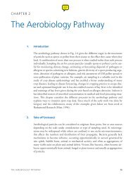
Figure 1. <strong>Shoot</strong> and Floral <strong>Meristem</strong> Development type (Ws-2), which enabled us to confirm <strong>the</strong>ir genotype<br />
(A) Organization <strong>of</strong> <strong>the</strong> shoot meristem. The tissues <strong>of</strong> <strong>the</strong> plant at <strong>the</strong> WUS locus by PCR for <strong>the</strong> closely l<strong>in</strong>ked markers<br />
shoot are derived from three germ layers <strong>of</strong> <strong>the</strong> shoot meristem,<br />
m216 and yUP3C8 right (Figure 2A). Roots taken from<br />
L1–L3 (arrows). Each layer presumably conta<strong>in</strong>s 1–3 stem cells (sc,<br />
darkly shaded) with<strong>in</strong> <strong>the</strong> apical part <strong>of</strong> <strong>the</strong> central zone (CZ, fa<strong>in</strong>tly 14 seedl<strong>in</strong>gs that were transformed with cosmids 6, 8,<br />
shaded). Organ primordia (p) are <strong>in</strong>itiated <strong>in</strong> <strong>the</strong> peripheral zone (PZ, or 9 (Figure 2A) gave rise to a total <strong>of</strong> 76 transgenic<br />
hatched). Underneath <strong>the</strong> CZ, <strong>the</strong> flat cells <strong>of</strong> <strong>the</strong> rib zone (RZ, plants that showed complementation <strong>of</strong> <strong>the</strong> wus defect<br />
horizontal stripes) form <strong>the</strong> central pith <strong>of</strong> <strong>the</strong> shoot axis. and formed flowers with a complete set <strong>of</strong> organs (Figure<br />
(B and D) Live seedl<strong>in</strong>gs. (B) Wild-type seedl<strong>in</strong>gs give rise to a<br />
2B). By contrast, all transgenic plants conta<strong>in</strong><strong>in</strong>g any <strong>of</strong><br />
rosette <strong>of</strong> leaves. (D) wus seedl<strong>in</strong>g apices discont<strong>in</strong>ue leaf formation<br />
<strong>the</strong> o<strong>the</strong>r cosmids showed <strong>the</strong> wus mutant phenotype.<br />
after two true leaves have been formed. c, cotyledon; l, leaf.<br />
(C and E) Scann<strong>in</strong>g electron micrographs <strong>of</strong> flowers. (C) Wild-type The progeny <strong>of</strong> <strong>the</strong> plants that showed complementation<br />
flowers conta<strong>in</strong> four whorls <strong>of</strong> organs: four sepals <strong>in</strong> <strong>the</strong> first, four segregated WUS and wus phenotypes. All <strong>of</strong> 338 kana-<br />
petals <strong>in</strong> <strong>the</strong> second, six stamens <strong>in</strong> <strong>the</strong> third, and two fused carpels myc<strong>in</strong>-resistant plants tested exhibited a wild-type phe<strong>in</strong><br />
<strong>the</strong> fourth whorl. (E) wus flowers, which are occasionally formed notype, <strong>in</strong>dicat<strong>in</strong>g that <strong>the</strong> T-DNA that conferred kanaon<br />
adventitious shoots, display normal organ numbers <strong>in</strong> <strong>the</strong> two<br />
myc<strong>in</strong> resistance also carried <strong>the</strong> WUS gene (data not<br />
outer whorls but term<strong>in</strong>ate <strong>in</strong> a s<strong>in</strong>gle central stamen.<br />
Scale bars, 1 mm (B and D) and 500 �m (C and E).<br />
shown).<br />
The three complement<strong>in</strong>g cosmids shared two H<strong>in</strong>dIII<br />
DNA fragments <strong>of</strong> 10 and 1.5 kb, respectively. With <strong>the</strong>se<br />
fragments, we screened 5 � 105 Recent genetic analysis <strong>in</strong> <strong>Arabidopsis</strong> suggests a<br />
clones <strong>of</strong> a cDNA library<br />
central role for <strong>the</strong> WUS gene <strong>in</strong> regulat<strong>in</strong>g stem cell fate made from young flowers (Weigel et al., 1992) and isothroughout<br />
development. Mutations <strong>in</strong> <strong>the</strong> WUS gene lated seven <strong>in</strong>dependent cDNA clones, which repre-<br />
specifically result <strong>in</strong> <strong>the</strong> failure <strong>of</strong> shoot and floral meri- sented a s<strong>in</strong>gle transcript. Two clones displayed one<br />
stems to self-ma<strong>in</strong>ta<strong>in</strong> (Figures 1B–1E) (Laux et al., long open read<strong>in</strong>g frame <strong>of</strong> 873 nucleotides. We con-<br />
1996). Although <strong>in</strong> <strong>the</strong> absence <strong>of</strong> WUS, stem cells ap- firmed that this open read<strong>in</strong>g frame represented <strong>the</strong><br />
pear misspecified, <strong>the</strong>y are not <strong>in</strong>corporated <strong>in</strong>to or- WUS gene by identify<strong>in</strong>g mutations <strong>in</strong> <strong>the</strong> cognate geno-<br />
gans, <strong>in</strong>dicat<strong>in</strong>g that WUS is required to specify stem mic DNA <strong>of</strong> four different wus alleles (see below). Stop<br />
cell fate ra<strong>the</strong>r than to repress organ formation. Genetic codons upstream <strong>of</strong> this open read<strong>in</strong>g frame <strong>in</strong>dicated<br />
analysis <strong>in</strong>dicated that WUS <strong>in</strong>teracts with several o<strong>the</strong>r that <strong>the</strong>se cDNAs conta<strong>in</strong>ed <strong>the</strong> complete cod<strong>in</strong>g region.
<strong>Role</strong> <strong>of</strong> <strong>WUSCHEL</strong> <strong>in</strong> <strong>Shoot</strong> and Floral <strong>Meristem</strong>s<br />
807<br />
The WUS Gene Encodes a Homeodoma<strong>in</strong> Prote<strong>in</strong> <strong>the</strong> loss <strong>of</strong> about one-third <strong>of</strong> <strong>the</strong> prote<strong>in</strong>, <strong>in</strong>clud<strong>in</strong>g <strong>the</strong><br />
The putative transcription start <strong>of</strong> <strong>the</strong> WUS gene was putative transactivation doma<strong>in</strong>. The weak wus-3 allele<br />
determ<strong>in</strong>ed by 5�-RACE. We analyzed <strong>the</strong> genomic se- conta<strong>in</strong>s a prol<strong>in</strong>e to leuc<strong>in</strong>e missense mutation <strong>in</strong> <strong>the</strong><br />
quence upstream <strong>of</strong> <strong>the</strong> WUS gene and identified a TATA<br />
box, a CCAAT box, and a GC box at 35 bp, 56 bp, and<br />
N-term<strong>in</strong>al part <strong>of</strong> <strong>the</strong> homeodoma<strong>in</strong>.<br />
80 bp upstream <strong>of</strong> <strong>the</strong> transcription start, respectively<br />
(data not shown). A putative polyadenylation signal (Wu<br />
et al., 1995) was identified 20 bp upstream <strong>of</strong> <strong>the</strong> poly(A)<br />
tail (data not shown). A comparison <strong>of</strong> genomic and<br />
cDNA sequences revealed <strong>the</strong> presence <strong>of</strong> two <strong>in</strong>trons<br />
with a length <strong>of</strong> 566 bp and 90 bp, respectively (Figure<br />
2C). The WUS gene encodes a conceptual prote<strong>in</strong> <strong>of</strong><br />
291 am<strong>in</strong>o acids, and sequence comparison revealed<br />
two putative functional doma<strong>in</strong>s (Figure 2C). The region<br />
between am<strong>in</strong>o acid residues 33–98 shows similarity to<br />
homeodoma<strong>in</strong>s (see below), and <strong>the</strong> region between<br />
<strong>Cell</strong>ular Localization <strong>of</strong> <strong>the</strong> WUS Prote<strong>in</strong><br />
In order to study <strong>the</strong> subcellular localization <strong>of</strong> <strong>the</strong> WUS<br />
prote<strong>in</strong>, we used a translational fusion between WUS<br />
and <strong>the</strong> Glucuronidase (GUS) reporter gene <strong>in</strong> transient<br />
expression assays (Varagona et al., 1992). Figure 3 shows<br />
that WUS targeted most GUS activity <strong>in</strong>to <strong>the</strong> nucleus<br />
<strong>of</strong> onion epidermis cells. The nuclear localization activity<br />
<strong>of</strong> WUS is consistent with its proposed role as a tran-<br />
scriptional regulator.<br />
residues 234–241 conta<strong>in</strong>s a cluster <strong>of</strong> acidic am<strong>in</strong>o acid WUS Expression dur<strong>in</strong>g Embryogenesis<br />
residues. Computer analysis predicted that <strong>the</strong> acidic Phenotypic analyses had suggested that WUS is re-<br />
cluster can form an amphipathic � helix, similar to known quired for pluripotent cell fate dur<strong>in</strong>g embryonic shoot<br />
transactivation doma<strong>in</strong>s <strong>of</strong> transcriptional regulators meristem development (Laux et al., 1996). To test this<br />
(Ptashne, 1988).<br />
hypo<strong>the</strong>sis, we analyzed WUS mRNA expression dur<strong>in</strong>g<br />
embryogenesis. Due to an <strong>in</strong>variant cell division pattern<br />
WUS Represents a Novel Subtype <strong>of</strong> <strong>the</strong><br />
Homeodoma<strong>in</strong> Prote<strong>in</strong> Family<br />
Homeodoma<strong>in</strong> prote<strong>in</strong>s have been identified <strong>in</strong> many<br />
organisms and generally appear to be <strong>in</strong>volved <strong>in</strong> devel-<br />
opmental or cell type regulation. The characteristic ho-<br />
meodoma<strong>in</strong> is a conserved prote<strong>in</strong> doma<strong>in</strong> <strong>of</strong> about 60<br />
am<strong>in</strong>o acids that functions <strong>in</strong> sequence-specific DNA<br />
b<strong>in</strong>d<strong>in</strong>g. The hallmarks are a helix-loop-helix-turn-helix<br />
structure and 12 highly conserved am<strong>in</strong>o acid residues<br />
(Gehr<strong>in</strong>g et al., 1990; Laughon, 1991). The WUS homeo-<br />
doma<strong>in</strong> has all <strong>of</strong> <strong>the</strong>se properties and was found to be<br />
about 30% identical to and 45%–50% similar to several<br />
homeodoma<strong>in</strong> sequences from plants, animals, and yeast<br />
(Figure 2D). To determ<strong>in</strong>e <strong>the</strong> relationship <strong>of</strong> WUS to<br />
o<strong>the</strong>r homeodoma<strong>in</strong> prote<strong>in</strong>s, we performed a similarity<br />
analysis us<strong>in</strong>g a heuristic tree-build<strong>in</strong>g program. This<br />
analysis revealed that <strong>the</strong> WUS homeodoma<strong>in</strong> cannot<br />
be significantly grouped toge<strong>the</strong>r with any o<strong>the</strong>r homeodoma<strong>in</strong><br />
(data not shown) and <strong>the</strong>refore represents a<br />
novel branch on <strong>the</strong> homeodoma<strong>in</strong> prote<strong>in</strong> tree.<br />
<strong>in</strong> <strong>in</strong>itial stages <strong>of</strong> <strong>Arabidopsis</strong> embryo development,<br />
morphological regions <strong>of</strong> <strong>the</strong> seedl<strong>in</strong>g can be traced<br />
back to cells <strong>in</strong> <strong>the</strong> early embryo (Mansfield and Briarty,<br />
1991; Jürgens and Mayer, 1994). The eight-cell embryo<br />
consists <strong>of</strong> an apical region <strong>of</strong> four cells, which will form<br />
<strong>the</strong> shoot meristem and most <strong>of</strong> <strong>the</strong> cotyledons, and a<br />
basal region <strong>of</strong> four cells, which will form <strong>the</strong> hypocotyl<br />
and most <strong>of</strong> <strong>the</strong> root. Up to <strong>the</strong> eight-cell embryo stage,<br />
no WUS expression was detected (Figure 4A).<br />
Each <strong>of</strong> <strong>the</strong> eight embryo cells divides pericl<strong>in</strong>ally to<br />
give an <strong>in</strong>ner cell and an outer protoderm cell. This was<br />
<strong>the</strong> earliest stage at which WUS mRNA was detected.<br />
It was conf<strong>in</strong>ed to <strong>the</strong> four <strong>in</strong>ner cells <strong>of</strong> <strong>the</strong> apical region<br />
(Figure 4B). Each <strong>of</strong> <strong>the</strong>se <strong>in</strong>ner cells <strong>the</strong>n divides longitud<strong>in</strong>ally,<br />
giv<strong>in</strong>g rise to a central and a peripheral daugh-<br />
ter. WUS mRNA was only detected <strong>in</strong> central daughter<br />
cells but was absent from peripheral daughters (Figures<br />
4C and 4D).<br />
The transition-stage embryo consists <strong>of</strong> about 100<br />
cells and is characterized by <strong>the</strong> outgrowth <strong>of</strong> cotyledon-<br />
ary primordia on each side <strong>of</strong> <strong>the</strong> presumptive shoot<br />
meristem region (Figure 4E). At this stage, WUS mRNA<br />
Mutations Disrupt<strong>in</strong>g <strong>the</strong> WUS Gene<br />
was detected <strong>in</strong> two subepidermal cells <strong>in</strong> <strong>the</strong> center <strong>of</strong><br />
Based on <strong>the</strong> average number <strong>of</strong> central stamens <strong>the</strong> apex (Figure 4E). Serial sections suggested that <strong>the</strong><br />
formed <strong>in</strong> mutant flowers, we classified <strong>the</strong> three mutant WUS expression doma<strong>in</strong> was only one cell deep (data<br />
alleles wus-1, wus-2, and wus-4 as strong (about one not shown).<br />
stamen per flower) and wus-3 as a weak allele (about At <strong>the</strong> heart stage, <strong>the</strong> basic body organization, com-<br />
three stamens per flower). The WUS gene was amplified pris<strong>in</strong>g cotyledonary primordia, shoot meristem primorand<br />
sequenced from genomic DNA <strong>of</strong> homozygous wus dium, hypocotyl primordium, and root pole, is histologi-<br />
mutants. In each allele a s<strong>in</strong>gle po<strong>in</strong>t mutation relative cally recognizable. The subepidermal cells with<strong>in</strong> <strong>the</strong><br />
to wild type was detected (Figure 2C). The mutations shoot meristem region have divided transversely, giv<strong>in</strong>g<br />
<strong>in</strong> <strong>the</strong> three strong wus alleles presumably lead to a rise to upper (L2) and lower (L3) daughter cells. WUS<br />
premature translational stop. In wus-4, a TAA stop co- mRNA was restricted to <strong>the</strong> L3 daughter cells but was<br />
don results <strong>in</strong> a translational stop after 44 am<strong>in</strong>o acid absent from <strong>the</strong> L2 daughters (Figure 4F). Longitud<strong>in</strong>al<br />
residues. In wus-1, <strong>the</strong> 5�-exon-<strong>in</strong>tron boundary <strong>of</strong> <strong>in</strong>tron sections perpendicular to <strong>the</strong> plane <strong>of</strong> <strong>the</strong> cotyledons<br />
2 is changed from GG to GA, replac<strong>in</strong>g a G that is highly revealed that <strong>the</strong> WUS expression doma<strong>in</strong> spans only<br />
conserved at plant gene splice sites (Brown, 1996). The one cell <strong>in</strong> depth (Figure 4G). Until this stage, we have<br />
failure to remove <strong>in</strong>tron 2 results <strong>in</strong> a translational stop not found a s<strong>in</strong>gle case (n � 60) <strong>in</strong> which after a cell<br />
after a few codons with<strong>in</strong> <strong>the</strong> <strong>in</strong>tron (data not shown). division both daughter cells conta<strong>in</strong>ed WUS mRNA, <strong>in</strong>di-<br />
wus-2 conta<strong>in</strong>s a TAG stop codon at am<strong>in</strong>o acid position cat<strong>in</strong>g that asymmetric WUS mRNA distribution was<br />
209. In wus-1 and wus-2, a translational stop results <strong>in</strong><br />
established dur<strong>in</strong>g or rapidly after cell division.
<strong>Cell</strong><br />
808<br />
Figure 2. Positional Clon<strong>in</strong>g and Structure <strong>of</strong> <strong>the</strong> WUS Gene<br />
(A) A 1.0 cM region between <strong>the</strong> RFLP marker locus pCitd84 and <strong>the</strong> right end <strong>of</strong> yUP3C8 was spanned with YAC, P1, and cosmid clones.<br />
Cosmids 4–6, 8–10, and 12 were tested for complementation <strong>of</strong> <strong>the</strong> wus-1 mutation. The complement<strong>in</strong>g region is <strong>in</strong>dicated by shad<strong>in</strong>g. The<br />
frequency <strong>of</strong> recomb<strong>in</strong>ants found with a given marker is <strong>in</strong>dicated.<br />
(B) Comparison <strong>of</strong> a flower from a wus-1 plant (wus) that term<strong>in</strong>ates <strong>in</strong> a s<strong>in</strong>gle stamen and does not form a gynoecium and a flower from a wus-1<br />
plant transformed with cosmid 9 (rescue), which formed a gynoecium and was <strong>in</strong>dist<strong>in</strong>guishable from wild-type flowers (compare with Figure 1C).<br />
(C) The WUS prote<strong>in</strong> sequence deduced from <strong>the</strong> longest open read<strong>in</strong>g frame <strong>of</strong> <strong>the</strong> WUS cDNA. The mutations <strong>in</strong> wus-2 and wus-4 result <strong>in</strong>
<strong>Role</strong> <strong>of</strong> <strong>WUSCHEL</strong> <strong>in</strong> <strong>Shoot</strong> and Floral <strong>Meristem</strong>s<br />
809<br />
expressed throughout <strong>the</strong> shoot meristem <strong>in</strong> late-stage<br />
wild-type embryos (Figure 4L). In wus <strong>the</strong>re are a few<br />
slightly enlarged cells <strong>in</strong> place <strong>of</strong> <strong>the</strong> shoot meristem at<br />
this stage (Laux et al., 1996). At least some <strong>of</strong> <strong>the</strong>se<br />
cells expressed STM (Figure 4M). However, we did not<br />
detect STM expression <strong>in</strong> term<strong>in</strong>ated apices <strong>of</strong> wus<br />
seedl<strong>in</strong>gs (data not shown). Therefore, expression <strong>of</strong><br />
both WUS and STM dur<strong>in</strong>g embryo development appears<br />
to be <strong>in</strong>itiated <strong>in</strong>dependently <strong>of</strong> <strong>the</strong> respective<br />
o<strong>the</strong>r gene, but it was no longer found <strong>in</strong> apparently<br />
differentiated apices <strong>of</strong> <strong>the</strong> correspond<strong>in</strong>g mutant seedl<strong>in</strong>gs.<br />
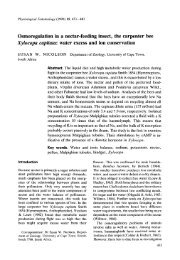
Figure 3. Nuclear Localization <strong>of</strong> <strong>the</strong> WUS-GUS Prote<strong>in</strong> WUS Expression <strong>in</strong> Postembryonic Development<br />
(A) WUS-GUS fusion prote<strong>in</strong> is localized <strong>in</strong> <strong>the</strong> nucleus <strong>of</strong> onion The wus phenotype suggested that dur<strong>in</strong>g postembryepidermis<br />
cells.<br />
(B) Negative control: GUS prote<strong>in</strong> is evenly distributed throughout<br />
<strong>the</strong> cell.<br />
onic development, WUS is required <strong>in</strong> central cells <strong>of</strong><br />
shoot and floral meristems (Laux et al., 1996). To test<br />
this hypo<strong>the</strong>sis, we studied WUS expression <strong>in</strong> postembryonic<br />
shoot meristems and dur<strong>in</strong>g flower development.<br />
In both <strong>the</strong> vegetative (Figure 5A) and <strong>the</strong> <strong>in</strong>flores-<br />
Dur<strong>in</strong>g subsequent development, <strong>the</strong> embryonic shoot<br />
meristem <strong>in</strong>creases <strong>in</strong> size and <strong>in</strong>itiates <strong>the</strong> first two<br />
leaf primordia. In late-stage embryos, WUS mRNA was<br />
detected <strong>in</strong> a small group <strong>of</strong> central cells <strong>in</strong> and underneath<br />
<strong>the</strong> L3 <strong>of</strong> <strong>the</strong> shoot meristem (Figure 4H). By contrast,<br />
WUS mRNA was absent from <strong>the</strong> two outer cell<br />
layers. In summary, WUS expression was <strong>in</strong>itiated <strong>in</strong> <strong>the</strong><br />
16-cell embryo and was gradually conf<strong>in</strong>ed to <strong>the</strong> center<br />
<strong>of</strong> <strong>the</strong> develop<strong>in</strong>g embryonic shoot meristem, but it was<br />
absent from all o<strong>the</strong>r parts <strong>of</strong> <strong>the</strong> embryo.<br />
cence shoot meristem (Figure 5B), WUS mRNA was<br />
conf<strong>in</strong>ed to a small group <strong>of</strong> cells <strong>in</strong> <strong>the</strong> center <strong>of</strong> <strong>the</strong><br />
shoot meristem underneath <strong>the</strong> three outermost cell<br />
layers.<br />
Floral meristems emerge as small bulges (stage 1 <strong>in</strong><br />
Figure 5B; stages accord<strong>in</strong>g to Bowman, 1994) at <strong>the</strong><br />
periphery <strong>of</strong> <strong>the</strong> <strong>in</strong>florescence meristem and develop<br />
<strong>in</strong>to a dome <strong>of</strong> cells (stage 2, Figure 5D). Subsequently,<br />
sepals, petals, and stamens are successively <strong>in</strong>itiated<br />
at <strong>the</strong> periphery <strong>of</strong> <strong>the</strong> floral meristem (Figures 5B and<br />
5E). The floral meristem term<strong>in</strong>ates when its central cells<br />
WUS and STM Gene Expression Are Initiated<br />
Independently <strong>of</strong> Each O<strong>the</strong>r<br />
In stm mutants <strong>the</strong> shoot meristem term<strong>in</strong>ates prema-<br />
turely <strong>in</strong> fused organs. Genetic analysis showed that<br />
mutations <strong>in</strong> <strong>the</strong> WUS gene did not lead to morphological<br />
changes <strong>in</strong> seedl<strong>in</strong>gs <strong>of</strong> strong stm alleles (Endrizzi et<br />
al., 1996). However, WUS expression is <strong>in</strong>itiated <strong>in</strong> shoot<br />
meristem precursor cells several embryo stages before<br />
STM is expressed. To exam<strong>in</strong>e <strong>the</strong> relation between<br />
WUS and STM at <strong>the</strong> transcriptional level, we used <strong>in</strong><br />
situ hybridization <strong>in</strong> wus and stm late-stage embryos,<br />
<strong>in</strong> which <strong>the</strong> mutant phenotypes could be scored unam-<br />
biguously. In late-stage stm embryos, <strong>the</strong>re is no wild-<br />
type shoot meristem, and <strong>the</strong> cotyledonary primordia<br />
jo<strong>in</strong> at an acute angle (Figure 4K). At this stage, we<br />
are consumed dur<strong>in</strong>g <strong>the</strong> <strong>in</strong>itiation <strong>of</strong> carpel primordia<br />
(Figure 5F). WUS mRNA was detected <strong>in</strong> stage 1 floral<br />
meristems <strong>in</strong> a region separate from <strong>the</strong> WUS expression<br />
doma<strong>in</strong> <strong>of</strong> <strong>the</strong> <strong>in</strong>florescence meristem, suggest<strong>in</strong>g<br />
that WUS expression was newly established dur<strong>in</strong>g<br />
flower development (Figures 5B and 5C). Subsequently,<br />
<strong>the</strong> strongest WUS expression was observed <strong>in</strong> stage<br />
2 flowers (Figures 5C and 5D), after which <strong>the</strong> hybridiza-<br />
tion signal decreased (Figure 5E) to become undetectable<br />
when <strong>the</strong> carpel primordia emerged (Figure 5F). In<br />
contrast to <strong>the</strong> shoot meristem, floral meristems accu-<br />
mulated WUS mRNA <strong>in</strong> a small group <strong>of</strong> central cells<br />
that <strong>in</strong>cluded <strong>the</strong> third cell layer. WUS mRNA was only<br />
excluded from <strong>the</strong> two outermost layers.<br />
observed strong WUS expression <strong>in</strong> apical cells correspond<strong>in</strong>g<br />
to <strong>the</strong> position <strong>of</strong> <strong>the</strong> shoot meristem <strong>in</strong> wild<br />
Discussion<br />
type, <strong>in</strong>dicat<strong>in</strong>g that <strong>the</strong>re are at least some cells <strong>in</strong> We have reported <strong>the</strong> clon<strong>in</strong>g <strong>of</strong> <strong>the</strong> WUS gene, a central<br />
<strong>the</strong> stm apex that have meristematic identity. We were, regulator <strong>of</strong> stem cell fate <strong>in</strong> shoot and floral meristems,<br />
however, unable to detect WUS expression <strong>in</strong> apices and have addressed its role dur<strong>in</strong>g embryonic and post-<br />
<strong>of</strong> stm seedl<strong>in</strong>gs (data not shown). The STM gene is embryonic meristem development.<br />
stop codons (designated *). The mutation <strong>in</strong> wus-1 changes an exon–<strong>in</strong>tron border and results <strong>in</strong> a predicted translational stop a few codons<br />
later (designated *). The mutation <strong>in</strong> wus-3 results <strong>in</strong> an am<strong>in</strong>o acid substitution as <strong>in</strong>dicated. The homeodoma<strong>in</strong> (solid l<strong>in</strong>e) and an acidic<br />
doma<strong>in</strong> (broken l<strong>in</strong>e) are underl<strong>in</strong>ed. The positions <strong>of</strong> <strong>in</strong>trons are <strong>in</strong>dicated by triangles.<br />
(D) Comparison <strong>of</strong> <strong>the</strong> WUS prote<strong>in</strong> region between residues 33 and 98 with homeodoma<strong>in</strong> sequences identified <strong>in</strong> <strong>the</strong> GenBank database.<br />
Residues identical or similar (Gribskov et al., 1986) to <strong>the</strong> WUS sequence are shaded. The highly conserved residues <strong>in</strong> homeodoma<strong>in</strong>s are<br />
<strong>in</strong>dicated (*) (Gehr<strong>in</strong>g et al., 1990). Gaps are given as dashes. GenBank accession numbers: LIM3, Z22702; ZFH1, M63449; ATBF1, D10250;<br />
Pho2, M24613; ZFH2, M63450; SHOXB, U82668; HOXA13, U82827; engrailed (en), U82487; AtHB8, Z50851; AtHB9, Y10922; AtHB14, Y11122.<br />
A, <strong>Arabidopsis</strong> thaliana; B,Branchiostoma floridae; D,Drosophila melanogaster; H, human; S, Saccharomyces cerevisiae; and X, Xenopus.
<strong>Cell</strong><br />
810<br />
Figure 4. Expression <strong>of</strong> WUS mRNA <strong>in</strong> <strong>Arabidopsis</strong><br />
Embryo Development<br />
(A) An 8-cell embryo. The four upper cells,<br />
two <strong>of</strong> which are seen <strong>in</strong> <strong>the</strong> plane <strong>of</strong> section,<br />
will give rise to cotyledons and shoot meristem,<br />
while <strong>the</strong> four lower cells will form hypocotyl<br />
and root. Tissue layers have not formed<br />
yet. WUS mRNA was not detected at or before<br />
this stage.<br />
(B) A 16-cell embryo. All cells <strong>of</strong> <strong>the</strong> eight-cell<br />
embryo shown <strong>in</strong> (A) have divided pericl<strong>in</strong>ally,<br />
separat<strong>in</strong>g <strong>the</strong> outer protoderm and <strong>the</strong> <strong>in</strong>ner<br />
cells. In <strong>the</strong> <strong>in</strong>ner cells <strong>of</strong> <strong>the</strong> apical region<br />
(white arrow), WUS mRNA is detected, but<br />
not <strong>in</strong> <strong>the</strong> basal region or <strong>in</strong> <strong>the</strong> protoderm<br />
(black arrow).<br />
(C) One <strong>of</strong> <strong>the</strong> <strong>in</strong>ner apical cells shown <strong>in</strong> (B)<br />
has divided longitud<strong>in</strong>ally. The daughter cell<br />
<strong>in</strong> <strong>the</strong> center (white arrow) cont<strong>in</strong>ues WUS<br />
expression, whereas <strong>the</strong> peripheral daughter<br />
has discont<strong>in</strong>ued WUS expression (black<br />
arrow).<br />
(D) A 32-cell embryo. All <strong>in</strong>ner apical cells<br />
shown <strong>in</strong> (B) have divided longitud<strong>in</strong>ally. WUS<br />
mRNA is detected <strong>in</strong> <strong>the</strong> central daughter<br />
cells (white arrow), but not <strong>in</strong> <strong>the</strong> peripheral<br />
daughters (black arrow).<br />
(E) Transition-stage embryo. The apical doma<strong>in</strong><br />
conta<strong>in</strong>s two cell layers, <strong>the</strong> protoderm<br />
and one subepidermal layer. Two subepidermal<br />
cells <strong>in</strong> <strong>the</strong> center express WUS (white<br />
arrow).<br />
(F) Heart-stage embryo. The subepidermal<br />
cells shown <strong>in</strong> (E) have divided transversely,<br />
result<strong>in</strong>g <strong>in</strong> a three-layered apical doma<strong>in</strong>.<br />
WUS mRNA was detected <strong>in</strong> <strong>the</strong> lower daughters<br />
but not <strong>in</strong> <strong>the</strong> upper daughters <strong>of</strong> <strong>the</strong><br />
WUS express<strong>in</strong>g cells shown <strong>in</strong> (E). The black<br />
l<strong>in</strong>e represents <strong>the</strong> plane <strong>of</strong> <strong>the</strong> section shown<br />
<strong>in</strong> (G).<br />
(G) Heart-stage embryo. Only a s<strong>in</strong>gle cell expresses<br />
WUS <strong>in</strong> a longitud<strong>in</strong>al section as <strong>in</strong>dicated<br />
<strong>in</strong> (F).<br />
(H) Late-stage embryo. A small cell group <strong>of</strong><br />
<strong>the</strong> shoot meristem expresses WUS. The outer<br />
two layers do not show WUS expression.<br />
(I) Late heart-stage embryo. Sense control.<br />
No sta<strong>in</strong><strong>in</strong>g detected.<br />
(K) Late-stage stm embryo. WUS expression<br />
is found <strong>in</strong> cells underneath <strong>the</strong> po<strong>in</strong>t where<br />
<strong>the</strong> cotyledons meet (arrow).<br />
(L) Late-stage wild-type embryo. STM is expressed<br />
throughout <strong>the</strong> shoot meristem bulge.<br />
(M) Late-stage wus embryo. STM is expressed<br />
<strong>in</strong> a few apical cells.<br />
WUS mRNA is <strong>in</strong>dicated by dark brown.<br />
*, cells <strong>of</strong> <strong>the</strong> <strong>in</strong>tegument show dark brown<br />
color <strong>in</strong>dependently <strong>of</strong> <strong>the</strong> <strong>in</strong> situ hybridization.<br />
c, cotyledon; v, vasculature; p, leaf primordium;<br />
sus, suspensor. Scale bars, 10 �m.<br />
WUS Represents a Novel Type phenotypic defects (Laux et al., 1996). The less severe<br />
<strong>of</strong> Homeodoma<strong>in</strong> Prote<strong>in</strong>s<br />
wus-3 phenotype results from an am<strong>in</strong>o acid exchange<br />
Our data <strong>in</strong>dicate that WUS represents a novel subtype <strong>in</strong> <strong>the</strong> N-term<strong>in</strong>al part <strong>of</strong> <strong>the</strong> homeodoma<strong>in</strong>. The analysis<br />
<strong>of</strong> homeodoma<strong>in</strong> prote<strong>in</strong>s and functions as a transcrip- <strong>of</strong> mutant homeodoma<strong>in</strong>s has suggested that this region<br />
tional regulator. The early nonsense mutation <strong>in</strong> wus-4 is essential for DNA b<strong>in</strong>d<strong>in</strong>g specificity (Laughon, 1991;<br />
<strong>in</strong>dicates a complete loss <strong>of</strong> function <strong>in</strong> this allele. From Kornberg, 1993). The phenotypic defects <strong>in</strong> <strong>the</strong> wus-3<br />
<strong>the</strong>ir <strong>in</strong>dist<strong>in</strong>guishable phenotypes compared to wus-4, mutant may thus result from a reduced aff<strong>in</strong>ity <strong>of</strong> WUS<br />
we conclude that wus-1 and wus-2 also represent null for its DNA target(s), consistent with its proposed func-<br />
alleles, support<strong>in</strong>g <strong>the</strong> previous <strong>in</strong>terpretation <strong>of</strong> <strong>the</strong>ir tion as a transcription factor.
<strong>Role</strong> <strong>of</strong> <strong>WUSCHEL</strong> <strong>in</strong> <strong>Shoot</strong> and Floral <strong>Meristem</strong>s<br />
811<br />
Figure 6. Model for <strong>the</strong> <strong>Role</strong> <strong>of</strong> WUS <strong>in</strong> <strong>the</strong> <strong>Shoot</strong> <strong>Meristem</strong><br />
WUS expression is conf<strong>in</strong>ed to a small group <strong>of</strong> cells (striped) <strong>in</strong><br />
<strong>the</strong> lower part <strong>of</strong> <strong>the</strong> central zone (CZ, black frame), underneath <strong>the</strong><br />
L1–L3 stem cells (sc, darkly shaded) and above <strong>the</strong> rib zone (RZ),<br />
a region <strong>of</strong> flat cells that will form <strong>the</strong> pith. WUS-express<strong>in</strong>g cells<br />
are required to specify <strong>the</strong> overly<strong>in</strong>g neighbors (arrow) as stem cells.<br />
l, leaf; p, leaf primordium; PZ, peripheral zone.<br />
a model <strong>in</strong> which WUS enables <strong>the</strong> cells express<strong>in</strong>g it<br />
to function as an organiz<strong>in</strong>g center that confers stem<br />
cell fate to its overly<strong>in</strong>g neighbors (Figure 6).<br />
This model suggests common mechanistic pr<strong>in</strong>ciples<br />
between shoot and root meristems. The root meristem<br />
conta<strong>in</strong>s a set <strong>of</strong> stem cells surround<strong>in</strong>g a small group<br />
<strong>of</strong> mitotically <strong>in</strong>active cells, <strong>the</strong> quiescent center. The<br />
results <strong>of</strong> ablation studies suggest that <strong>the</strong> cells <strong>of</strong> <strong>the</strong><br />
quiescent center <strong>in</strong>hibit differentiation <strong>of</strong> neighbor<strong>in</strong>g<br />
stem cells (van den Berg et al., 1997). Thus, although<br />
Figure 5. Expression <strong>of</strong> WUS mRNA <strong>in</strong> Postembryonic Development <strong>the</strong>re is ample evidence that shoot and root meristems<br />
(A) Seedl<strong>in</strong>g apex. A small group <strong>of</strong> cells <strong>in</strong> <strong>the</strong> center <strong>of</strong> <strong>the</strong> shoot employ different sets <strong>of</strong> regulatory genes, <strong>in</strong> both stem<br />
meristem, underneath <strong>the</strong> L3, expresses WUS. cells may be specified by a central organiz<strong>in</strong>g center.<br />
(B) Inflorescence. In <strong>the</strong> <strong>in</strong>florescence meristem (im), a small group <strong>of</strong><br />
Both meristems may <strong>the</strong>refore represent modifications<br />
central cells underneath <strong>the</strong> L3 expresses WUS. In floral meristems,<br />
<strong>of</strong> a basic meristem organization that function <strong>in</strong> differstages<br />
1 (1) and 3 (3), a small group <strong>of</strong> cells underneath <strong>the</strong> L2<br />
expresses WUS. Note that <strong>the</strong> expression doma<strong>in</strong> <strong>in</strong> floral meristems ent developmental contexts to give rise to <strong>the</strong> shoot<br />
is shifted one cell layer upward <strong>in</strong> comparison to vegetative shoot and <strong>the</strong> root, respectively. Our f<strong>in</strong>d<strong>in</strong>gs also suggest<br />
meristems (shown <strong>in</strong> [A]) and <strong>in</strong>florescence meristems. The black similar mechanisms <strong>of</strong> stem cell specification <strong>in</strong> plants<br />
l<strong>in</strong>e represents <strong>the</strong> plane <strong>of</strong> section shown <strong>in</strong> (C). and animals. For example, <strong>in</strong> C. elegans, <strong>the</strong> stem cells<br />
(C) Inflorescence. Cross section through an <strong>in</strong>florescence (im), a <strong>of</strong> <strong>the</strong> germl<strong>in</strong>e are ma<strong>in</strong>ta<strong>in</strong>ed by signal<strong>in</strong>g from a neighstage<br />
1 (1), and a stage 2 (2) floral meristem all show<strong>in</strong>g WUS expresbor<strong>in</strong>g<br />
somatic cell, <strong>the</strong> distal tip cell (Henderson et<br />
sion <strong>in</strong> <strong>the</strong> center.<br />
(D) Stage 2 flower. Strong WUS expression is detected <strong>in</strong> a cell<br />
al., 1994). Thus, <strong>in</strong>teractions between neighbor<strong>in</strong>g cells<br />
group below <strong>the</strong> L2.<br />
appear to be a common <strong>the</strong>me <strong>in</strong> how different organ-<br />
(E) Stage 6 flower. Sepals (s), petal (pt), and stamen (st) primordia isms specify <strong>the</strong> pluripotent state <strong>of</strong> stem cells. It reare<br />
visible. WUS expression seems to be reduced compared to ma<strong>in</strong>s to be determ<strong>in</strong>ed whe<strong>the</strong>r such cellular <strong>in</strong>teracearlier<br />
stages as shown <strong>in</strong> (D). tions rely on similar molecular mechanisms.<br />
(F) Stage 10 flower. Carpel primordia (ca) occupy <strong>the</strong> center <strong>of</strong> <strong>the</strong> The phenotypic defects <strong>of</strong> <strong>the</strong> respective mutants and<br />
flower. No WUS expression is detected.<br />
<strong>the</strong> expression patterns <strong>of</strong> <strong>the</strong> genes <strong>in</strong>dicate that WUS<br />
(G) Seedl<strong>in</strong>g apex. Sense control. No sta<strong>in</strong><strong>in</strong>g detected.<br />
(H) Stage 2 and 3 flowers. Sense control. No sta<strong>in</strong><strong>in</strong>g detected.<br />
and STM act at different regulatory levels <strong>in</strong> <strong>the</strong> shoot<br />
WUS mRNA is <strong>in</strong>dicated by dark brown. p, leaf primordium; pe, meristem. WUS is expressed <strong>in</strong> a small subset <strong>of</strong> meripedicel;<br />
cl, caul<strong>in</strong>e leaf; s, sepal. Scale bars, 30 �m.<br />
stem cells and specifically affects <strong>the</strong> fate <strong>of</strong> cells <strong>in</strong><br />
<strong>the</strong> meristem center. By contrast, STM is expressed<br />
throughout <strong>the</strong> meristem dome but is downregulated <strong>in</strong><br />
WUS Affects <strong>Stem</strong> <strong>Cell</strong> <strong>Fate</strong> <strong>in</strong> a <strong>the</strong> <strong>in</strong>cipient organ primordia (Long et al., 1996). To-<br />
Non–<strong>Cell</strong>-Autonomous Manner ge<strong>the</strong>r with its antagonist CLV1, it appears to regulate<br />
WUS expression def<strong>in</strong>es a novel functional doma<strong>in</strong> <strong>of</strong> <strong>the</strong> transition <strong>of</strong> cells toward differentiation and organ<br />
<strong>the</strong> shoot meristem that has not been previously recog- formation, with STM prevent<strong>in</strong>g premature organ formanized<br />
as a functional or morphological unit. WUS is ex- tion (Clark et al., 1996; Endrizzi et al., 1996). Thus, we<br />
pressed <strong>in</strong> a group <strong>of</strong> cells underneath <strong>the</strong> stem cells, suggest a model <strong>in</strong> which WUS is required to specify<br />
but not <strong>in</strong> <strong>the</strong> stem cells <strong>the</strong>mselves, which appear to stem cells, while STM activity allows <strong>the</strong>ir progeny to<br />
be misspecified <strong>in</strong> <strong>the</strong> wus mutant. This f<strong>in</strong>d<strong>in</strong>g suggests<br />
proliferate before be<strong>in</strong>g partitioned <strong>in</strong>to organ primordia.
<strong>Cell</strong><br />
812<br />
<strong>the</strong> WUS expression pattern suggests that both meristems<br />
are <strong>in</strong> part organized differently.<br />
<strong>Role</strong> <strong>of</strong> WUS dur<strong>in</strong>g Embryonic <strong>Shoot</strong><br />
<strong>Meristem</strong> Initiation<br />
The expression <strong>of</strong> WUS <strong>in</strong> <strong>the</strong> 16-cell embryo demonstrates<br />
that <strong>the</strong> shoot meristem orig<strong>in</strong>ates dur<strong>in</strong>g early<br />
embryonic pattern<strong>in</strong>g long before it can be recognized<br />
histologically (Figure 7). Our data show that at this stage,<br />
cell type differences along <strong>the</strong> apical-basal as well as<br />
<strong>the</strong> radial (<strong>in</strong>ner vs. outer cell layers) axes are already<br />
established and that WUS expression responds to <strong>the</strong>se<br />
differences.<br />
The subsequent changes <strong>in</strong> <strong>the</strong> WUS expression pattern<br />
<strong>in</strong>dicate that embryonic shoot meristem formation<br />
is a prolonged dynamic process (Figure 7), <strong>the</strong> earliest<br />
step <strong>of</strong> which may be <strong>the</strong> <strong>in</strong>itiation <strong>of</strong> a group <strong>of</strong> cells<br />
able to specify stem cells. By asymmetric cell divisions,<br />
this ability becomes progressively directed to its prospective<br />
doma<strong>in</strong> <strong>of</strong> function <strong>in</strong> <strong>the</strong> embryonic shoot meristem<br />
primordium. Dur<strong>in</strong>g this process, WUS could primarily<br />
act <strong>in</strong> <strong>the</strong> WUS-express<strong>in</strong>g cells <strong>the</strong>mselves, for<br />
example shield<strong>in</strong>g <strong>the</strong>m aga<strong>in</strong>st differentiation. Alternatively,<br />
<strong>the</strong>se cells could already function <strong>in</strong> a manner<br />
similar to <strong>the</strong> situation <strong>in</strong> active meristems from <strong>the</strong><br />
earliest embryo stages on, conferr<strong>in</strong>g to overly<strong>in</strong>g neighbors<br />
a pluripotent state. However, although <strong>in</strong> part also<br />
derived from <strong>the</strong> early WUS-express<strong>in</strong>g cells, <strong>the</strong> cotyledons<br />
are not affected <strong>in</strong> wus mutants (Laux et al., 1996).<br />
Likewise, <strong>the</strong> onset <strong>of</strong> STM expression <strong>in</strong> descendants<br />
<strong>of</strong> <strong>the</strong> early WUS-express<strong>in</strong>g cells does not require WUS<br />
Figure 7. Dynamics <strong>of</strong> <strong>the</strong> WUS Expression Pattern dur<strong>in</strong>g Early activity. These two observations <strong>in</strong>dicate that, although<br />
Embryogenesis WUS is expressed very early <strong>in</strong> embryogenesis, its re-<br />
Schematic representation <strong>of</strong> <strong>the</strong> WUS expression pattern at different quirement is restricted to <strong>the</strong> development <strong>of</strong> <strong>the</strong> proembryo<br />
stages (left panel) and a diagram <strong>of</strong> <strong>the</strong> asymmetric cell<br />
posed organiz<strong>in</strong>g center and <strong>the</strong> specification <strong>of</strong> stem<br />
divisions that lead to <strong>the</strong> localization <strong>of</strong> <strong>the</strong> WUS doma<strong>in</strong> at <strong>the</strong> base<br />
cells.<br />
<strong>of</strong> <strong>the</strong> shoot meristem primordium. WUS expression (red) is <strong>in</strong>itiated<br />
<strong>in</strong> <strong>the</strong> four subepidermal cells <strong>of</strong> <strong>the</strong> apical embryo region at <strong>the</strong> Asymmetric cell division is a well-known mechanism<br />
16-cell stage. These cells divide longitud<strong>in</strong>ally, but only <strong>the</strong> central to establish different cell fates. For example, <strong>in</strong> <strong>the</strong> C.<br />
daughter cells cont<strong>in</strong>ue WUS expression. Two mechanisms are de- elegans embryo, asymmetric localization <strong>of</strong> <strong>the</strong> PIE-1<br />
picted: <strong>in</strong>itially all daughter cells <strong>in</strong>herit WUS mRNA, which is subse- prote<strong>in</strong> marks <strong>the</strong> germl<strong>in</strong>e precursor cells and prevents<br />
quently degraded <strong>in</strong> <strong>the</strong> peripheral daughter cells, whereas <strong>the</strong> cen<strong>the</strong>ir<br />
differentiation (Mello et al., 1996). In <strong>the</strong> Arabitral<br />
daughters susta<strong>in</strong> WUS expression. Alternatively, WUS mRNA<br />
dopsis root meristem, <strong>the</strong> asymmetric divisions estabcould<br />
already be distributed asymmetrically before cell division and<br />
thus would be <strong>in</strong>herited by only one daughter cell. WUS-express<strong>in</strong>g lish<strong>in</strong>g endodermal and cortical cell layers from one set<br />
cells eventually divide horizontally, sett<strong>in</strong>g up <strong>the</strong> L2 and L3 cell <strong>of</strong> <strong>in</strong>itial cells require <strong>the</strong> activity <strong>of</strong> <strong>the</strong> SCARECROW<br />
layers at <strong>the</strong> late heart stage. In this case, WUS expression is con- gene, which is expressed <strong>in</strong> <strong>the</strong> <strong>in</strong>itials and <strong>the</strong>ir end<strong>of</strong><strong>in</strong>ed<br />
to <strong>the</strong> basal daughter cells. The orientation <strong>of</strong> <strong>the</strong> follow<strong>in</strong>g dermal daughters (Di Laurenzio et al., 1996). The dycell<br />
division is <strong>in</strong>dicated by broken l<strong>in</strong>es.<br />
namic pattern <strong>of</strong> WUS expression is <strong>in</strong>trigu<strong>in</strong>g <strong>in</strong> this<br />
context, because it may exemplify a more general strategy<br />
<strong>of</strong> how plant embryos direct a specific cell type to<br />
Differences <strong>in</strong> <strong>the</strong> Organization <strong>of</strong> <strong>Shoot</strong><br />
its appropriate position, s<strong>in</strong>ce cell migration, which<br />
and Floral <strong>Meristem</strong>s<br />
plays an important role <strong>in</strong> animal embryogenesis, is pre-<br />
In contrast to <strong>in</strong>determ<strong>in</strong>ate shoot meristems, floral mercluded<br />
<strong>in</strong> plants.<br />
istems term<strong>in</strong>ate due to <strong>the</strong> recruitment <strong>of</strong> <strong>the</strong> central<br />
cells for organogenesis. This step is preceded by a<br />
downregulation <strong>of</strong> WUS expression, consistent with a Conclusions<br />
role <strong>of</strong> WUS-express<strong>in</strong>g cells <strong>in</strong> conferr<strong>in</strong>g stem cell fate. The analysis <strong>of</strong> <strong>the</strong> WUS gene leads to novel concepts<br />
It is <strong>in</strong>terest<strong>in</strong>g to note, however, that <strong>in</strong> floral meristems <strong>of</strong> how meristems are formed and ma<strong>in</strong>ta<strong>in</strong>ed <strong>in</strong> plant<br />
WUS expression is shifted up by one cell layer <strong>in</strong> com- development. Our data <strong>in</strong>dicate that <strong>the</strong> formation <strong>of</strong> a<br />
parison to vegetative and <strong>in</strong>florescence meristems. shoot meristem is <strong>the</strong> outcome <strong>of</strong> a successive pat-<br />
Thus, although <strong>in</strong>determ<strong>in</strong>ate shoot meristems and detern<strong>in</strong>g process <strong>in</strong>itiated very early <strong>in</strong> embryo developterm<strong>in</strong>ate<br />
floral meristems are thought to be homolo- ment. Once <strong>the</strong> shoot meristem is established, WUS<br />
gous structures and share regulatory mechanisms (En- expression def<strong>in</strong>es a group <strong>of</strong> cells that function to specdrizzi<br />
et al., 1996; Laux et al., 1996; Clark et al., 1997), ify overly<strong>in</strong>g neighbors as stem cells.
<strong>Role</strong> <strong>of</strong> <strong>WUSCHEL</strong> <strong>in</strong> <strong>Shoot</strong> and Floral <strong>Meristem</strong>s<br />
813<br />
Experimental Procedures 0.3% Triton X-100 <strong>in</strong> TBS), 120 �l applied to each slide with a cover<br />
slip, and <strong>in</strong>cubated for 2 hr. Slides were <strong>the</strong>n washed 4� 20 m<strong>in</strong><br />
Mutant L<strong>in</strong>es, Growth Conditions, Histology, and Scann<strong>in</strong>g<br />
with BXT. One hundered microliters <strong>of</strong> fresh sta<strong>in</strong><strong>in</strong>g solution (220<br />
Electron Microscopy �g/ml NBT and 80 �g/ml BCIP <strong>in</strong> 100 mM Tris [pH 9.5], 50 mM<br />
wus-1, -2, and -3 were <strong>in</strong>duced <strong>in</strong> <strong>the</strong> Landsberg erecta ecotype. MgCl2, 100 mM NaCl) and cover slips were applied daily for 40–60<br />
The isolation <strong>of</strong> wus-1 and -2 has been previously described (Laux hr. For microscopy, 50% glycerol and a cover slip were applied.<br />
et al., 1996); wus-3 was k<strong>in</strong>dly provided by Ulrike Mayer and wus-4 Photographs were made us<strong>in</strong>g a Zeiss Axiophot with Normarski<br />
by Jennifer Fletcher. Plant growth conditions, histology, and scan- optics and Kodak Ektachrome 64T film. Probes were labeled us<strong>in</strong>g<br />
n<strong>in</strong>g electron microscopy were done as previously described (Laux Digoxigen<strong>in</strong> label<strong>in</strong>g mix (Boehr<strong>in</strong>ger) accord<strong>in</strong>g to <strong>the</strong> manufactur-<br />
et al., 1996). er’s protocol. An antisense probe from a full-length WUS cDNA<br />
clone was generated us<strong>in</strong>g T7 RNA polymerase, and a sense probe<br />
Molecular Clon<strong>in</strong>g <strong>of</strong> WUS<br />
was syn<strong>the</strong>sized us<strong>in</strong>g T3 RNA polymerase. A 200 bp probe (DraI-<br />
For RFLP mapp<strong>in</strong>g, 1575 plants from a cross <strong>of</strong> wus-1 to Niederzenz XbaI) that conta<strong>in</strong>ed nei<strong>the</strong>r homeobox nor putative transactivation<br />
(Nd-0) were exam<strong>in</strong>ed. DNA was prepared as previously described doma<strong>in</strong> led to similar results. In order to exclude cross hybridization<br />
(Lukowitz et al., 1996). PCR fragments were subcloned us<strong>in</strong>g ei<strong>the</strong>r with o<strong>the</strong>r homeobox genes, we performed low-str<strong>in</strong>gency Sou<strong>the</strong>rn<br />
a T-clon<strong>in</strong>g kit (MBI Fermentas, Vilnius, Lithuania) or <strong>the</strong> pMOS Blue blot hybridization <strong>of</strong> <strong>Arabidopsis</strong> genomic DNA with WUS full-length<br />
Vector kit (Amersham). We used <strong>the</strong> follow<strong>in</strong>g YAC libraries: yUP cDNA as a probe. In all cases we detected only a s<strong>in</strong>gle band,<br />
(Ecker, 1990), CIC (Creusot et al., 1995), and EW (Ward and Jen, <strong>in</strong>dicat<strong>in</strong>g that WUS cDNA hybridized to a s<strong>in</strong>gle locus (data not<br />
1990) and a pBIC20 cosmid library (Meyer et al., 1994). YAC end shown). A detailed protocol is available upon request from <strong>the</strong> auprobes<br />
were generated by modified vectorette PCR (Matallana et<br />
al., 1992) and by TAIL-PCR (Liu and Whittier, 1995). Sequences <strong>of</strong><br />
thor. Protocol modified after Jackson (1991).<br />
CAPS markers, PCR reaction conditions, and primers suitable for<br />
yUP, CIC, and EW-YACs are available upon request from <strong>the</strong> author.<br />
The primer pair 5�-TCT CTT GTT CCT CTC TAA GTC TTG-3� and<br />
5�-CAT AGT TGT GAA CAT ACG AGT ACG-3� was used to amplify<br />
<strong>the</strong> WUS genomic sequence <strong>of</strong> <strong>the</strong> mutant alleles. For each allele<br />
two products from <strong>in</strong>dependent PCR reactions were subcloned and<br />
sequenced.<br />
Computer Analysis and Image Process<strong>in</strong>g<br />
Predictions <strong>of</strong> <strong>the</strong> secondary structure were made us<strong>in</strong>g <strong>the</strong> Mac-<br />
Vector s<strong>of</strong>tware (Kodak Scientific Imag<strong>in</strong>g Systems, New Haven,<br />
CT). Database searches were done us<strong>in</strong>g <strong>the</strong> BLAST and BEAUTY<br />
algorithms (Altschul et al., 1990; Whorley et al., 1995). A Neighbor-<br />
hood-jo<strong>in</strong><strong>in</strong>g dendrogram was created us<strong>in</strong>g <strong>the</strong> complete-deletion<br />
option with <strong>the</strong> MEGA 1.01 program (Kumar et al., 1994). Image<br />
process<strong>in</strong>g was done us<strong>in</strong>g <strong>the</strong> Adobe Photoshop program (Adobe<br />
Complementation <strong>of</strong> wus-1<br />
wus-1/WUS plants were crossed twice <strong>in</strong>to <strong>the</strong> Ws-2 background.<br />
Systems Inc., Mounta<strong>in</strong> View, CA).<br />
Cosmids conta<strong>in</strong><strong>in</strong>g parts <strong>of</strong> <strong>the</strong> WUS genomic region were transformed<br />
<strong>in</strong>to Agrobacterium tumefaciens stra<strong>in</strong> AGL1, and homozy- Acknowledgments<br />
gous wus-1 root explants were transformed as described (Valvekens<br />
et al., 1988).<br />
We thank Rita Groß-Hardt and Dagmar Neubüser for help with se-<br />
quenc<strong>in</strong>g <strong>the</strong> wus alleles and Tim Golds for help with nuclear local-<br />
5�-RACE-PCR<br />
ization experiments. We are grateful to Knut Meyer, Elliot Meyero-<br />
5�-RACE-PCR was performed with a 5�-3�-RACE-Kit (Boehr<strong>in</strong>ger witz, Detlef Weigel, José Mart<strong>in</strong>ez-Zapater, and <strong>the</strong> Ohio Stock<br />
Mannheim) accord<strong>in</strong>g to <strong>the</strong> manufacturer’s <strong>in</strong>structions. Primers center for provid<strong>in</strong>g material and <strong>in</strong>formation for <strong>the</strong> clon<strong>in</strong>g <strong>of</strong> WUS,<br />
used for reverse transcription and PCR reactions were as follows: and to Ulrike Mayer and Jennifer Fletcher for provid<strong>in</strong>g seeds. We<br />
5�-GCC TTA TGG TTC TGG AAC CAG-3�, 5�-CTC AAT CTT TCC thank Jeff Dangl, Elliot Meyerowitz, Kathr<strong>in</strong> Schrick, and members<br />
GAA CTG TCT CAG CC-3�, and 5�-TGA TCG GCT GTT GGT GAC <strong>of</strong> <strong>the</strong> Laux laboratory for helpful comments on <strong>the</strong> manuscript. This<br />
CGG-3�.<br />
study has been supported by grants from <strong>the</strong> Deutsche Forschungsgeme<strong>in</strong>schaft<br />
to T. L., by a Leibniz award to G. J., and by stipends<br />
Transient Transformation <strong>of</strong> Onion Epidermis <strong>Cell</strong>s<br />
The construct for transient expression was generated by <strong>in</strong>sert<strong>in</strong>g<br />
a PCR-amplified WUS cDNA fragment compris<strong>in</strong>g <strong>the</strong> full-length<br />
from <strong>the</strong> Konrad Adenauer Stiftung to H.S. and from <strong>the</strong> Boehr<strong>in</strong>ger<br />
Ingelheim Fonds to M. L.<br />
cod<strong>in</strong>g region <strong>in</strong>-frame with <strong>the</strong> GUS reporter gene moiety <strong>in</strong>to <strong>the</strong><br />
vector pNT160 <strong>in</strong> place <strong>of</strong> <strong>the</strong> Ac moiety (Boehm et al., 1995). For<br />
Received July 7, 1998; revised October 27, 1998.<br />
<strong>the</strong> negative control, <strong>the</strong> Ac moiety was excised from pNT160 and<br />
<strong>the</strong> vector religated. Transformation <strong>of</strong> onion epidermis cells us<strong>in</strong>g a<br />
References<br />
PDS1000 helium particle gun (Bio-Rad, Hercules) and histochemical<br />
sta<strong>in</strong><strong>in</strong>g were performed as described (Varagona et al., 1992).<br />
Altschul, S.F., Gish, W., Miller, W., Myers, E.W., and Lipman, D.J.<br />
(1990). Basic local alignment search tool. J. Mol. Biol. 215, 403–410.<br />
In Situ Hybridization<br />
Barlow, P.W. (1978). The concept <strong>of</strong> <strong>the</strong> stem cell <strong>in</strong> <strong>the</strong> context <strong>of</strong><br />
Plant material was fixed with 4% paraformaldehyde (Sigma) <strong>in</strong> PBS<br />
plant growth and development. In <strong>Stem</strong> <strong>Cell</strong>s and Tissue Homeostafor<br />
8 hr after vacuum <strong>in</strong>filtration. The tissue was dehydrated and<br />
sis, B.I. Lord, C.S. Potten, and R.J. Cole, eds. (Cambridge, MA:<br />
embedded <strong>in</strong> Paraplast Plus (Oxford Labware). Eight-micrometer<br />
Cambridge University Press), pp. 87–113.<br />
sections were placed on SuperFrost/Plus slides (Menzel Gläser). Barton, M.K., and Poethig, R.S. (1993). Formation <strong>of</strong> <strong>the</strong> shoot apical<br />
Paraplast was removed by immersion <strong>in</strong> Histoclear. Sections were meristem <strong>in</strong> <strong>Arabidopsis</strong> thaliana: an analysis <strong>of</strong> development <strong>in</strong> <strong>the</strong><br />
rehydrated, <strong>in</strong>cubated 10 m<strong>in</strong> with 0.125 mg/ml Pronase (Sigma) <strong>in</strong> wild type and <strong>in</strong> <strong>the</strong> shoot meristemless mutant. Development 119,<br />
TE (50 mM Tris-HCl [pH 7.5], 5 mM EDTA), 10 m<strong>in</strong> <strong>in</strong> 4% paraformal- 823–831.<br />
dehyde <strong>in</strong> PBS, and 10 m<strong>in</strong> <strong>in</strong> 0.5% acetic anhydride <strong>in</strong> 0.1 M trietha- Boehm, U., He<strong>in</strong>le<strong>in</strong>, M., Behrens, U., and Kunze, R. (1995). One <strong>of</strong><br />
nolam<strong>in</strong>e (pH 8). After dehydration by an ethanol series, slides were three nuclear localization signals <strong>of</strong> maize Activator (Ac) transpoair<br />
dried before application <strong>of</strong> <strong>the</strong> hybridization solution. Per slide, sase overlaps <strong>the</strong> DNA-b<strong>in</strong>d<strong>in</strong>g doma<strong>in</strong>. Plant J. 7, 441–451.<br />
50–200 ng probe was applied <strong>in</strong> 80 �l hybridization solution. After<br />
<strong>in</strong>cubation <strong>in</strong> a humid box at 50�C overnight, slides were washed<br />
twice <strong>in</strong> 0.2� SSCfor1hrat55�C. After <strong>in</strong>cubation with 20 �g/ml<br />
RNAse A for 20 m<strong>in</strong> at 37�C, slides were aga<strong>in</strong> washed <strong>in</strong> 0.2� SSC<br />
for1hrat55�C. Slides were <strong>in</strong>cubated <strong>in</strong> 0.5% block<strong>in</strong>g reagent<br />
Bowman, J. (1994). ARABIDOPSIS: An Atlas <strong>of</strong> Morphology and<br />
Development. (New York: Spr<strong>in</strong>ger-Verlag).<br />
Brown, J.W.S. (1996). <strong>Arabidopsis</strong> <strong>in</strong>tron mutations and pre-mRNA<br />
splic<strong>in</strong>g. Plant J. 10, 771–780.<br />
(Boehr<strong>in</strong>ger) <strong>in</strong> TBS (100 mM Tris [pH 7.5], 150 mM NaCl) and gently Clark, S.E. (1997). Organ formation at <strong>the</strong> vegetative shoot meristem.<br />
agitated for 45 m<strong>in</strong>. Anti-Digoxigen<strong>in</strong>-alkal<strong>in</strong>e-phosphatase-cou- Plant <strong>Cell</strong> 9, 1067–1076.<br />
pled antibody (Boehr<strong>in</strong>ger) was diluted 1:1250 <strong>in</strong> BXT (1% BSA, Clark, S.E., Runn<strong>in</strong>g, M.P., and Meyerowitz, E.M. (1993). CLAVATA1,
<strong>Cell</strong><br />
814<br />
a regulator <strong>of</strong> meristem and flower development <strong>in</strong> <strong>Arabidopsis</strong>. <strong>WUSCHEL</strong> gene is required for shoot and floral meristem <strong>in</strong>tegrity<br />
Development 119, 397–418.<br />
<strong>in</strong> <strong>Arabidopsis</strong>. Development 122, 87–96.<br />
Clark, S.E., Jacobsen, S.E., Lev<strong>in</strong>, J.Z., and Meyerowitz, E.M. (1996). Laux, T., and Scho<strong>of</strong>, H. (1997). Ma<strong>in</strong>ta<strong>in</strong><strong>in</strong>g <strong>the</strong> shoot meristem—<strong>the</strong><br />
The CLAVATA and SHOOT MERISTEMLESS loci competitively regu- role <strong>of</strong> CLAVATA1. Trends Plant Sci. 2, 325–327.<br />
late meristem activity <strong>in</strong> <strong>Arabidopsis</strong>. Development 122, 1565–1575. Liu, Y.G., and Whittier, R.F. (1995). Thermal asymmetric <strong>in</strong>terlaced<br />
Clark, S.E., Williams, R.W., and Meyerowitz, E.M. (1997). The CLA- PCR: automatable amplification and sequenc<strong>in</strong>g <strong>of</strong> <strong>in</strong>sert end frag-<br />
VATA1 gene encodes a putative receptor-k<strong>in</strong>ase that controls shoot ments from P1 and YAC clones for chromosome walk<strong>in</strong>g. Genomics<br />
and floral meristem size <strong>in</strong> <strong>Arabidopsis</strong>. <strong>Cell</strong> 89, 575–585.<br />
25, 674–681.<br />
Creusot, F., Fouilloux, E., Dron, M., Fafleuriel, J., Picard, G., Billault, Long, J.A., and Barton, M.K. (1998). The development <strong>of</strong> apical<br />
A., Paslier, D.L., Cohen, D., Chabouté, M.-E., Durr, A., et al. (1995). embryonic pattern <strong>in</strong> <strong>Arabidopsis</strong>. Development 125, 3027–3035.<br />
The CIC library: a large <strong>in</strong>sert YAC library for genome mapp<strong>in</strong>g <strong>in</strong> Long, J.A., Moan, E.I., Medford, J.I., and Barton, M.K. (1996). A<br />
<strong>Arabidopsis</strong> thaliana. Plant J. 8, 763–770.<br />
member <strong>of</strong> <strong>the</strong> KNOTTED class <strong>of</strong> homeodoma<strong>in</strong> prote<strong>in</strong>s encoded<br />
Di Laurenzio, L., Wysocka-Diller, J., Malamy, J.E., Pysh, L., Helari- by <strong>the</strong> STM gene <strong>of</strong> <strong>Arabidopsis</strong>. Nature 379, 66–69.<br />
utta, Y., Freshour, G., Hahn, M.G., Feldmann, K.A., and Benfey, P.N. Lukowitz, W., Mayer, U., and Jürgens, G. (1996). Cytok<strong>in</strong>esis <strong>in</strong> <strong>the</strong><br />
(1996). The SCARECROW gene regulates an asymmetric cell division <strong>Arabidopsis</strong> embryo <strong>in</strong>volves <strong>the</strong> syntax<strong>in</strong>-related KNOLLE gene<br />
that is essential for generat<strong>in</strong>g <strong>the</strong> radial organization <strong>of</strong> <strong>the</strong> Arabi- product. <strong>Cell</strong> 84, 61–71.<br />
dopsis root. <strong>Cell</strong> 86, 423–433. Mansfield, S.G., and Briarty, L.G. (1991). Early embryogenesis <strong>in</strong><br />
Ecker, J.R. (1990). PFGE and YAC analysis <strong>of</strong> <strong>the</strong> <strong>Arabidopsis</strong> ge- <strong>Arabidopsis</strong> thaliana: II. <strong>the</strong> develop<strong>in</strong>g embryo. Can. J. Bot. 69,<br />
nome. Methods Enzymol. 1, 186–194.<br />
461–476.<br />
Endrizzi, K., Moussian, B., Haecker, A., Lev<strong>in</strong>, J., and Laux, T. (1996). Matallana, E., Bell, C.J., Dunn, P.J., Lu, M., and Ecker, J.R. (1992).<br />
The SHOOT MERISTEMLESS gene is required for ma<strong>in</strong>tenance <strong>of</strong> Genetic and physical l<strong>in</strong>kage <strong>of</strong> <strong>the</strong> <strong>Arabidopsis</strong> genome: methods<br />
undifferentiated cells <strong>in</strong> <strong>Arabidopsis</strong> shoot and floral meristems and for anchor<strong>in</strong>g yeast artifical chromosomes. In Methods <strong>in</strong> Arabi-<br />
acts at a different regulatory level than <strong>the</strong> meristem genes dopsis Research, C. Koncz, N.H. Chua, and J. Schell, eds. (S<strong>in</strong>gapur:<br />
<strong>WUSCHEL</strong> and ZWILLE. Plant J. 10, 967–979.<br />
World Scientific Publish<strong>in</strong>g), pp. 144–169.<br />
Gehr<strong>in</strong>g, W.J., Muller, M., Affolter, M., Percival-Smith, A., Billeter, Mello, C.C., Schubert, C., Draper, B., Zhang, W., Lobel, R., and<br />
M., Qian, Y.Q., Ott<strong>in</strong>g, G., and Wuthrich, K. (1990). The structure <strong>of</strong> Priess, J.R. (1996). The PIE-1 prote<strong>in</strong> and germl<strong>in</strong>e specification <strong>in</strong><br />
<strong>the</strong> homeodoma<strong>in</strong> and its functional implications. Trends Genet. 6, C. elegans embryos. Nature 382, 710–713.<br />
323–329.<br />
Meyer, K., Leube, M.P., and Grill, E. (1994). A prote<strong>in</strong> phosphatase 2C<br />
Gribskov, M., Burgess, R.R., and Devereux, J. (1986). PEPPLOT, <strong>in</strong>volved <strong>in</strong> ABA signal transduction <strong>in</strong> <strong>Arabidopsis</strong> thaliana. Science<br />
a prote<strong>in</strong> secondary structure analysis program for <strong>the</strong> UWGCG 264, 1452–1455.<br />
sequence analysis s<strong>of</strong>tware package. Nucleic Acids Res. 14, Meyerowitz, E.M. (1997). Genetic control <strong>of</strong> cell division patterns <strong>in</strong><br />
327–334.<br />
develop<strong>in</strong>g plants. <strong>Cell</strong> 88, 299–308.<br />
Hake, S., Char, B.R., Chuck, G., Foster, T., Long, J., and Jackson, Moussian, B., Scho<strong>of</strong>, H., Haecker, A., Jürgens, G., and Laux, T.<br />
D. (1995). Homeobox genes <strong>in</strong> <strong>the</strong> function<strong>in</strong>g <strong>of</strong> plant meristems. (1998). <strong>Role</strong> <strong>of</strong> <strong>the</strong> ZWILLE gene <strong>in</strong> <strong>the</strong> regulation <strong>of</strong> central shoot<br />
Philosophical Transactions <strong>of</strong> <strong>the</strong> Royal Society <strong>of</strong> London, Series meristem cell fate dur<strong>in</strong>g <strong>Arabidopsis</strong> embryogenesis. EMBO J. 17,<br />
B 350, 45–51.<br />
1799–1809.<br />
Henderson, S.T., Gao, D., Lambie, E.J., and Kimble, J. (1994). lag-2 Newman, I.V. (1965). Patterns <strong>in</strong> <strong>the</strong> meristems <strong>of</strong> vascular plants:<br />
may encode a signal<strong>in</strong>g ligand for <strong>the</strong> GLP-1 and LIN-12 receptors III. pursu<strong>in</strong>g <strong>the</strong> patterns where no cell is a permanent cell. J. L<strong>in</strong>n.<br />
<strong>of</strong> C. elegans. Development 120, 2913–2924.<br />
Soc. Bot. 59, 185–214.<br />
Jackson, D.P. (1991). In situ hybridization <strong>in</strong> plants. In Molecular Ptashne, M. (1988). How eukaryotic transcriptional activators work.<br />
Plant Pathology: A Practical Approach, D.J. Bowles, S.J. Gurr, and Nature 335, 683–689.<br />
M. McPhereson, eds. (Oxford: Oxford University Press), pp. 163–174. Ruth, J., Klekowski, E.J., and Ste<strong>in</strong>, O.L. (1985). Impermanent <strong>in</strong>itials<br />
Jürgens, G., and Mayer, U. (1994). <strong>Arabidopsis</strong>. In A Colour Atlas <strong>of</strong> <strong>of</strong> <strong>the</strong> shoot apex and diplontic selection <strong>in</strong> a juniper chimera. Am.<br />
Develop<strong>in</strong>g Embryos, J. Bard, ed. (London: Wolfe Publish<strong>in</strong>g), pp. J. Bot. 72, 1127–1135.<br />
7–21.<br />
Sat<strong>in</strong>a, S., Blakeslee, A.F., and Avery, A. (1940). Demonstration <strong>of</strong><br />
Kaplan, D. (1969). Seed development <strong>in</strong> Down<strong>in</strong>gia. Phytomorph. three germ layers <strong>in</strong> <strong>the</strong> shoot apex <strong>of</strong> Datura by means <strong>of</strong> <strong>in</strong>duced<br />
19, 253–278.<br />
polyploidy <strong>in</strong> pericl<strong>in</strong>al chimeras. Am. J. Bot. 27, 895–905.<br />
Kaplan, D.R., and Cooke, T.J. (1997). Fundamental concepts <strong>in</strong> <strong>the</strong> Spurr, A.R. (1949). Histogenesis and organization <strong>of</strong> <strong>the</strong> embryo <strong>in</strong><br />
embryogenesis <strong>of</strong> dicotyledons: a morphological <strong>in</strong>terpretation <strong>of</strong> P<strong>in</strong>us strobus L. Am. J. Bot. 36, 629–641.<br />
embryo mutants. Plant <strong>Cell</strong> 9, 1903–1919.<br />
Steeves, T.A., and Sussex, I.M. (1989). Patterns <strong>in</strong> Plant Develop-<br />
Kerstetter, R., Vollbrecht, E., Lowe, B., Veit, B., Yamaguchi, J., and<br />
ment (Cambridge, MA: Cambridge University Press).<br />
Hake, S. (1994). Sequence analysis and expression patterns divide Stewart, R.N., and Dermen, H. (1970). Determ<strong>in</strong>ation <strong>of</strong> number and<br />
<strong>the</strong> maize knotted1-like homeobox genes <strong>in</strong>to two classes. Plant mitotic activity <strong>of</strong> shoot apical <strong>in</strong>itial cells by analysis <strong>of</strong> mericl<strong>in</strong>al<br />
<strong>Cell</strong> 6, 1877–1887.<br />
chimeras. Am. J. Bot. 57, 816–826.<br />
Kornberg, T.B. (1993). Understand<strong>in</strong>g <strong>the</strong> homeodoma<strong>in</strong>. J. Biol. Valvekens, D., Montagu, M.V., and Lijsebettens, M.V. (1988). Agro-<br />
Chem. 268, 26813–26816.<br />
bacterium tumefaciens-mediated transformation <strong>of</strong> <strong>Arabidopsis</strong><br />
Kumar, S., Tamura, K., and Nei, M. (1994). MEGA: molecular evolutionary<br />
genetics analysis s<strong>of</strong>tware for microcomputers. Comput.<br />
Appl. Biosci. 10, 189–191.<br />
Laufs, P., Grandjean, O., Jonak, C., Kieu, K., and Traas, J. (1998).<br />
<strong>Cell</strong>ular parameters <strong>of</strong> <strong>the</strong> shoot apical meristem <strong>in</strong> <strong>Arabidopsis</strong>.<br />
Plant <strong>Cell</strong> 10, 1375–1390.<br />
Laughon, A. (1991). DNA b<strong>in</strong>d<strong>in</strong>g specificity <strong>of</strong> homeodoma<strong>in</strong>s. Bio-<br />
chemistry 30, 11357–11367.<br />
Laux, T., and Jürgens, G. (1997). Embryogenesis: a new start <strong>in</strong> life.<br />
Plant <strong>Cell</strong> 9, 989–1000.<br />
thaliana root explants by us<strong>in</strong>g kanamyc<strong>in</strong> selection. Proc. Natl.<br />
Acad. Sci. USA 85, 5536–5540.<br />
van den Berg, C., Willemsen, V., Hendriks, G., Weisbeek, P., and<br />
Scheres, B. (1997). Short-range control <strong>of</strong> cell differentiation <strong>in</strong> <strong>the</strong><br />
<strong>Arabidopsis</strong> root meristem. Nature 390, 287–289.<br />
Varagona, M.J., Schmidt, R.J., and Raikhel, N.V. (1992). Nuclear<br />
localization signal(s) required for nuclear target<strong>in</strong>g <strong>of</strong> <strong>the</strong> maize regulatory<br />
prote<strong>in</strong> Opaque-2. Plant <strong>Cell</strong> 4, 1213–1227.<br />
Ward, E.R., and Jen, G.C. (1990). Isolation <strong>of</strong> s<strong>in</strong>gle-copy-sequence<br />
clones from a yeast artificial chromosome libary <strong>of</strong> randomlysheared<br />
<strong>Arabidopsis</strong> thaliana DNA. Plant Mol. Biol. 14, 561–568.<br />
Laux, T., and Mayer, K.F.X. (1998). <strong>Cell</strong> fate regulation <strong>in</strong> <strong>the</strong> shoot<br />
meristem. Sem. <strong>Cell</strong> Dev. Biol. 9, 195–200.<br />
Weigel, D., Alvarez, J., Smyth, D.R., Yan<strong>of</strong>sky, M.F., and Meyerowitz,<br />
E.M. (1992). LEAFY controls floral meristem identity <strong>in</strong> <strong>Arabidopsis</strong>.<br />
Laux, T., Mayer, K.F.X., Berger, J., and Jürgens, G. (1996). The<br />
<strong>Cell</strong> 69, 843–859.
<strong>Role</strong> <strong>of</strong> <strong>WUSCHEL</strong> <strong>in</strong> <strong>Shoot</strong> and Floral <strong>Meristem</strong>s<br />
815<br />
Whorley, K.C., Wiese, B.A., and Smith, R.F. (1995). BEAUTY: an<br />
enhanced BLAST-based search tool that <strong>in</strong>tegrates multiple biological<br />
<strong>in</strong>formation resources <strong>in</strong>to sequence similarity search results.<br />
Genome Res. 5, 173–184.<br />
Williams, R.W., Wilson, J.M., and Meyerowitz, E.M. (1997). A possible<br />
role for k<strong>in</strong>ase-associated prote<strong>in</strong> phosphatase <strong>in</strong> <strong>the</strong> <strong>Arabidopsis</strong><br />
CLAVATA1 signal<strong>in</strong>g pathway. Proc. Natl. Acad. Sci. USA 94, 10467–<br />
10472.<br />
Wu, L., Ueda, T., and Mess<strong>in</strong>g, J. (1995). The formation <strong>of</strong> RNA<br />
3�-ends <strong>in</strong> plants. Plant J. 8, 323–329.<br />
GenBank Accession Number<br />
The WUS sequence has been deposited under accession number<br />
AJ012310.

![Estructuras secretoras internas [4.64 MB]](https://img.yumpu.com/14294979/1/190x143/estructuras-secretoras-internas-464-mb.jpg?quality=85)
![anatomía y exomorfología [7.14 MB]](https://img.yumpu.com/12744163/1/190x143/anatomia-y-exomorfologia-714-mb.jpg?quality=85)