REVIEW
REVIEW
REVIEW

You also want an ePaper? Increase the reach of your titles
YUMPU automatically turns print PDFs into web optimized ePapers that Google loves.
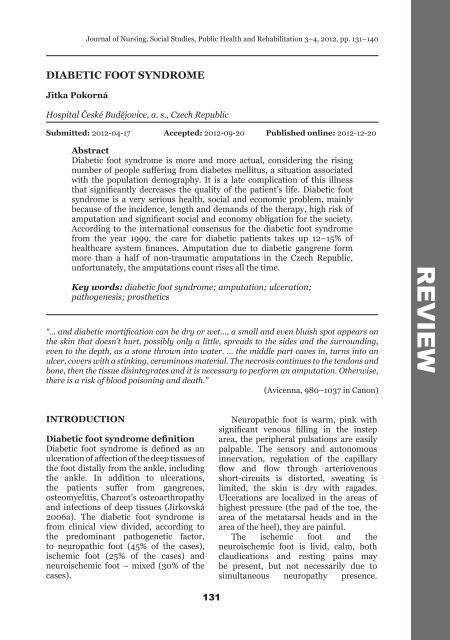
DIABETIC FOOT SYNDROME<br />
Jitka Pokorná<br />
Journal of Nursing, Social Studies, Public Health and Rehabilitation 3–4, 2012, pp. 131–140<br />
Hospital České Budějovice, a. s., Czech Republic<br />
Submitted: 2012-04-17 Accepted: 2012-09-20 Published online: 2012-12-20<br />
Abstract<br />
Diabetic foot syndrome is more and more actual, considering the rising<br />
number of people suffering from diabetes mellitus, a situation associated<br />
with the population demography. It is a late complication of this illness<br />
that significantly decreases the quality of the patient’s life. Diabetic foot<br />
syndrome is a very serious health, social and economic problem, mainly<br />
because of the incidence, length and demands of the therapy, high risk of<br />
amputation and significant social and economy obligation for the society.<br />
According to the international consensus for the diabetic foot syndrome<br />
from the year 1999, the care for diabetic patients takes up 12–15% of<br />
healthcare system finances. Amputation due to diabetic gangrene form<br />
more than a half of non-traumatic amputations in the Czech Republic,<br />
unfortunately, the amputations count rises all the time.<br />
Key words: diabetic foot syndrome; amputation; ulceration;<br />
pathogenesis; prosthetics<br />
“… and diabetic mortification can be dry or wet..., a small and even bluish spot appears on<br />
the skin that doesn’t hurt, possibly only a little, spreads to the sides and the surrounding,<br />
even to the depth, as a stone thrown into water. … the middle part caves in, turns into an<br />
ulcer, covers with a stinking, ceruminous material. The necrosis continues to the tendons and<br />
bone, then the tissue disintegrates and it is necessary to perform an amputation. Otherwise,<br />
there is a risk of blood poisoning and death.”<br />
(Avicenna, 980–1037 in Canon)<br />
INTRODUCTION<br />
Diabetic foot syndrome definition<br />
Diabetic foot syndrome is defined as an<br />
ulceration of affection of the deep tissues of<br />
the foot distally from the ankle, including<br />
the ankle. In addition to ulcerations,<br />
the patients suffer from gangrenes,<br />
osteomyelitis, Charcot’s osteoarthropathy<br />
and infections of deep tissues (Jirkovská<br />
2006a). The diabetic foot syndrome is<br />
from clinical view divided, according to<br />
the predominant pathogenetic factor,<br />
to neuropathic foot (45% of the cases),<br />
ischemic foot (25% of the cases) and<br />
neuroischemic foot – mixed (30% of the<br />
cases).<br />
131<br />
Neuropathic foot is warm, pink with<br />
significant venous filling in the instep<br />
area, the peripheral pulsations are easily<br />
palpable. The sensory and autonomous<br />
innervation, regulation of the capillary<br />
flow and flow through arteriovenous<br />
short-circuits is distorted, sweating is<br />
limited, the skin is dry with ragades.<br />
Ulcerations are localized in the areas of<br />
highest pressure (the pad of the toe, the<br />
area of the metatarsal heads and in the<br />
area of the heel), they are painful.<br />
The ischemic foot and the<br />
neuroischemic foot is livid, calm, both<br />
claudications and resting pains may<br />
be present, but not necessarily due to<br />
simultaneous neuropathy presence.<br />
<strong>REVIEW</strong>
Jitka Pokorná<br />
Atherosclerosis of the lower extremity arteries<br />
is present, the most often from the popliteal<br />
artery.<br />
The diabetic foot syndrome classification<br />
according to Wagner-Meggitt (Table 1) is a<br />
Level Lesion description<br />
0 foot with a high risk of ulcerations<br />
1 superficial ulceration<br />
2 deep ulceration without an inflammation<br />
3 deep ulceration, phlegmone, abscess, osteomyelitis<br />
4 localized gangrene<br />
5 gangrene of the whole foot<br />
Diabetic foot syndrome incidence<br />
The registered diabetics number in the Czech<br />
Republic rises from year to year, in the year<br />
2010, 806,230 patients were treated due to<br />
diabetes. When comparing with the year 2000,<br />
the number was increased by 150 thousand.<br />
The number of patients treated only with diet<br />
132<br />
clinical classification based on assessment<br />
of the ulceration depth and the infection<br />
presence. It correlates well with the clinical<br />
severity, it is a method of amputation need<br />
estimation.<br />
Table 1. The diabetic foot syndrome classification according to Wagner-Meggitt<br />
amendments decreases, however, the number<br />
of the patients treated with medicaments rises.<br />
Chronic complications chronically appear in<br />
27% of treated diabetic patients. In the year<br />
2010, 45,118 patients were treated due to the<br />
diabetic foot, out of those 8,501 reached the<br />
amputation stage (Table 2).<br />
Table 2. The diabetic foot syndrome incidence and the number of amputations<br />
2000 2005 2010<br />
Diabetic patients number 654 164 739 305 (113%) 806 230 (123%)<br />
Patients with the diabetic foot syndrome 37 764 38 090 (101%) 45 118 (119%)<br />
Number of amputation 5 865 7 303 (124%) 8 501 (144%)<br />
Source: ÚZIS (Institute for healhtcare information and statistics of the Czech Republic)<br />
In the year 2010, 1.05% of all the diabetic<br />
patients in the Czech Republic had to undergo<br />
amputation (ÚZIS [Institute for healthcare<br />
information and statistics of the Czech<br />
Republic]).<br />
The incidence of amputations under<br />
the knee level is 12–15times higher in<br />
diabetic patients, when compared with<br />
individuals without diabetes. Transmetatarsal<br />
amputations are 400times more common in<br />
diabetic patients. More than 60% of patients<br />
undergoes the amputation of the second foot<br />
within 4 years of losing the first extremity<br />
(Rušavý et al. 1998). Individual living alone,<br />
with insufficient education and individuals<br />
with low social and economy level have<br />
an increased risk of amputations. Other<br />
groups with high risks consist of patients<br />
with renal insufficiency, as the nitrous<br />
substance retention worsens wound healing;<br />
patients after a renal transplantation due to<br />
immunosuppressive therapy and patients<br />
with sight disorders (International working<br />
group for the diabetic foot syndrome 2000).<br />
Various studies repeatedly state that the<br />
pacients with high risks aren’t provided with<br />
a podiagric preventive care of a sufficient level<br />
(Lavery et al. 2010).
Risk factors for ulcerations<br />
appearance<br />
Hyperglycemia<br />
Diabetic foot emergence reasons are multifactorial.<br />
The first place in the list holds the<br />
hyperglycemia, the connection between the<br />
hyperglycemia and the late complications<br />
emergence is proven. A short-term hyperglycemia<br />
influences microcirculation, decreases<br />
the immunity of the organism and the<br />
reologic blood characteristics, increases the<br />
oxidative stress, agregabitility and adhesivity<br />
of the platelets.<br />
Diabetic microangiopathy – specific changes<br />
of arterioles, precapillaries and capillaries<br />
due to long-term hyperglycemia – is very<br />
serious. The thickened basal membrane with<br />
distorted permeability, resulting in decreased<br />
oxygen supply to the nutritive skin capillaries<br />
that can be even lower than 10%. At the same<br />
time, the total blood flow through the diabetic<br />
foot is increased, the endangered feet are<br />
warm, reddish, with widened venous system,<br />
can be even edematous. The edema worsens<br />
the skin hypoxia.<br />
Atherosclerosis<br />
Atherosclerosis is another very important<br />
factor. Ischemic illness of the lower<br />
extremities appears 2–4times more often<br />
in the diabetic patients than in the nondiabetic<br />
individuals. Typically, the crural<br />
and foot arteries are damaged, up to 80%<br />
of the changes on the arteries is distally of<br />
the popliteal artery. Another typical factor<br />
is the mediocalcinosis – calcification of the<br />
arterial media. 5–10% of diabetic patients<br />
suffer from this condition; it doesn’t have to<br />
worsen the peripheral circulation. The vessel<br />
wall damaged by mediocalcinosis is difficult<br />
to compress, therefore peripheral pressure<br />
in Doppler method measurement are falsely<br />
too high. Quicker atherosclerotic changes in<br />
the diabetic patients are caused by risk factors<br />
accumulation (dyslipidemia, hypertension,<br />
hyperglycemia, hyperinsulinemia, glycation<br />
of the LDH-cholesterol and collagen, hypercoagulation<br />
condition and endothelial dysfunction).<br />
Ischemic illness of the lower extremities is<br />
often without any clinical signs, claudication<br />
pain appears often in the ankle area and more<br />
distally, moreover, the claudication pain<br />
133<br />
Diabetic food syndrome<br />
sensing may be modified by the neuropathy<br />
presence (Piťhová 2006).<br />
Diabetic neuropathy<br />
Diabetic neuropathy is the most serious factor<br />
of the diabetic foot development. Diabetic<br />
neuropathy is a diffuse, non-inflammatory<br />
distortion of the function and structure of<br />
peripheral motor, sensitive and vegetative<br />
nerves. It leads to slowing the stimuli<br />
conductivity through the nerve fiber.<br />
The most common sign of the sensory<br />
neuropathy is the feeling of cold feet,<br />
hyperesthesia, paresthesia – burning, whiplike,<br />
stinging feet pain. On the other hand,<br />
the patients lose the ability to sense warm,<br />
cold and vibrations. As the diabetic patient<br />
doesn’t feel the pain in his/her feet, he doesn’t<br />
pay attention to them and he/she won’t treat<br />
small injuries on time.<br />
Motor neuropathy in a diabetic patients<br />
leads to an atrophy of small muscles of the<br />
foot, dysbalance of the flexors and extensors<br />
leading to stronger flexor action and chronic<br />
toes flexion, resulting in trigger fingers and the<br />
architecture of the foot changes. The pressure<br />
is overly transferred to the metatarsal and<br />
toe area, the gait biomechanics in general is<br />
distorted. Neuropathy leads to the loss of deep<br />
perception, loss of the tendon reflexes and<br />
intercostal muscles atrophy.<br />
A joint movement limitation appears in<br />
up to 30% diabetics. It is caused by collagen<br />
glycation leading to skin and joint sheath<br />
thickening and rigidity, appears mainly in<br />
subtalar joints. It results in plantar pressure<br />
increase during walking. Hyperkeratosis<br />
formation in higher plantar pressure and<br />
autoimmune neuropathy can have the same<br />
effect as a foreign body and increase the<br />
plantar pressure even more. It culminates<br />
in Charcot’s arthropathy development – it<br />
stands for very advanced foot bone structure<br />
deformities.<br />
Autonomous neuropathy leads to sweating<br />
decrease or even to anhidrosis. Dry skin is<br />
more prone to injury and hyperkeratosis<br />
formation. Losing of tonus in sympatic<br />
nervous fibers of small vessels results in<br />
decreasing of peripheral vessel resistance<br />
and opening of arteriovenous short-cuts and<br />
further decrease of flow through the nutritive<br />
capillaries. The whole condition leads to tissue<br />
hypoxia (Piťhová 2008).
Jitka Pokorná<br />
Charcot’s osteoarthropathy can appear in<br />
up to 10% of patients with diabetic neuropathy<br />
and 16% of patients with neuropathic<br />
ulcerations. The highest risk is associated<br />
with the patients between 50 and 60 years<br />
of age with the diabetes duration for longer<br />
time than 10 years (Jirkovská 2003). It is a<br />
progressive destructive illness of foot bones<br />
and joints in the patients with a neuropathy.<br />
The exact pathogenetic mechanism isn’t<br />
known.<br />
The causes of diabetic defect emergence:<br />
• chronic low pressure for many hours<br />
(when using improper shoes);<br />
• direct injury (nail, a foreign body in the<br />
shoe);<br />
• chronic slight pressure leading to a blister<br />
and then to an ulcus;<br />
• scald, burns;<br />
• radages, mycoses.<br />
In addition to the above mentioned causes,<br />
some additional illnesses (hypertension,<br />
hyperlipidemia) and the life style of the patient<br />
– smoking – contribute to the pathogenesis.<br />
motor<br />
foot muscles<br />
atrophy<br />
decreased<br />
joint<br />
movability<br />
neuropathy<br />
sensitive<br />
small injury<br />
after stepping<br />
on an object<br />
insensitivity,<br />
painlessness<br />
visceral<br />
defect<br />
diabetes<br />
mellitus<br />
anhydrosis,<br />
edema<br />
skin<br />
fissures<br />
134<br />
An infection in the diabetic foot is<br />
defined as an invasion and multiplication<br />
of microorganisms in the tissue, tissue<br />
destruction and inflammatory response. The<br />
division to 1. no risk for the extremity; 2. risk<br />
for the extremity and 3. risk for the patient<br />
is really practical. Antibiotic therapy of the<br />
infection must always be supplemented with<br />
a complex care, relieve of pressure on the<br />
ulceration, surgery intervention (incisions,<br />
drainages, infected tissue removal via resection<br />
or amputation), débridement of necrotic<br />
tissue, vascular blood supply and metabolic<br />
compensation (Jirkovská 2006b). Infection<br />
in the diabetic foot is often polymicrobial,<br />
but the most common microbial agent is<br />
Staphylococcus aureus that is included in the<br />
common skin flora in the healthy population.<br />
Unfortunately, MRSA (methicilin resistant<br />
Staphylococcus aureus) infections rate increases.<br />
The microbe is resistant to nearly all<br />
the other antibiotics, except of vankomycin<br />
(Kratochvíl et al. 2006).<br />
A-V shunt<br />
edema<br />
blood flow<br />
disorder<br />
hyperviscosity<br />
hyperglycemia<br />
capillary<br />
flow<br />
disorder<br />
chronic<br />
skin<br />
hypoxia<br />
small<br />
injury<br />
infection<br />
atherosclerosis,<br />
smoking,<br />
lipids<br />
Source: Rušavý et al. (1998)<br />
Fig. 1. Pathogenesis of diabetic ulcerations
Examination methods<br />
In addition to clinical condition assessment,<br />
many laboratory and imaging methods are<br />
used for precise diagnostics to determine the<br />
level and extent of the patient’s disease with<br />
the diabetic foot syndrome.<br />
Basic medical history data, we try to find<br />
out, is the diabetes type and its duration.<br />
Diabetes duration isn‘t decisive for the<br />
extremity amputation, as in 15–19%, diabetes<br />
mellitus is diagnosed only in a situation<br />
leading to an extremity loss (Piťhová 2008).<br />
Furthermore, we determine the presence<br />
of the specific chronic complications<br />
(neuropathy, retinopathy), presence of<br />
cardial and vascular disease, possibly the<br />
undergone interventions. Further important<br />
medical history data are: smoking, lipid<br />
metabolism disorder, other serious general<br />
illnesses (liver, kidney illnesses, rheumatoid<br />
arthritis, psoriasis, even depressions). Social<br />
situation of the patient is of considerable<br />
importance, too – loneliness, low social level,<br />
homelessness, previous non-compliance.<br />
Neuropathic problems are the subjectively<br />
serious ones (fornication, prickling,<br />
feet sweating disorders), resting pain or<br />
claudication pain, gait problems.<br />
Clinical examinations: Via palpation, the<br />
physician determines the pulsation on the<br />
arteries, via auscultation, the presence of<br />
murmurs over big arteries. Then, the anklearm<br />
index is determined. Using the Doppler<br />
method, the blood pressure is measured on<br />
both arms in horizontal position, then on<br />
dorsal pedis artery and posterior tibial artery.<br />
Peripheral systolic blood pressure > 100 mm<br />
Hg indicates good blood perfusion of the<br />
extremities, values in the range of 50–100<br />
mm Hg indicate a medium perfusion disorder.<br />
Chronic critical extremity ischemia is a<br />
disorder significant due to hematodynamics<br />
changes that developed gradually. It is defined<br />
by resting pain lasting for more than 2 weeks<br />
or ulceration or gangrene presence. Systolic<br />
ankle blood pressure is < than 50 mm Hg,<br />
systolic pressure on the thumb < than 30<br />
mm Hg and the TcpO 2 < 10 mm Hg doesn‘t<br />
increase after oxygen inhalation.<br />
If the absolute value of the ankle blood<br />
pressure is higher than 220 mm Hg or the<br />
ankle-arm index is > 1.25, medicalcinosis is<br />
present.<br />
135<br />
Diabetic food syndrome<br />
Laboratory examination: used in the<br />
diabetic patients with a diabetic foot can be<br />
divided into 3 groups:<br />
1) Examinations monitoring the diabetes<br />
mellitus<br />
Glycemia is the basic monitored value,<br />
the target fasting glycemia is 4–6 mmol/l,<br />
postprandial glycemia 5–7.5 mmol/l.<br />
Glycated hemoglobin is a basic<br />
examination monitoring the long-term<br />
compensation of diabetes. The percent<br />
of the glycated hemoglobin reflects the<br />
glycemia for the last 2–3 months (target<br />
values are less than 4.5%).<br />
Further laboratory examinations are<br />
a detailed lipidogram, mineralogram,<br />
creatinine, urea, uric acid, hepatic tests<br />
(Haluzík 2009).<br />
2) Examinations for inflammation monitoration<br />
The default objectification method is<br />
measuring of the body temperature, then<br />
blood count (leukocytosis, typical shift in<br />
the white cells differential), sedimentation<br />
– FW, C-reactive protein (a component of<br />
the non-specific humoral immunity) and<br />
procalcitonin (Kolář 2008).<br />
In diabetic foot syndrome, microbiology<br />
examination is very important. It is<br />
necessary to collect the material from<br />
the festering focuses for cultivation and<br />
sensitivity assessment repeatedly and<br />
carefully to avoid contamination.<br />
3) Laboratory examinations performed in<br />
the pre-operative examination (Skalická<br />
2007).<br />
Imaging methods<br />
An X-ray image of the foot in two projections to<br />
show osteoporosis, subchondral pseudocysts,<br />
spontaneous fractures or soft tissue edema.<br />
Complicating osteomyelitis leads to osteolyses<br />
and deformations in the bone structure and to<br />
subluxations (Lacman et al. 2005).<br />
Ultrasonography examination allows both<br />
function (blood pressure measurement) and<br />
morphology examination of the arterial system.<br />
Using a two-dimensional ultrasonography<br />
imaging of the anatomy structures, it is<br />
possible to assess the arterial lumen and to<br />
find the stenoses and obstructions. Doppler<br />
techniques provide hemodynamic data about
Jitka Pokorná<br />
the direction, speed and quality of the blood<br />
flow in the artery.<br />
Arteriography of lower extremities is<br />
an invasive vascular examination indicated<br />
in clinical signs of the ischemic disease of<br />
lower extremities of IIb level (according to<br />
Fontaine) and more. The second indication<br />
for the invasive vascular examination is an<br />
ulceration without healing tendencies for 2–4<br />
weeks of a complex therapy. Furthermore,<br />
every time before a planned amputation<br />
(Bartoš and Pelikánová 2000). Angiography<br />
is more and more replaced with the CT AG (CT<br />
angiography) and MRI (magnetic resonance<br />
imaging) angiography. After a diagnostic<br />
angiography, there is often a therapeutic<br />
procedure – percutaneous transluminal<br />
angioplasty (Chlup et al. 2005).<br />
Scintigraphy using radionuclide-marked<br />
leukocytes is a suitable method. This method<br />
uses the fact that in the inflammation area of<br />
the organism, there is higher concentration of<br />
leukocytes. It is used to diagnose osteomyelitis<br />
in the patients with the diabetic foot syndrome.<br />
If a radionuclide is bound to the leukocytes,<br />
it is possible to determine the extent of the<br />
inflammation and extent of the tissue damage<br />
using a scintigraphy camera (differentiate the<br />
osteomyelitis from a soft tissue affection), to<br />
differentiate the Charcot’s osteoarthropathy<br />
from an inflammation and to monitor the<br />
results of the treatment by repeating the<br />
examination (Kuníková et al. 2010).<br />
Transcutaneous oxymetry is an effective<br />
non-invasive examination method using<br />
the polarography principle to determine<br />
the transcutaneous partial oxygen pressure<br />
(TcpO 2). The measured TcpO 2 value is a<br />
complex function of the skin blood circulation,<br />
metabolic activity, dissociation of the<br />
oxyhemoglobine and tissue oxygen perfusion<br />
(Gašpar et al. 2007).<br />
In the patients with the diabetic foot<br />
syndrome, it is possible to objectify, using<br />
this method, the extremities ischemia level,<br />
as the other blood pressure measurement<br />
methods can indicate artificially high blood<br />
pressure values in the lower extremities in<br />
mediocalcinosis (Fife et al. 2009).<br />
TcpO 2 value over 60 mm Hg is considered<br />
normal, the value under 30 mm Hg indicates a<br />
severe ischemia. It is not possible to assess only<br />
the absolute TcpO 2 value, you have to analyze<br />
the measured values ratio in a reference area<br />
136<br />
and on the foot. According to the Transatlantic<br />
Inter-Society Consensus (TASC), the value<br />
30 mm Hg stands for ischemia diagnosis<br />
and worse healing prognosis. However, the<br />
tolerance is 10 mm Hg, i.e. the healing is<br />
improbable in values under 20 mm Hg and<br />
almost sure in a value over 40 mm Hg.<br />
Diabetic foot prevention<br />
Many studies have shown that a complex feetcare<br />
can reduce the feet ulcerations amount<br />
by up to 50%.<br />
The basics of diabetic foot prevention is<br />
regular checking of the feet and the shoes in<br />
every patient’s visit at the treating physician<br />
and education of the patient every time.<br />
A detailed screening examination<br />
according to the guidelines is to be performed<br />
at least 1× per year, in high risk patients more<br />
often.<br />
During the regular feet inspection, it<br />
is necessary to focus on the skin defects –<br />
pressure sores, hyperkeratosis, blisters,<br />
ragades, mycoses, skin color and temperature<br />
changes, further on deformities and<br />
deformations. Shoes check should be a part of<br />
the examination!<br />
Every inspection includes medical history<br />
data collection summing up, among others,<br />
a question about the previous education<br />
aimed at the feet care, question about<br />
walking barefoot, availability of the care,<br />
social situation, question aimed to detect<br />
neuropathy (prickling, sensitivity loss) and<br />
extremity ischemia (question about possible<br />
claudications).<br />
Every year complete examination consists<br />
of arteries palpation on the lower extremities,<br />
including the dorsal pedis artery and anterior<br />
tibial artery, auscultation over the big arteries<br />
of the lower extremities.<br />
Another examination using Semmes-<br />
Weinstein’s monofilaments. It ise a 38 mm<br />
long nylon filament bending under the weight<br />
of 10 g. This very simple tool can help reveal<br />
a loss of the feet sensitivity. The filament<br />
is applied in 4 feet areas, insensitivity is<br />
proven, if the patient doesn’t feel the touch<br />
in 6 points of the total 8 on both the feet.<br />
Further examination is using a tuning for or a<br />
biothesiometer that are able to reveal the loss<br />
of the vibration sensitivity. Dopplerometric<br />
examination of the pressure index – dorsal<br />
pedis artery/brachial artery is more and more
common. The norm is 1.0–1.4. The index<br />
lower than 0.9 indicates extremity ischemia<br />
and lower than 0.6 a critical ischemia.<br />
The patients with a proven sensitivity loss,<br />
i.e. advanced neuropathy, are dispensed to<br />
a high risk group and can have the diabetic<br />
shoes prescribed. Patients with diagnosed<br />
extremities ischemia are indicated for<br />
angiography examination and the following<br />
intervention solution (committee of the Czech<br />
diabetologic organization 2012a).<br />
Education<br />
The education of the patient in prevention<br />
of the diabetic foot development is a neverending<br />
process. Education that is not repeated<br />
loses its effectiveness. The education of the<br />
diabetic patients in prevention of the diabetic<br />
foot development consists of some basic<br />
points.<br />
1) Wear corrects shoes, never walk barefoot,<br />
not even at home.<br />
2) Check your feet daily. If you are not able<br />
to see there, use a mirror or ask a family<br />
member for help.<br />
3) Maintain correct hygiene standards –<br />
daily feet bath in lukewarm water.<br />
4) Remove the rough skin with a pumice<br />
stone, use files when taking care of your<br />
feet, cut your nails straight after soaking<br />
in lukewarm water.<br />
5) Do not forget that your feet don’t feel<br />
warm, cold and pain well and protect<br />
them before possible injuries.<br />
The main target of the education is reaching<br />
a change in the patient’s behavior. Education<br />
must be comprehensible, lively discussion is<br />
more suitable than a monotonous lecture. The<br />
best option is the combination of presented<br />
information with a written text allowing the<br />
patients to revise the obtained information at<br />
home. It is necessary to adapt the education<br />
for the target group, naturally in blind, poorly<br />
mobile diabetic patients, it is necessary to<br />
work with the family members. It is necessary<br />
to win the active cooperation of the patients<br />
so that they have their feet checked by the<br />
physician, because no checking of a diabetic<br />
patient can be considered as complete if the<br />
feet weren’t checked (committee of the Czech<br />
diabetologic organization 2012b).<br />
In the everyday contact with the patients<br />
who have lost their foot/leg, it is possible<br />
determine a mistake, easily to be seen a mile<br />
137<br />
Diabetic food syndrome<br />
off, which, however, was the fatal mistake<br />
leading in its consequences to the extremity<br />
loss and that could be prevented in many<br />
cases if the care was correct.<br />
Diabetic foot syndrome economy<br />
The care of the patients with a diabetic<br />
foot syndrome is financially demanding<br />
for both the out-patient and the in-patient<br />
departments of the health care institutions.<br />
The treatment expenses can be generally<br />
divided into the direct, health care expenses<br />
(the work of the physician and using<br />
treatment products), direct non-healthcare<br />
expenses (activities of other kind than<br />
healthcare, connected with the treatment –<br />
e.g. the transport) and the indirect expenses,<br />
i.e. potential sources lost due to the disease<br />
(productivity loss due to inability, workload<br />
change, premature retirement, premature<br />
death). Not only antibiotics are financially<br />
demanding, but also the hospitalization and<br />
the diagnostic methods, or even seemingly<br />
low expenses as bandages of the defects by<br />
the nurses of the homecare services, transport<br />
with ambulance cars etc., if the problems last<br />
for months. The diabetology center in Plzeň<br />
tried to retrospectively quantify the half-year<br />
direct costs for health and social care (only the<br />
bandages by a homecare service are included)<br />
of a patient with the diabetic foot syndrome<br />
for the year 2000. It was on average 34,500<br />
Czech crowns per patient per half a year.<br />
In the diabetology center IKEM in the same<br />
year, the costs per a patient with a diabetic<br />
foot were about 2,800 Czech crowns per a day<br />
of hospitalization (without the angiography<br />
and vascular reconstruction procedures).<br />
Hospital treatment of the defect cost<br />
22,500 Czech crowns for a hospitalization<br />
of 26 days length on average. The total outpatient<br />
department care for one patient cost<br />
7,250 Czech crowns (monitored for 8 months).<br />
The amputation cost 22,000 Czech crowns for<br />
20 days of hospitalization on averages. Costs<br />
of compensation tools and social allowances<br />
were up to 400,000 Czech crowns in the first<br />
year after the amputation (Bruner and Anděl<br />
2002).<br />
Amputation<br />
Periphery amputation<br />
Toes amputation, even multiple, disturbs<br />
the stability of standing only minimally. In
Jitka Pokorná<br />
the prosthetic care, shoe filling is sufficient.<br />
In toes amputation, at least a part of the<br />
proximal phalanx is preserved if possible,<br />
so that the other toes do not migrate to<br />
the emptied space. The hallux amputation<br />
doesn’t significantly disrupt the foot stability<br />
as well. The load center is moved from the<br />
head of the 2 nd metatarsus to the head of the<br />
3 rd toe metatarsus and the patient after the<br />
amputation tends to walk on the outer side of<br />
the foot.<br />
High amputations<br />
Crural amputations are performed at the<br />
border of the middle and the upper third of<br />
the shank. When correctly indicated and<br />
performed, total healing can be expected in<br />
80% of the patients. The rehabilitation with<br />
a prosthesis is very good, up to 90% of the<br />
patients learn to walk.<br />
Amputation over the knee joint is indicated<br />
in necroses passing the shank, in patients with<br />
polymorbidity leading to conclusion that they<br />
will not walk in future and in severe infections.<br />
It is possible to assume total healing in 90% of<br />
the patients after the amputation even in severe<br />
ischemia. Optimal level for the amputation,<br />
considering the seating of the prosthesis, is<br />
the border of the lower and the middle third of<br />
the femur. The longer the stump is, the higher<br />
the probability for a successful rehabilitation<br />
(Greppe and Kremer 1993).<br />
Amputation stump adaptation is very<br />
important. An osteomyoplastic correction is<br />
performed. It is based on nerves preparation,<br />
extraction, preparation and resection as high<br />
over the osteotomy as possible so that they are<br />
out of the loaded part of the stump. This way,<br />
it is possible to prevent the “phantom pain”.<br />
Technically correct adaptation of the resected<br />
bone is necessary too – the femur or the<br />
shank bones. The bone stump is then covered<br />
by a muscle-skin lobe that is turned from the<br />
back side in the thigh and shank area. After<br />
the surgery, the stump is formed in diabetic<br />
patients with a soft bandage. After complete<br />
healing, edema and pain decline, prosthetic<br />
treatment can be performed (Zeman et al.<br />
1993).<br />
Prosthetics<br />
The prosthesis is a function and an esthetic<br />
replacement of the missing part of the<br />
extremity.<br />
138<br />
In the recent years, development of<br />
new technologies and materials used during<br />
production of artificial extremities<br />
replacements has significantly accelerated.<br />
It contributes to simpler, more reliable and<br />
comfortable usage of the prosthetic tools.<br />
Social impact of the diabetic foot<br />
syndrome<br />
Bio-psycho-social diabetes model extends<br />
the traditional biologic attitude to the illness<br />
and adds the personal and social aspects. A<br />
chronic diabetes complication – the diabetic<br />
foot syndrome – is a typical protracted illness,<br />
often requiring a long hospitalization or very<br />
often out-patient department visits. The<br />
diabetic foot treatment burdens not only the<br />
patients, but also the ones taking care of the<br />
patient. The quality of life of the patients (and<br />
not only of them) is mostly decreased by the<br />
wound care, poor mobility, often health care<br />
institutions visits and the fear of amputation<br />
(Nabuurs-Franssen et al. 2005). In a high<br />
amputation, working capability is not the<br />
only thing lost, often the patients are not<br />
self-sufficient anymore. Up to 20% of the<br />
patients worked and had a very active life at<br />
the moment of the diabetic foot syndrome<br />
development. Then, within a few weeks they<br />
became lost self-sufficiency and became<br />
dependent on social services.<br />
With the illness onset:<br />
– the working people lose their income;<br />
– the expenses associated with the therapy<br />
increase;<br />
– the travelling expenses (out-patient<br />
department bandages) increase;<br />
– social expenses appear (due to inability to<br />
take care of himself/herself).<br />
The most commonly applied form of social<br />
help are the allowances that can be divided<br />
into material and financial allowances and<br />
according to the length of the allowance<br />
provision – one-time or repeated (Kozlová<br />
2005).<br />
Luckily, the majority of the patients after<br />
the amputation have a family background<br />
(about 80 %) who can help the patient<br />
cope with the difficult situation (Krajcová<br />
2005). Those patients who have no family<br />
background and are often people with a low<br />
social and economy level or seniors of high<br />
age use the homecare service and institutions<br />
of social care.
CONCLUSION<br />
Diabetic foot syndrome is an illness affecting<br />
more and more patients. It is a cause for long<br />
suffering, long and expensive hospitalizations,<br />
furthermore a cause of invalidity, selfsufficiency<br />
loss and social care need.<br />
It is a multidisciplinary problem that<br />
involves not only diabetologists-podiatres,<br />
but alos surgeons, angiologists, invasive<br />
radiologists, vascular surgeons and prosthetists<br />
and many other specialists, and last<br />
REFERENCES<br />
139<br />
Diabetic food syndrome<br />
but not least all the diabetologists, education<br />
diabetologic nurses and general practitioners,<br />
since only education, prevention, active search<br />
for initial stages and their consistent therapy<br />
can avert the high amputation need.<br />
The diabetic foot syndrome significantly<br />
influences the life quality of the patient in a<br />
negative way. Despite many preventive and<br />
therapeutic procedures, this complication<br />
increases morbidity, invalidity and mortality<br />
of the population.<br />
1. Bartoš V, Pelikánová T (2000). Praktická diabetologie [Pediatric diabetology]. Praha: Maxdorf. 473 p.<br />
ISBN 80-85912-17-1 (Czech).<br />
2. Brunerová L, Anděl M (2002). Ekonomické náklady syndromu diabetická noha v mezinárodním<br />
srovnání [Ekonomic expenses of the diabetic foot syndrom in the internation comparison]. DMEV.<br />
3: 153–156, ISSN 1211-9326 (Czech).<br />
3. Chlup R, Holá J, Kudlová P, Kočí A, Hrubá V (2005). Co může vykonat diabetolog pro úspěšnou léčbu<br />
syndromu diabetické nohy [What a diabetologist can do for a successful treatment of the diabetic foot<br />
syndrome]. Trendy v medicíně. 6: 41–51. ISSN 1212-9046 (Czech).<br />
4. Czech diabetology organization committee (2012a). Standardy léčby pacientů se syndromem<br />
diabetické nohy [Therapy guidelines for the patients with the diabetic foot syndrome]. [online] [2012-<br />
04-22]. Available from http://www.diab.cz/standardy (Czech).<br />
5. Czech diabetology organization committee (2012b). Doporučení k edukaci diabetika [Guidelines for<br />
a diabetic patient education]. [online] [2012-04-22]. Available from: http://www.diab.cz/standardy<br />
(Czech).<br />
6. Fife CE, Smart DR, Sheffield PJ, Fopf HW, Hawkins G, Clarke D (2009). Trancutaneous oximetry<br />
in Clinical Practice: Consensus statements from an expert panel based on evidence. Undersea &<br />
Hyperbaric Medicine. 36: 43–53.<br />
7. Gašpar Ľ, Ambrózy E, Štrbová L, Gavorník P, Štvrtinová V (2007). Transkutánna oxymetria a jej<br />
význam v diagnostike syndrómu diabetickém nohy [Transcutaneous oxymetry and its significance in<br />
the diagnostics of the diabetic foot syndrome]. Praktická flebologie. 16/1: 3–5 (Slovak).<br />
8. Grepe H-E, Kremer K (1993). Atlas chirurgických operací [Surgical operations atlas]. Praha: Grada, p.<br />
724. ISBN 80-7169-028-7 (Czech).<br />
9. Haluzík M (2009). Praktická léčba diabetu [Practical diabetes treatment]. Praha: Mladá fronta, p. 361.<br />
ISBN 978-80-204-2071-8 (Czech).<br />
10. International working group for the diabetic foot syndrome (2000). Syndrom diabetické nohy<br />
[Diabetic foot syndrome]. Praha: Galén, p. 102. ISBN 80-7262-051-7 (Czech).<br />
11. Jirkovská A (2003). Diagnostika a terapie Charcotovy osteoartropatie [Diagnostics and therapy of<br />
Charcot’s osteoarthropathy]. Bulletin HPB. 11/1. ISSN 1210-6755. [online] [cit. 2012-04-24]. Available<br />
from: http://www.hpb.cz/index.php?pId=03-1-01 (Czech).<br />
12. Jirkovská A (2006a). Syndrom diabetické nohy [Diabetic foot syndrome]. Maxdorf, p. 397. ISBN 80-<br />
7345-095-X (Czech).<br />
13. Jirkovská A (2006b). Zásady terapie infekce u syndromu diabetické nohy [Infections therapy principles<br />
in the diabetic foot syndrome]. Bulletin HPB. 14/4: 141–143. ISSN 1210-6755 151 (Czech)<br />
14. Kolář M (2008). Infekce kriticky nemocných [Infections in critical patients]. Praha: Galén, p. 379.<br />
ISBN 978-80-7262-488-1 (Czech).<br />
15. Kozlová L (2005). Sociální služby [Social care services]. 1st ed. Praha: Triton, p. 79. ISBN 80-7254-<br />
662-7 (Czech).
Jitka Pokorná<br />
16. Krajcová M (2005). Terapeutická zátěž a psychosociální aspekty nemocných s gangrénou dolní<br />
končetiny. Diplomová práce [Therapeutic load and psychosocial aspects of the patients with a<br />
gangrena of a lower extremity. Diploma thesis]. Univerzita Karlova v Praze, 1. lékařská fakulta, Ústav<br />
teorie a praxe ošetřovatelství, 95 p. (Czech).<br />
17. Kratochvíl A, Treflová L, Šilhová E, Petrová K (2006). Meticilin rezistentní Staphylococcus aureus –<br />
problém nejen podiatrické ambulance [Meticilin resistant Staphylococcus aureus – a problem in not<br />
only the podiatric out-patient department]. Bulletin HPB. 14/4: 151–155. ISSN 1210-6755 (Czech).<br />
18. Kuníková I, Lang O, Beznosková A (2010). Vyšetření pacienta se syndromem diabetické nohy pomocí<br />
radionuklidem značených leukocytů [Examination of a patient with the diabetic foot syndrome using<br />
radionuclide-marked leukocytes]. DMEV. 13/4: 174–182. ISSN 1211-9326 (Czech).<br />
19. Lacman J, Mašková J, Rejchrt P (2005). Možnosti diagnostiky a léčby diabetické nohy na radiologickém<br />
pracovišti [Diagnostics and therapy possibilities in diabetic foot at a radiology department]. Bulletin<br />
HPB. 13/3–4: 111–113. ISSN 1210-6755 (Czech).<br />
20. Lavery LA, Hunt NA, Lafontaine J, Baxter Cl, Ndip A, Boulton AJ (2010). Diabetic foot prevention: a<br />
neglected opportunity in high-risk patients. Diabetes Care. 33/7: 1460–1462. [online] [cit. 2012-04-<br />
24]. Available from: http://www.ncbi.nlm.nih.gov/pubmed/2042422<br />
21. Nabuurs-Franssen MH, Huijberts MS, Nieuwenhuijzen Kruseman AC, Willems J, Schaper NC (2005).<br />
Health-related quality of life of diabetic foot ulcer patients and their caregivers. Diabetologia. 48/9:<br />
1906–1910.<br />
22. Piťhová P (2006). Syndrom diabetické nohy – závažná komplikace u pacientů s diabetes mellitus<br />
[Diabetic foot syndrome – serious complication in patients with diabetes mellitus]. Diabetologie,<br />
p. 97–132. ISBN 80-7254-882-4 (Czech).<br />
23. Piťhová P (2008). Syndrom diabetické nohy – závažná komplikace diabetes mellitus [Diabetic foot<br />
syndrome – serious complication of diabetes mellitus]. Medicína pro praxi. 5/3. [online] [cit. 2010-11-<br />
07]. Available from: http://www.medicinapropraxi.cz/artkey/med-200803-0008.php (Czech).<br />
24. Rušavý Z et al. (1998). Diabetická noha [Diabetic foot]. Praha: Galén. 189 p. ISBN 80-85824-73-6<br />
(Czech).<br />
25. Skalická H (2007). Předoperační vyšetření [Preoperative examination]. Praha: Galén, p. 152. ISBN<br />
978-80-247-1079-2 (Czech).<br />
26. Tošenovský P, Edmonds ME (2004). Moderní léčba syndromu diabetické nohy [Modern therapy of<br />
the diabetic foot syndrome]. Praha: Galén, p. 207. ISBN 80-7262-261-7 (Czech).<br />
27. ÚZIS (Institute for healtcare information and statistics of the Czech Republic) (2011). Péče o nemocné<br />
cukrovkou [Care of the patients with diabetes]. ISBN 978-80-7280-945-5. [online] [2011-11-07].<br />
Available from: http://www.uzis.cz/publikace/pece-nemocne-cukrovkou-2010 (Czech).<br />
28. Zeman M et al. (1993). Chirurgická propedeutika [Surgery propedeutics]. Praha: Grada, p. 488. ISBN<br />
80-85623-45-5 (Czech).<br />
Contact:<br />
Jitka Pokorná, Hospital České Budějovice, a. s., Czech Republic<br />
E-mail: pokjitka@seznam.cz<br />
140