2005 Proceedings - ASNR
2005 Proceedings - ASNR
2005 Proceedings - ASNR

Create successful ePaper yourself
Turn your PDF publications into a flip-book with our unique Google optimized e-Paper software.
Thursday<br />
Paper 366 Starting at 10:24 AM, Ending at 10:32 AM<br />
Comparison of High-Resolution 3D Flash Contrast-<br />
Enhanced MR Angiography Using Parallel Acquisition<br />
Techniques to Conventional 3D Time-of-Flight MR<br />
Angiography in Carotid Artery Stenosis<br />
Sunenshine, P. J. · Pramanik, B. · Law, E. M. · Hecht, E. M.<br />
New York University Medical Center<br />
New York, NY<br />
PURPOSE<br />
Three-dimensional (3D) time-of-flight (TOF) MR angiography<br />
(MRA) is a well established high-resolution noninvasive<br />
way to assess carotid artery stenosis. Although accurate, this<br />
method is prone to artifacts as a result of long acquisition<br />
times and saturation effects. Contrast-enhanced MRA techniques<br />
have become increasingly popular to counter the saturation<br />
effects of slow flow and decrease acquisition times.<br />
However, spatial resolution of contrast-enhanced MRA is<br />
relatively low compared to conventional 3D TOF imaging.<br />
With the advent of parallel acquisition techniques and multichannel<br />
neck coils, spatial resolution can be increased significantly<br />
without sacrificing temporal resolution. The purpose<br />
of this study is to compare conventional 3D TOF MRA<br />
and 3D FLASH contrast-enhanced MRA with parallel imaging<br />
to determine if parallel imaging can be used to improve<br />
the spatial resolution of dynamic contrast-enhanced MRA to<br />
that comparable with conventional 3D TOF MRA.<br />
MATERIALS & METHODS<br />
Prospectively, four consecutive patients presenting for<br />
carotid MRA underwent 3D TOF MRA and 3D contrastenhanced<br />
MRA with parallel acquisition techniques (GRAP-<br />
PA, acceleration factor 3) at 1.5 T using a 4-channel phasedarray<br />
neck coil and a multiple-channel head coil.<br />
Conventional 3D imaging was performed using a voxel size<br />
of 0.7 x 0.5 x 0.9 mm, a 288 x 384 matrix, and an acquisition<br />
time of 6:21 minutes. Dynamic contrast-enhanced MRA<br />
with parallel imaging was performed using a 3D FLASH<br />
sequence using a voxel size of 0.8 x 0.8 x 0.8, a 384 x 512<br />
matrix, and an acquisition time of 24 seconds following<br />
intravenous injection of gadolinium (0.2 mmol/kg) and a 20<br />
cc saline bolus via power injector. Timing was determined<br />
using a time-resolved echo-sharing angiographic technique<br />
(TREAT) using a 3 cc contrast bolus and 20 cc saline flush.<br />
Quantitative assessment was performed by calculating signal-to-noise<br />
ratio (SNR) and contrast-to-noise ratios (CNR)<br />
in the distal common carotid artery for each MRA sequence.<br />
Qualitative assessment was performed by two neuroradiologists<br />
blinded to the imaging method used. Subjective assessment<br />
of image quality, noise, degree of stenosis using the<br />
NASCET criteria, and degree of confidence in diagnosis was<br />
performed using a 4-point scale (1 = excellent, 4 = nondiagnostic).<br />
RESULTS<br />
Signal-to-noise ratio and CNR were both increased with contrast-enhanced<br />
MRA as shown in the table. Qualitative<br />
parameters such as overall quality was better in the contrastenhanced<br />
MRA sequences. Image quality of the contrastenhanced<br />
MRA was rated the same or higher on all patients<br />
when compared with conventional noncontrast-enhanced 3D<br />
TOF imaging.<br />
198<br />
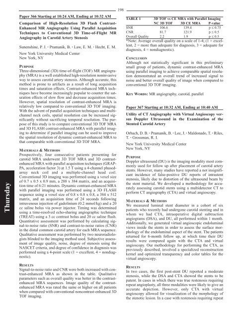
TABLE 1 3D TOF vs CE MRA with Parallel Imaging<br />
NC 3D TOF 3D CE MRA P-value<br />
SNR 106.6 139.4 p ≤ 0.75<br />
CNR 81.7 121.9 p ≤ 0.5<br />
Overall Quality 2.2 1.9 p ≤ 0.5<br />
*Note: Average overall quality on a scale of 1-4; (1 = excellent,<br />
2 = more than adequate for diagnosis, 3 = adequate for<br />
diagnosis, 4 = nondiagnostic).<br />
CONCLUSION<br />
Although not statistically significant in this preliminary<br />
small group of patients, dynamic contrast-enhanced MRA<br />
using parallel imaging to achieve comparable spatial resolution<br />
demonstrated an overall trend of increased signal to<br />
noise and better overall quality of image when compared to<br />
conventional 3D TOF imaging.<br />
KEY WORDS: MR angiography, carotid, parallel<br />
Paper 367 Starting at 10:32 AM, Ending at 10:40 AM<br />
Utility of CT Angiography with Virtual Angioscopy versus<br />
Doppler Ultrasound in the Examination of the<br />
Stented Carotid Artery<br />
Orbach, D. B. · Pramanik, B. · Lee, J. · Maldonado, T. · Riles,<br />
T. · Grossman, R. I.<br />
New York University Medical Center<br />
New York, NY<br />
PURPOSE<br />
Doppler ultrasound (DU) is the imaging modality most commonly<br />
used for follow up after placement of carotid artery<br />
stents. However, many studies have reported a not insignificant<br />
incidence of false-positive DU reports of intrastent<br />
stenosis, likely due to distortion of the ultrasound beam by<br />
the stent material. We developed a methodology for accurately<br />
assessing carotid stents using a multidetector CT to<br />
perform CT angiography (CTA) and virtual angioscopy.<br />
MATERIALS & METHODS<br />
We measured luminal stent diameter in a cohort of six<br />
patients who recently had undergone carotid stenting and in<br />
whom we had CTA, intraoperative digital subtraction<br />
angiograms (DSA), and DU, all performed within 1 month.<br />
Additionally, we generated virtual angioscopic endoluminal<br />
views inside the stents in order to assess the surface morphology<br />
of the endoluminal aspect of the stent. The patients<br />
returned for 6-month follow up, at which time their DU<br />
results were compared again with the CTA and virtual<br />
angioscopy. Our methodology for performing the CTA, as<br />
previously described, involved a specialized reconstruction<br />
kernel and optimized transparency and color tables for the<br />
virtual angioscopy.<br />
RESULTS<br />
In two cases, the first post-stent DU reported a moderate<br />
stenosis, while the DSA and CTA showed the stents to be<br />
patent. In cases in which there was true restenosis requiring<br />
repeat angioplasty, all three modalities were likely to give an<br />
accurate depiction. However, only CTA with virtual<br />
angioscopy allowed for visualization of the morphology of<br />
the stenotic lesion. In a case with restenosis requiring repeat