- Page 1 and 2:
2005 PROCEEDINGS 43 rd Annual Meeti
- Page 3 and 4:
How to use your Proceedings CD-Rom
- Page 5 and 6:
TABLE OF CONTENTS Table of Contents
- Page 7 and 8:
COMMITTEES 2004-2005 Meeting Arrang
- Page 9 and 10:
INVITED SPEAKERS 43rd Annual Meetin
- Page 11 and 12:
SEATTLE, WA About Toronto, Ontario,
- Page 13 and 14:
EXIT STAIRS eception Hall 104 B C D
- Page 15 and 16:
FRONT STREET EXIT STAIRS 12'h drape
- Page 17 and 18:
Printed and Electronic Scientific E
- Page 19 and 20:
401 301 Cardinal Health 407 406 307
- Page 21 and 22:
TECHNICAL EXHIBITS Technical Exhibi
- Page 23 and 24:
General GENERAL Information INFORMA
- Page 25 and 26:
OPTIONAL TOURS Optional Tour Desk H
- Page 27 and 28:
FUTURE MEETINGS Future ASNR Annual
- Page 29 and 30:
Past ASNR PAST Annual Meetings MEET
- Page 31 and 32:
AWARDS & HONORS Awards and Honors P
- Page 33 and 34:
AWARDS & Awards and Honors HONORS 2
- Page 35 and 36:
AWARDS & Awards and Honors HONORS P
- Page 37 and 38:
AWARDS & HONORS Awards and Honors N
- Page 39 and 40:
CME SCIENTIFIC PROGRAM AND MEETING
- Page 41 and 42:
Electronic ELC Learning WORKSHOPS C
- Page 43 and 44:
NLM • SEMINARS Business Center
- Page 45 and 46:
Scientific PROGRAM Program Overview
- Page 47 and 48:
Scientific PROGRAM Program Overview
- Page 49 and 50:
PROGRAM OVERVIEW Scientific Program
- Page 51 and 52:
Notes:
- Page 53 and 54:
Monday Morning 8:00 AM - 8:05 AM Th
- Page 55 and 56:
Carotid Revascularization Endartere
- Page 57 and 58:
vention with concept of transcathet
- Page 59 and 60:
CONCLUSION The described method of
- Page 61 and 62:
provide real-time 3D navigation in
- Page 63 and 64:
CONCLUSION Accessing the posterior
- Page 65 and 66:
to observed on direct otoscopy. It
- Page 67 and 68:
CONCLUSION Sixty-four-slice CT imag
- Page 69 and 70:
2. Angiographic compartments and se
- Page 71 and 72:
centage of small aneurysms at risk
- Page 73 and 74:
Photomicrographs of human brain ane
- Page 75 and 76:
high in aneurysms, whereas expressi
- Page 77 and 78:
Monday Morning 10:00 AM - 11:30 AM
- Page 79 and 80:
CONCLUSION This study, comprising a
- Page 81 and 82:
Paper 55 Starting at 10:42 AM, Endi
- Page 83 and 84:
Carotid measurement and example of
- Page 85 and 86:
neck vascular malformations with et
- Page 87 and 88:
each nerve as well as muscles inner
- Page 89 and 90:
Paper 75 Starting at 3:00 PM, Endin
- Page 91 and 92:
KEY WORDS: Mesothelioma/*diagnosis/
- Page 93 and 94:
CONCLUSION Carotid wall imaging ben
- Page 95 and 96:
priately as the phase and frequency
- Page 97 and 98:
CONCLUSION A relatively high percen
- Page 99 and 100:
Paper 94 Starting at 3:53 PM, Endin
- Page 101 and 102:
nous embolization, 4 patients, who
- Page 103 and 104:
Paper 101 Starting at 3:16 PM, Endi
- Page 105 and 106:
and minimal inflammatory changes in
- Page 107 and 108:
MATERIALS & METHODS A 60-year-old m
- Page 109 and 110:
the hemorrhage was seen on MR imagi
- Page 111 and 112:
CONCLUSION In this series of 117 co
- Page 113 and 114:
CONCLUSION CT perfusion is used wid
- Page 115 and 116:
structures that belong in the orbit
- Page 117 and 118:
anatomical regions which contain la
- Page 119 and 120:
Tuesday Morning 8:00 AM - 12:00 PM
- Page 121 and 122:
Angioplasty for Intracranial Arteri
- Page 123 and 124:
PRESENTATION SUMMARY Buccal Space L
- Page 125 and 126:
Tuesday Morning 10:00 AM - 11:36 AM
- Page 127 and 128:
Paper 146 Starting at 10:24 AM, End
- Page 129 and 130:
GDC®-treated aneurysms was atypica
- Page 131 and 132:
CONCLUSION Therefore, while our ana
- Page 133 and 134:
CONCLUSION Thrombolysis of intracra
- Page 135 and 136:
Patient’s age ranged from 31-82 y
- Page 137 and 138:
Paper 164 Starting at 11:12 AM, End
- Page 139 and 140:
Tuesday Morning 10:00 AM - 11:44 AM
- Page 141 and 142:
CONCLUSION These initial clinical r
- Page 143 and 144:
Paper 175 Starting at 11:04 AM, End
- Page 145 and 146:
CONCLUSION The results demonstrate
- Page 147 and 148:
were calculated in each case. Minim
- Page 149 and 150:
in the regions of HIF-1α expressio
- Page 151 and 152:
addition, a capsule-like rim of in-
- Page 153 and 154:
Tuesday Morning 10:15 AM - 11:45 AM
- Page 155 and 156:
achieving Category I designation wi
- Page 157 and 158:
turing on all aspects of Head and N
- Page 159 and 160:
sible or tenuous. The current repor
- Page 161 and 162:
stents alone demonstrated vascular
- Page 163 and 164:
CONCLUSION MR digital subtraction a
- Page 165 and 166:
CONCLUSION HARDI represents a major
- Page 167 and 168:
MATERIALS & METHODS Forty dyslexic
- Page 169 and 170:
sion measure, which was calculated
- Page 171 and 172:
deliberately lie and deny their inv
- Page 173 and 174:
using lactate edited 1H MRSI, and t
- Page 175 and 176:
CONCLUSION We evaluated the benefit
- Page 177 and 178:
values in the pathologic areas to A
- Page 179 and 180:
CONCLUSION MR imaging-based volumet
- Page 181 and 182:
RESULTS No significant abnormalitie
- Page 183 and 184:
MCI and mild AD was 85% and 100%, r
- Page 185 and 186:
FLAIR signal of the periaqueductal
- Page 187 and 188:
Tuesday Afternoon 4:45 PM - 6:15 PM
- Page 189 and 190:
Tuesday Afternoon 5:00 PM - 6:00 PM
- Page 191 and 192:
Wednesday Morning 8:00 AM - 9:30 AM
- Page 193 and 194:
and provide direct costs for the 2-
- Page 195 and 196:
Functional Imaging of Neurodegenera
- Page 197 and 198:
2002, Dr. Rutka was listed by Globe
- Page 199 and 200:
CONCLUSION The apparatus shows exce
- Page 201 and 202:
four-point scale regarding distorti
- Page 203 and 204:
Paper 287 Starting at 3:32 PM, Endi
- Page 205 and 206:
131 cases with cerebral pathology t
- Page 207 and 208:
Wednesday Afternoon 3:00 PM - 4:32
- Page 209 and 210:
Paper 297 Starting at 3:21 PM, Endi
- Page 211 and 212:
Regions of interest (ROIs) were dra
- Page 213 and 214:
cerebellum was diffusely swollen. T
- Page 215 and 216:
approximately 2.5 years later demon
- Page 217 and 218:
had been involved in accidents clai
- Page 219 and 220:
images, and 4) T1-weighted, T2-weig
- Page 221 and 222:
MATERIALS & METHODS MR imaging of t
- Page 223 and 224:
Paper 321 Starting at 3:16 PM, Endi
- Page 225 and 226:
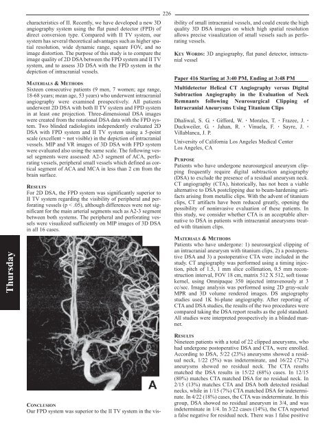
dimeglumine had been administered o
- Page 227 and 228: eduction in fractional anisotropy o
- Page 229 and 230: Wednesday Afternoon 4:40 PM - 6:10
- Page 231 and 232: mation technology from Aberdeen Uni
- Page 233 and 234: Wednesday Afternoon 5:40 PM - 6:10
- Page 235 and 236: Thursday Morning 8:00 AM - 9:00 AM
- Page 237 and 238: ventional MR imaging and can be ide
- Page 239 and 240: Thursday Morning 8:00 AM - 9:30 AM
- Page 241 and 242: RESULTS Sagittal diffusion-weighted
- Page 243 and 244: whole body scanner) with a 12-eleme
- Page 245 and 246: RESULTS Of 31 patients (age range 1
- Page 247 and 248: RESULTS In each subject, not only t
- Page 249 and 250: accepted prospective series of DWI
- Page 251 and 252: angioplasty, virtual angioscopy sho
- Page 253 and 254: liver disease, Child-Pugh stages B
- Page 255 and 256: using an axial single-shot spin-ech
- Page 257 and 258: Paper 378 Starting at 10:32 AM, End
- Page 259 and 260: MATERIALS & METHODS Four dogs under
- Page 261 and 262: MATERIALS & METHODS Routine CT angi
- Page 263 and 264: Paper 388 Starting at 10:10 AM, End
- Page 265 and 266: MATERIALS & METHODS We retrospectiv
- Page 267 and 268: CONCLUSION The patient underwent a
- Page 269 and 270: Paper 398 Starting at 11:00 AM, End
- Page 271 and 272: pressure headaches had undergone an
- Page 273 and 274: mon in older children and adults. T
- Page 275 and 276: 4. Bulte JW, Duncan ID, Frank JA. I
- Page 277: studies suggest that MR imaging als
- Page 281 and 282: MATERIALS & METHODS Eleven patients
- Page 283 and 284: sive procedure. High-resolution con
- Page 285 and 286: CONCLUSION Small volume epidural in
- Page 287 and 288: cation of vertebroplasty although l
- Page 289 and 290: need for routine MR imaging were en
- Page 291 and 292: CONCLUSION Increased flow accompani
- Page 293 and 294: also was assessed using regression
- Page 295 and 296: ependymomas, and pilocytic astrocyt
- Page 297 and 298: RESULTS Of the total 188 angiograms
- Page 299 and 300: Comparison of metabolite concentrat
- Page 301 and 302: CONCLUSION In low-grade astrocytoma
- Page 303 and 304: Thursday Afternoon 4:40 PM - 6:10 P
- Page 305 and 306: PRESENTATION SUMMARY The goal of th
- Page 307 and 308: Friday Morning 8:00 AM - 9:43 AM Ro
- Page 309 and 310: of vertebroplasty, we consider thes
- Page 311 and 312: that the T2 values for L1/2 and L2/
- Page 313 and 314: 261 Paper 475A Starting at 9:35 AM,
- Page 315 and 316: the studies, and decisions regardin
- Page 317 and 318: Paper 481 Starting at 8:40 AM, Endi
- Page 319 and 320: seizures. No stigmata of any neuroe
- Page 321 and 322: CONCLUSION Desmoplastic infantile g
- Page 323 and 324: ise to often dramatic clinical impr
- Page 325 and 326: tion of DNET. All patients had MR s
- Page 327 and 328: CONCLUSION Atypical teratoid/rhabdo
- Page 329 and 330:
277 Paper 501 Starting at 8:21 AM,
- Page 331 and 332:
Paper 505 Starting at 8:47 AM, Endi
- Page 333 and 334:
whether there is a higher prevalenc
- Page 335 and 336:
Friday Morning 10:00 AM - 11:30 AM
- Page 337 and 338:
Scientific Posters 1-184 Exhibit Ha
- Page 339 and 340:
CONCLUSION A significant reduction
- Page 341 and 342:
Poster 8 Mineralization of the Basa
- Page 343 and 344:
difficult to assess. Quite recently
- Page 345 and 346:
isons. To investigate and compare t
- Page 347 and 348:
discussed include congenital malfor
- Page 349 and 350:
297 Poster 23 Poster 24 Hippocampal
- Page 351 and 352:
MATERIALS & METHODS Two patients wi
- Page 353 and 354:
MATERIALS & METHODS We retrospectiv
- Page 355 and 356:
of 0.2 mmol/kg provides additional
- Page 357 and 358:
MR imaging and dynamic contrast-enh
- Page 359 and 360:
pre- and postdose T1-weighted image
- Page 361 and 362:
patients with radiation necrosis. T
- Page 363 and 364:
Beside that, the rCBV and rCBF valu
- Page 365 and 366:
MATERIALS & METHODS In order to inv
- Page 367 and 368:
tive condition is 20% at the poster
- Page 369 and 370:
Poster 58 Quantitative Analysis of
- Page 371 and 372:
Poster 62 Increased Signal in the S
- Page 373 and 374:
CONCLUSION For 3D TOF MRA, the long
- Page 375 and 376:
CONCLUSION Intracranial arteries we
- Page 377 and 378:
adiation, beam hardening, and ring
- Page 379 and 380:
angiograms using the most current C
- Page 381 and 382:
Poster 80 Three-Dimensional Dynamic
- Page 383 and 384:
RESULTS The mean corrected Cho/Cr a
- Page 385 and 386:
333 Poster 87 Poster 88 Automated F
- Page 387 and 388:
335 Poster 90 strate a possible nee
- Page 389 and 390:
CONCLUSION Line scan diffusion tens
- Page 391 and 392:
CONCLUSION This study suggests that
- Page 393 and 394:
CONCLUSION Relevant information fro
- Page 395 and 396:
CONCLUSION A highly motion-sensitiv
- Page 397 and 398:
CONCLUSION Although images from 1.5
- Page 399 and 400:
CONCLUSION Parathyroid tumors show
- Page 401 and 402:
sion at the time of initial treatme
- Page 403 and 404:
gap: 1.2 mm, FOV: 22 x 22 cm). In a
- Page 405 and 406:
CONCLUSION To the best of our knowl
- Page 407 and 408:
A 53-year-old woman with unruptured
- Page 409 and 410:
357 Poster 133 Poster 134 Multiple
- Page 411 and 412:
Poster 136 Endovascular Treatment o
- Page 413 and 414:
MATERIALS & METHODS Twenty-six pati
- Page 415 and 416:
imaging. In cases where the pre-IPS
- Page 417 and 418:
ty. 1 indicated subtle evidence of
- Page 419 and 420:
hypothesize that diffusion tensor i
- Page 421 and 422:
Poster 156 Evaluation of Anterior P
- Page 423 and 424:
RESULTS A correlation was attempted
- Page 425 and 426:
DWI and ADC maps calculated. Diffus
- Page 427 and 428:
MATERIALS & METHODS Volumetric MR i
- Page 429 and 430:
this study was to determine whether
- Page 431 and 432:
Poster 175 Let the Gravity Work to
- Page 433 and 434:
CONCLUSION In this severely immunoc
- Page 435 and 436:
Poster 183 Poster 184 Atlanto-Occip
- Page 437 and 438:
Scientific Exhibits 1-91 Exhibit Ha
- Page 439 and 440:
Scientific Exhibit 4 Brainstem and/
- Page 441 and 442:
FINDINGS Twenty-seven patients with
- Page 443 and 444:
FINDINGS Gangliogliomas were locate
- Page 445 and 446:
lesions include hereditary entities
- Page 447 and 448:
Scientific Exhibit 23 Comprehensive
- Page 449 and 450:
397 Scientific Exhibit 27 Scientifi
- Page 451 and 452:
sue disorders. History of a minor p
- Page 453 and 454:
401 Scientific Exhibit 36 Scientifi
- Page 455 and 456:
APPROACH/METHODS Ten healthy volunt
- Page 457 and 458:
structure similar to the precommiss
- Page 459 and 460:
CONCLUSION MR imaging is excellent
- Page 461 and 462:
APPROACH/METHODS Following a discus
- Page 463 and 464:
APPROACH/METHODS Five patients from
- Page 465 and 466:
tion in relation to the deep cervic
- Page 467 and 468:
415 Scientific Exhibit 67 Scientifi
- Page 469 and 470:
Interventional 71-73 Scientific Exh
- Page 471 and 472:
noid cyst (n = 2), choroidal plexus
- Page 473 and 474:
abnormality correlated with patient
- Page 475 and 476:
Scientific Exhibit 85 Expanding Rol
- Page 477 and 478:
Scientific Exhibit 90 Imaging of Sp
- Page 479 and 480:
Electronic Scientific Exhibits (eSE
- Page 481 and 482:
een discussed. The onset of morphol
- Page 483 and 484:
tional surgical or radiologic inter
- Page 485 and 486:
Electronic Scientific Exhibit 14 El
- Page 487 and 488:
anterior neck: thyroglossal duct cy
- Page 489 and 490:
CONCLUSION For the uninitiated, eva
- Page 491 and 492:
APPROACH/METHODS From a retrospecti
- Page 493 and 494:
Stand Alone 32-41 Electronic Scient
- Page 495 and 496:
opacification of the middle ear spa
- Page 497 and 498:
Electronic Scientific Exhibit 41 Th
- Page 499 and 500:
A A. Boukai, A. S-83 Aagaard-Kienti
- Page 501 and 502:
Cowsill, C. O-94 Craig, E. O-38 Cra
- Page 503 and 504:
Harrington, D. L. P-94 Harris, F. S
- Page 505 and 506:
Layton, K. F. O-475A Layzer, R. O-4
- Page 507 and 508:
Nouri-Mahdavi, K. P-63, P-66, P-109
- Page 509 and 510:
Shreiber, R. O-214, O-377, O-384 Sh
- Page 511 and 512:
Yakes, W. F. I-64, O-15, O-87 Yakin
- Page 513 and 514:
ASNR 43 rd Annual Meeting May 23-27
- Page 515 and 516:
Paper NN3 Starting at 1:16 PM, Endi
- Page 517 and 518:
dopamine replacement medications [1
- Page 519 and 520:
Figure 2: Visualization of single m
- Page 521 and 522:
Paper NN9 Starting at 2:04 PM, Endi
- Page 523 and 524:
Paper NN11 Starting at 2:14 PM, End
- Page 525 and 526:
Paper NN14 Starting at 2:27 PM, End
- Page 527 and 528:
Monday, May 23 PROGRAM ADDENDUM As
- Page 529 and 530:
2005 Proceedings Book Addendum Scie
- Page 531:
PLATINUM LEVEL An educational grant


