Interesting Case Series Pedicled Anterolateral Thigh Flap ... - ePlasty
Interesting Case Series Pedicled Anterolateral Thigh Flap ... - ePlasty
Interesting Case Series Pedicled Anterolateral Thigh Flap ... - ePlasty

Create successful ePaper yourself
Turn your PDF publications into a flip-book with our unique Google optimized e-Paper software.
<strong>Interesting</strong> <strong>Case</strong> <strong>Series</strong><br />
<strong>Pedicled</strong> <strong>Anterolateral</strong> <strong>Thigh</strong> <strong>Flap</strong> for Vaginal and<br />
Perineal Reconstruction<br />
Sachin M. Shridharani, MD, Howard D. Wang, BA, and Justin M. Sacks, MD<br />
Department of Plastic and Reconstructive Surgery, The Johns Hopkins University School of<br />
Medicine, Baltimore, Md<br />
DESCRIPTION<br />
Correspondence: sshridh1@jhmi.edu<br />
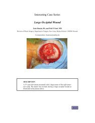
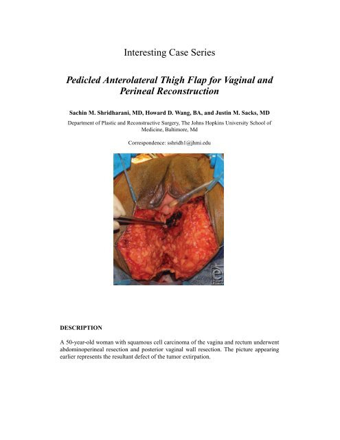
A 50-year-old woman with squamous cell carcinoma of the vagina and rectum underwent<br />
abdominoperineal resection and posterior vaginal wall resection. The picture appearing<br />
earlier represents the resultant defect of the tumor extirpation.
QUESTIONS<br />
1. What is the classification of the ALT flap when harvested as a fasciocutaneous<br />
flap?<br />
2. What is the classification of the ALT flap when harvested as a myocutaneous<br />
flap with the vastus lateralis muscle?<br />
3. What is the dominant blood supply to the ALT flap?<br />
4. How much pedicle length is available for rotation of the pedicled ALT flap<br />
and which areas can be reached?<br />
5. Where are the cutaneous perforators most commonly located?<br />
6. After elevating the pedicled ALT flap, what are the key anatomical landmarks<br />
during its transposition to the perineum?
DISCUSSION<br />
This patient had a large defect with exposure of the perineum and loss of posterior vaginal<br />
wall. To reconstruct this defect, the pedicled anterolateral thigh (ALT) flap was selected.<br />
The ALT flap is a versatile flap that can be utilized in its pedicled form or as a free tissue<br />
transfer. The surgeon can choose to harvest it as either a fasciocutaneous or a myocutaneous<br />
flap. When harvested as a fasciocutaneous flap, it is generally considered a type B or type<br />
C according the Mathes-Nahai classification for fasciocutaneous flaps. It falls under these<br />
categories because of the perforators being either septocutaneous or musculocutaneous. 1<br />
Under the Cormack-Lamberty classification system, the ALT flap is considered a type C<br />
fasciocutaneous flap, because it has segmental perforators. 2 When it is harvested with the<br />
vastus lateralis muscle as a myocutaneous flap, its single dominant vascular pedicle makes<br />
it a type I Mathes-Nahai myocutaneous flap. 3<br />
The dominant blood supply to the anterolateral skin and vastus lateralis muscle is<br />
the descending branch of the lateral circumflex femoral artery. The mean arterial and<br />
venous calibers of the pedicle are 2.1 mm and 2.3 mm, respectively. The pedicle length is<br />
generally reported as 12 cm; however, this number may vary depending on the flap design<br />
and perforator selection. 4 The branches of the lateral femoral cutaneous nerve provide the<br />
sensory innervations to the skin of the anterolateral thigh and should be identified and<br />
preserved during dissection of the flap. In its pedicled form, the ALT flap is a versatile<br />
flap that may be employed to reconstruct the inferior posterior trunk, inferior abdomen,<br />
ipsilateral groin, ischium, anterior thigh, posterior thigh, and perineum.<br />
When the flap is used for perineal reconstruction, it is raised with the patient in the<br />
lithotomy position. However, surface markings should be performed with the patient in the<br />
supine position with the hip joint in a neutral position and the knee extended. In our patient,<br />
a straight line was drawn from the anterior superior iliac spine to the most lateral superior<br />
point on the patella. This line represents the cleavage plane between the rectus femoris<br />
and vastus lateralis muscle. Perforator B is located 1.5 cm lateral to the half-way point of<br />
this line (Fig 1). Peforator A and C are located 5 cm proximal and distal to perforator B,<br />
respectively. 5 These perforator locations were confirmed with a transcutaneous Doppler.<br />
The planned skin paddle was marked in the shape of an ellipse using a template of the<br />
wound. The ellipse should be centered as distally as possible over a reliable perforator as<br />
confirmed by Doppler to reach the defect.<br />
To assist with the obliteration of dead-space in the perineum, a cuff of vastus lateralis<br />
muscle is routinely included with the pedicled ALT. By maintaining the perforating arteries,<br />
the muscle will be sustained on the same pedicle as the ALT flap. Donor-site morbidity has<br />
been shown to be minimal when harvesting part of or all of the vastus lateralis muscle. 6<br />
Once the flap is islandized and elevated to the pedicle origin, the surgeon may inset the<br />
flap in nearly any direction by taking advantage of a large arc of rotation. For perineal<br />
reconstruction in our case, the ALT flap was tunneled below both the rectus femoris muscle<br />
(Fig 2a) and the sartorius muscle (Fig 2b). During this maneuver, the surgeon must take care<br />
to preserve the dominant blood supply to the rectus femoris muscle. The sartorius muscle<br />
has a Mathes-Nahai Type IV blood supply, thus segmental perforators may be ligated to<br />
create an adequate submuscular tunnel for the flap. A subcutaneous tunnel along the medial<br />
thigh was then created to gain access to the perineum for reconstruction of this area (Fig 2c).<br />
When the flap is transposed onto the perineum, it is critical that the flap’s pedicle is not
on any tension. The immediate postoperative image of the reconstructed perineum of our<br />
patient is shown below (Fig 3). The donor site was then primarily closed, and a small<br />
split-thickness skin graft was applied to the area of the thigh that would not close in a<br />
tension-free manner.<br />
Using a submuscular window under the rectus femoris and sartorius and a wide<br />
subcutaneous medial thigh window, this robust flap can be successfully transferred to the<br />
perineum. By harvesting the vastus lateralis muscle along with the skin paddle, the pedicled<br />
ALT myocutaneous flap is an important tool in reconstruction of composite defects of the<br />
perineum including the posterior and lateral walls of the vagina.<br />
Figure 1. Skin markings of the ALT flap with three perforators<br />
labeled as a, b and c.<br />
Figure 2. (a) Tunneling of the flap below the rectus femoris muscle. (b) Tunneling of the flap below<br />
the sartorius muscle. (c) Subcutaneous tunnel along the medial thigh for transposition of flap to<br />
perineum.<br />
Figure 3. Postoperative image of the reconstructed<br />
perineum.
REFERENCES<br />
1. Mathes SJ, Nahai F. Reconstructive Surgery: Principles, Anatomy, and Technique. NewYork,NY:<br />
Churchill Livingstone; 1997.<br />
2. Cormack GC, Lamberty BG. A classification of fascio-cutaneous flaps according to their patterns of<br />
vascularisation. Br J Plast Surg. 1984;37:80-7.<br />
3. Mathes SJ, Nahai F. Classification of the vascular anatomy of muscles: experimental and clinical correlation.<br />
Plast Reconstr Surg. 1981;67:177-87.<br />
4. Wei FC, Jain V, Celik N, Chen HC, Chuang DC, Lin CH. Have we found an ideal soft-tissue flap? An<br />
experience with 672 anterolateral thigh flaps. Plast Reconstr Surg. 2002;109:2219-26; discussion 27-30.<br />
5. Yu P, Youssef A. Efficacy of the handheld Doppler in preoperative identification of the cutaneous<br />
perforators in the anterolateral thigh flap. Plast Reconstr Surg. 2006;118:928-33; discussion 934-5.<br />
6. Hanasono MM, Skoracki RJ, Yu P. A prospective study of donor-site morbidity after anterolateral thigh<br />
fasciocutaneous and myocutaneous free flap harvest in 220 patients. Plast Reconstr Surg. 2010;125:209-<br />
14.<br />
Shridharani et al. <strong>Pedicled</strong> <strong>Anterolateral</strong> <strong>Thigh</strong> <strong>Flap</strong> for Vaginal and Perineal Reconstruction. www.<strong>ePlasty</strong>.com,<br />
<strong>Interesting</strong> <strong>Case</strong>, January 21, 2013