Atomic Force Microscopy in Cancer Cell Research - Clarkson ...
Atomic Force Microscopy in Cancer Cell Research - Clarkson ...
Atomic Force Microscopy in Cancer Cell Research - Clarkson ...

Create successful ePaper yourself
Turn your PDF publications into a flip-book with our unique Google optimized e-Paper software.
10 <strong>Atomic</strong> <strong>Force</strong> <strong>Microscopy</strong> <strong>in</strong> <strong>Cancer</strong> <strong>Cell</strong> <strong>Research</strong><br />
modulus of rigidity from the force curves measured as<br />
described above. To f<strong>in</strong>d Young’s modulus, one needs to<br />
know the tip geometry and structure of the sample. The<br />
tip geometry can be found as described above. However,<br />
the structure of the sample is too complicated to be treated<br />
without any assumptions. Here we will assume that the cell<br />
is a homogeneous, isotropic medium. Obviously this is an<br />
approximation. There is a plethora of literature show<strong>in</strong>g that<br />
such an approximation is plausible [19, 20, 26–29, 40, 134,<br />
136–139]. We will demonstrate this <strong>in</strong> more detail later <strong>in</strong><br />
this section.<br />
Configuration of a spherical probe over a flat homogeneous<br />
surface is described by the Hertz–Sneddon model<br />
[107, 154, 155]. This model was developed for either a spherical<br />
probe of radius R or a parabolic tip over a plane surface.<br />
In the case of a parabolic tip described by equation<br />
Z = b · X 2 , the effective radius R can be found as 1/2b. In<br />
this model, the Young’s modulus E is given by<br />
E = 3 1 − <br />
4<br />
2 dF<br />
√<br />
R dp3/2 (1)<br />
<br />
where F is the load force, R is the radius of the ball, p is the<br />
probe penetration <strong>in</strong>to the cell. The Poisson ratio of the<br />
cells is unknown. For the majority of biological materials,<br />
= 05. Nonetheless, we will treat the Poisson ratio to be<br />
unknown. To remove this uncerta<strong>in</strong>ty, we will speak about<br />
an “apparent” Young’s modulus, to be def<strong>in</strong>ed as follows<br />
Eapp = E<br />
1 − 2 (2)<br />
In the derivation of Eq. (1) it was assumed that the rigidity<br />
of the probe is much higher than the rigidity of the sample.<br />
This is generally true for the majority of AFM probes. The<br />
Hertz–Sneddon model has also two additional assumptions.<br />
The first one is that the probe penetration <strong>in</strong>to the sample<br />
should be much smaller than the radius of the probe. This<br />
can easily be controlled. The second assumption is about<br />
the lack of adhesion between the probe and sample surface.<br />
Look<strong>in</strong>g at the force curves, one can easily identify the<br />
adhesion (Fadd <strong>in</strong> Fig. 2). If the adhesion is high, one has<br />
to use another model, the so-called JKR (Johnson–Kandall–<br />
Roberts) model [156–161] to f<strong>in</strong>d the Young’s modulus. The<br />
material of the probe can be chosen, or altered to ensure low<br />
adhesion. Typically the adhesion between an AFM probe<br />
(for example, a clean silica sphere) and a cell is low. Therefore,<br />
we will not describe the JKR model here, referr<strong>in</strong>g the<br />
reader to the orig<strong>in</strong>al literature [156–161].<br />
The majority of standard AFM cantilevers have a conical<br />
shape. It is useful to present a result of application of the<br />
Hertz–Sneddon model to a conical probe. For a conical tip<br />
with a semivertical (open<strong>in</strong>g) angle , the apparent Young’s<br />
modulus can be given by the follow<strong>in</strong>g formula<br />
Eapp = 1 dF<br />
tan <br />
2 dp2 (3)<br />
<br />
Let us show an example of the application of the Hertz–<br />
Sneddon model to an epithelial cell. It is well known that<br />
the cell is a very complicated object from a structural<br />
po<strong>in</strong>t of view. Nevertheless as we have already mentioned,<br />
it is surpris<strong>in</strong>gly well described by the approximation of<br />
a homogeneous medium. From the general po<strong>in</strong>t of view<br />
such an assumption is not that unexpected if we deal with<br />
small deformations. However, for larger deformations this<br />
assumption is obviously broken. Accord<strong>in</strong>g to Eq. (1), a plot<br />
of the load force versus the probe penetration to the 3/2th<br />
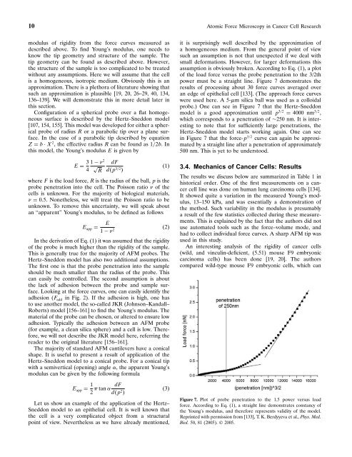
power must be a straight l<strong>in</strong>e. Figure 7 demonstrates the<br />
results of process<strong>in</strong>g about 30 force curves averaged over<br />
an edge of epithelial cell [133]. (The approach force curves<br />
were used here. A 5-m silica ball was used as a colloidal<br />
probe.) One can see <strong>in</strong> Figure 7 that the Hertz–Sneddon<br />
model is a good approximation until p 3/2 = 4000 nm 3/2 ,<br />
which corresponds to a penetration of ∼250 nm. It is <strong>in</strong>terest<strong>in</strong>g<br />
to note that for sufficiently large penetrations, the<br />
Hertz–Sneddon model starts work<strong>in</strong>g aga<strong>in</strong>. One can see<br />
<strong>in</strong> Figure 7 that the force-p 3/2 curve can aga<strong>in</strong> be approximated<br />
by a straight l<strong>in</strong>e after a penetration of approximately<br />
500 nm. This is yet to be understood.<br />
3.4. Mechanics of <strong>Cancer</strong> <strong>Cell</strong>s: Results<br />
The results we discuss below are summarized <strong>in</strong> Table 1 <strong>in</strong><br />
historical order. One of the first measurements on a cancer<br />
cell l<strong>in</strong>e was done on human lung carc<strong>in</strong>oma cells [134].<br />
It showed quite a variation <strong>in</strong> the measured Young’s modulus,<br />
13–150 kPa, and was essentially a demonstration of<br />
the method. Such variability <strong>in</strong> the modulus is presumably<br />
a result of the few statistics collected dur<strong>in</strong>g these measurements.<br />
This is expla<strong>in</strong>ed by the fact that the authors did not<br />
use automated tools such as the force–volume mode, and<br />
had to collect <strong>in</strong>dividual force curves. A sharp AFM tip was<br />
used <strong>in</strong> this study.<br />
An <strong>in</strong>terest<strong>in</strong>g analysis of the rigidity of cancer cells<br />
(wild, and v<strong>in</strong>cul<strong>in</strong>-deficient, (5.51) mouse F9 embryonic<br />
carc<strong>in</strong>oma cells) has been done [19, 20]. The authors<br />
compared wild-type mouse F9 embryonic cells, which can<br />
Figure 7. Plot of probe penetration to the 1.5 power versus load<br />
force. Accord<strong>in</strong>g to Eq. (1), a straight l<strong>in</strong>e demonstrates constancy of<br />
the Young’s modulus, and therefore represents validity of the model.<br />
Repr<strong>in</strong>ted with permission from [133], T. K. Berdyyeva et al., Phys. Med.<br />
Biol. 50, 81 (2005). © 2005.