The Bioartificial Pancreas: Progress and Challenges Review
The Bioartificial Pancreas: Progress and Challenges Review
The Bioartificial Pancreas: Progress and Challenges Review

You also want an ePaper? Increase the reach of your titles
YUMPU automatically turns print PDFs into web optimized ePapers that Google loves.
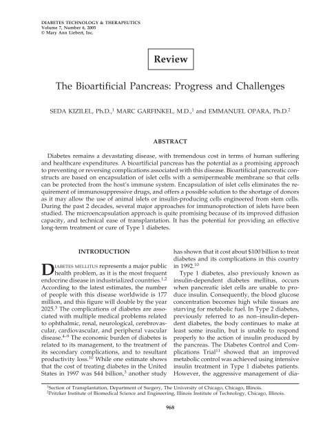
DIABETES TECHNOLOGY & THERAPEUTICS<br />
Volume 7, Number 6, 2005<br />
© Mary Ann Liebert, Inc.<br />
<strong>The</strong> <strong>Bioartificial</strong> <strong>Pancreas</strong>: <strong>Progress</strong> <strong>and</strong> <strong>Challenges</strong><br />
SEDA KIZILEL, Ph.D., 1 MARC GARFINKEL, M.D., 1 <strong>and</strong> EMMANUEL OPARA, Ph.D. 2<br />
ABSTRACT<br />
Diabetes remains a devastating disease, with tremendous cost in terms of human suffering<br />
<strong>and</strong> healthcare expenditures. A bioartificial pancreas has the potential as a promising approach<br />
to preventing or reversing complications associated with this disease. <strong>Bioartificial</strong> pancreatic constructs<br />
are based on encapsulation of islet cells with a semipermeable membrane so that cells<br />
can be protected from the host’s immune system. Encapsulation of islet cells eliminates the requirement<br />
of immunosuppressive drugs, <strong>and</strong> offers a possible solution to the shortage of donors<br />
as it may allow the use of animal islets or insulin-producing cells engineered from stem cells.<br />
During the past 2 decades, several major approaches for immunoprotection of islets have been<br />
studied. <strong>The</strong> microencapsulation approach is quite promising because of its improved diffusion<br />
capacity, <strong>and</strong> technical ease of transplantation. It has the potential for providing an effective<br />
long-term treatment or cure of Type 1 diabetes.<br />
INTRODUCTION<br />
DIABETES MELLITUS represents a major public<br />
health problem, as it is the most frequent<br />
endocrine disease in industrialized countries. 1,2<br />
According to the latest estimates, the number<br />
of people with this disease worldwide is 177<br />
million, <strong>and</strong> this figure will double by the year<br />
2025. 3 <strong>The</strong> complications of diabetes are associated<br />
with multiple medical problems related<br />
to ophthalmic, renal, neurological, cerebrovascular,<br />
cardiovascular, <strong>and</strong> peripheral vascular<br />
disease. 4–9 <strong>The</strong> economic burden of diabetes is<br />
related to its management, to the treatment of<br />
its secondary complications, <strong>and</strong> to resultant<br />
productivity loss. 10 While one estimate shows<br />
that the cost of treating diabetes in the United<br />
States in 1997 was $44 billion, 3 another study<br />
<strong>Review</strong><br />
968<br />
has shown that it cost about $100 billion to treat<br />
diabetes <strong>and</strong> its complications in this country<br />
in 1992. 10<br />
Type 1 diabetes, also previously known as<br />
insulin-dependent diabetes mellitus, occurs<br />
when pancreatic islet cells are unable to produce<br />
insulin. Consequently, the blood glucose<br />
concentration becomes high while tissues are<br />
starving for metabolic fuel. In Type 2 diabetes,<br />
previously referred to as non–insulin-dependent<br />
diabetes, the body continues to make at<br />
least some insulin, but is unable to respond<br />
properly to the action of insulin produced by<br />
the pancreas. <strong>The</strong> Diabetes Control <strong>and</strong> Complications<br />
Trial 11 showed that an improved<br />
metabolic control was achieved using intensive<br />
insulin treatment in Type 1 diabetes patients.<br />
However, the aggressive management of dia-<br />
1 Section of Transplantation, Department of Surgery, <strong>The</strong> University of Chicago, Chicago, Illinois.<br />
2 Pritzker Institute of Biomedical Science <strong>and</strong> Engineering, Illinois Institute of Technology, Chicago, Illinois.
THE BIOARTIFICIAL PANCREAS 969<br />
betes with exogenous insulin only delayed the<br />
progression <strong>and</strong> development of complications<br />
of this disease. Also, intensive insulin treatment<br />
could only be applied to fewer than 10%<br />
of patients due an increased risk of severe<br />
episodes of hypoglycemia. Furthermore, it appears<br />
that C-peptide, a cleavage product from<br />
the processing of pro-insulin to insulin, prevents<br />
diabetes <strong>and</strong> hyperglycemia-induced<br />
vascular <strong>and</strong> neural dysfunction in animal <strong>and</strong><br />
clinical models of diabetes <strong>and</strong> should therefore<br />
be used in combination with insulin to<br />
maintain vascular <strong>and</strong> neural functions as well<br />
as blood glucose levels. 12–15<br />
Advances in transplantation <strong>and</strong> in immunosuppression<br />
have made pancreas transplantation<br />
another treatment option for Type 1<br />
diabetes. <strong>The</strong> main objective for pancreas transplantation<br />
is to achieve normal level of glucose<br />
in the blood <strong>and</strong> to free the patient from exogenous<br />
insulin requirements based on multiple<br />
finger stick glucose measurements. 16 Even<br />
though successful pancreas transplantation<br />
provides normal glucose homeostasis, the<br />
requirement of lifelong immunosuppression<br />
makes it unclear whether transplantation is advantageous<br />
over continued insulin treatment. 17<br />
Also, because of the need for immunosuppression,<br />
most pancreas transplantations have been<br />
done simultaneously with a kidney transplant<br />
in patients who have advanced nephropathy. 18<br />
In contrast to pancreas transplantation, islet<br />
transplantation requires no major surgery.<br />
Also, islet transplantation offers an alternative<br />
to exogenous insulin treatment, as it can result<br />
in better glycemic control 19 <strong>and</strong> potentially<br />
avoids surgical complications associated with<br />
whole-organ pancreas transplantation. It is<br />
known that islet cell transplantation can prevent,<br />
<strong>and</strong>, in some cases, reverse existing complications<br />
of diabetes probably because of the<br />
role played by C-peptide. 20 However, major<br />
challenges remain to be addressed before islet<br />
transplantation can be used routinely <strong>and</strong> applied<br />
more widely to patients with diabetes.<br />
One major barrier is the shortage of human<br />
pancreas as a source of islet cells. Also, lifelong<br />
use of immunosuppressive drugs to overcome<br />
the rejection of transplanted tissue poses a significant<br />
risk to patients, as the use of glucocorticoids<br />
<strong>and</strong> cyclosporine have dose-dependent<br />
deleterious effects on glucose homeostasis <strong>and</strong><br />
-cell function, <strong>and</strong> result in increased incidences<br />
of infection <strong>and</strong> cancer. 20–24 <strong>The</strong> introduction<br />
of the steroid-free immunosuppressive<br />
drug regimen has resulted in long-term graft<br />
survival of islets with concomitant control of<br />
blood glucose levels. 25 However, this immunosuppressive<br />
drug regimen is also not without<br />
risks to graft recipients. 26<br />
<strong>The</strong> routine application of islet transplantation<br />
would be tremendously enhanced by an<br />
unlimited supply of donor tissue, a st<strong>and</strong>ard<br />
implantation procedure, <strong>and</strong> the ability to<br />
maintain the transplanted tissue without the requirement<br />
of immunosuppressive drugs. 27 To<br />
achieve these goals, immunoisolation (encapsulation)<br />
of islet cells has been proposed to protect<br />
cells from attack by the host immune system.<br />
Using immunoisolation of the islets,<br />
chronic administration of immunosuppressive<br />
drugs can be theoretically eliminated or minimized,<br />
as transplanted cells are separated from<br />
the host immune system by a biocompatible<br />
<strong>and</strong> semipermeable membrane. 28,29 Moreover,<br />
immunoisolation of pancreatic islet cells for<br />
transplantation into patients with diabetes theoretically<br />
permits grafting of xenogenic islets,<br />
thus exp<strong>and</strong>ing the potential supply of donor<br />
islet tissue. 25 Hence, immunoisolation could<br />
open up the possibility of using islets harvested<br />
from animals, or insulin-producing cells engineered<br />
from stem cells without the requirement<br />
of immunosuppression of transplant recipients.<br />
Porcine <strong>and</strong> bovine insulin amino acid sequences<br />
have considerable sequence homology<br />
with human insulin, 30 <strong>and</strong> therefore pigs <strong>and</strong><br />
cows are considered attractive as options for<br />
xenogeneic donors.<br />
One important consideration in using microencapsulated<br />
islets is defining the optimal<br />
site for transplantation. To date, most research<br />
has considered peritoneal cavity as the optimal<br />
site for implantation of encapsulated islets, as<br />
it is possible to transplant microcapsules into<br />
the peritoneal cavity using a simple procedure<br />
that could be suitable for routine outpatient<br />
use. 29,31–34 Also, this site allows for implantation<br />
of high numbers of islets, which can be retrieved<br />
by peritoneal lavage 35 if necessary. 36<br />
However, this site limits graft viability because<br />
of low blood supply, oxygen tension, <strong>and</strong> con-
970<br />
centration of essential minerals. 34 In addition,<br />
the release of insulin from the peritoneal cavity<br />
to the blood stream is delayed compared<br />
with hormones released to the portal vein. 37<br />
Also, peritoneal macrophages have a high toxicity<br />
against encapsulated islets. 38<br />
Several researchers have also explored the<br />
portal vein of the liver as an alternative site for<br />
transplantation of immunoisolated islets. 39<br />
Based on its double vascularization (hepatic<br />
artery <strong>and</strong> portal vein), the intraportal location<br />
has a high oxygen <strong>and</strong> nutrient supply, which<br />
could be beneficial for the performance <strong>and</strong> the<br />
longevity of microencapsulated cells. 39 Also,<br />
the liver is considered to be an immunologically<br />
privileged site. 40 But, even more clinically<br />
relevant, is the use of percutaneous transhepatic<br />
catheterization, which provides relatively<br />
simple, inexpensive, <strong>and</strong> non-surgical access to<br />
the liver. 41 In addition, <strong>The</strong> International Islet<br />
Registry reports more C-peptide-positive cases<br />
(more than 0.5 ng/mL) 1 year after transplantation<br />
into the portal vein than from any other<br />
site. 42<br />
This article reviews the recent progress <strong>and</strong><br />
challenges remaining for the successful development<br />
of a bioartificial pancreas, <strong>and</strong> discusses<br />
the requirements to bring this technology<br />
closer to clinical application.<br />
HISTORICAL DEVELOPMENT<br />
<strong>The</strong> pancreatic origin of diabetes was discovered<br />
in 1889 by Mering <strong>and</strong> Minkowski,<br />
when surgical removal of the pancreas caused<br />
dogs to develop diabetes. In 1913, Murlin <strong>and</strong><br />
Kramer prepared extracts of bovine pancreas<br />
to lower the blood glucose in a dog with diabetes.<br />
However, the benefit of the extract was<br />
explained by the presence of lactate. Finally, in<br />
1922, Banting <strong>and</strong> Best developed a method for<br />
preparing a pancreatic extract, <strong>and</strong> a few years<br />
later, injection of the active factor in this extract,<br />
insulin, became the main therapy for Type 1 diabetes.<br />
As of today, the treatment options for Type<br />
1 diabetes are exogenous insulin with external<br />
glucose monitoring, whole-organ pancreas<br />
transplantation, islet cell transplantation, the artificial<br />
pancreas, <strong>and</strong> the bioartificial pancreas.<br />
KIZILEL ET AL.<br />
Among these treatments, the bioartificial<br />
pancreas avoids the use of immunosuppressive<br />
drugs while providing moment-to-moment<br />
glucose homeostasis consistent with a wellfunctioning<br />
native pancreas. It is important in<br />
this context to distinguish between the terms<br />
“artificial pancreas” (sometimes called “mechanical<br />
artificial pancreas”) <strong>and</strong> “bioartificial<br />
pancreas.”<br />
<strong>The</strong> mechanical artificial pancreas consists of<br />
a glucose sensor, an insulin pump, <strong>and</strong> a computer,<br />
which determines the rate of insulin delivery.<br />
<strong>The</strong> sensor may be implanted in the<br />
vena cava, <strong>and</strong> may require periodic replacement.<br />
<strong>The</strong> development of a glucose biosensor<br />
has been challenged by the difficulty of creating<br />
a sensitive, stable, <strong>and</strong> accurate sensor, despite<br />
the capability of a mechanical artificial<br />
pancreas to regulate glucose levels. An error in<br />
sensing, computation, or delivery could result<br />
in insulin overdosing, which could be potentially<br />
risky with life-threatening hypoglycemia,<br />
43–46 an unacceptable potential side effect<br />
of an artificial pancreas. It is also very difficult<br />
to produce a mechanical artificial pancreas that<br />
responds quickly enough to changes in blood<br />
sugar (islets respond in less than 10 min). 47<br />
In contrast, a bioartificial pancreas is a device<br />
that substitutes for the endocrine portion<br />
of the pancreas. Devices in this category contain<br />
synthetic materials <strong>and</strong> functional islets<br />
encapsulated within a semipermeable membrane<br />
to protect cells from the host immune response.<br />
<strong>The</strong> semipermeable membrane permits<br />
exchange of nutrients, glucose, <strong>and</strong> insulin<br />
with the host, but excludes the diffusion of immunoglobulins,<br />
complement, <strong>and</strong> white blood<br />
cells. <strong>The</strong> device may be implanted into a vascularized<br />
site 48,49 or into the peritoneal cavity. 36<br />
<strong>The</strong> idea of using ultrathin polymer membrane<br />
microcapsules was proposed by Chang 50<br />
in 1964 for the immunoprotection of transplanted<br />
cells. Nearly 2 decades later, the concept<br />
of bioencapsulation was successfully<br />
shown to maintain glucose homeostasis in rats<br />
with diabetes using encapsulated allogeneic<br />
islets. 32 When alginate microcapsules containing<br />
islets were implanted in rats, normal blood<br />
glucose levels were achieved for 13 weeks. <strong>The</strong><br />
microcapsule used in that study consisted of an<br />
inner alginate core surrounded by a polylysine
THE BIOARTIFICIAL PANCREAS 971<br />
membrane, which was then surrounded by an<br />
outer polyethyleneimine coating. In later studies,<br />
the outer layer was replaced by the more<br />
biocompatible alginate coating because of an<br />
inflammatory reaction induced by polyethyleneimine.<br />
51 Since then, a variety of studies have<br />
been performed to underst<strong>and</strong> the requirements<br />
for successful transplantation of encapsulated<br />
islet cells, but there is still a need for<br />
convincing evidence of success of encapsulated<br />
islet transplantation in either non-human primates<br />
or humans. 49<br />
IMPEDIMENTS TO THE PROGRESS OF<br />
CELL ENCAPSULATION TECHNOLOGY<br />
Fibrotic overgrowth of the implanted microcapsules<br />
is one of the major obstacles to progress<br />
in cell encapsulation technology. Invariably,<br />
the materials used for encapsulation are<br />
not completely inert, <strong>and</strong> can induce foreign<br />
body <strong>and</strong> inflammatory reactions. As a result of<br />
this fibrous tissue overgrowth, the diffusion of<br />
nutrients, oxygen, hormones, <strong>and</strong> waste products<br />
through the capsule is diminished, <strong>and</strong> encapsulated<br />
islets are destroyed because of hypoxia,<br />
starvation, <strong>and</strong> the secretion of nitric<br />
oxide by the stimulated macrophages. 52 This fibrotic<br />
reaction to implanted microcapsules had<br />
previously been associated with commercially<br />
available alginate with high mannuronic acid<br />
content, which activates macrophages in vivo,<br />
resulting in fibroblast proliferation <strong>and</strong> eventual<br />
overgrowth. 53 However, later studies did<br />
not support that notion. 33 It has also been<br />
shown that polylysine may cause a fibrotic response<br />
through the induction of cytokines. 54<br />
Another important component of capsular<br />
design is permselectivity. Most capsule designs<br />
are based on the presumption that the effectiveness<br />
of immunoisolation of a polymer capsule<br />
is closely related to the pore size of the capsule<br />
membrane. 55–57 However, pore sizes of<br />
polymer membranes are not homogeneous by<br />
nature, <strong>and</strong> consist of a broad spectrum of<br />
sizes. 58 Total immunoprotection of encapsulated<br />
cells can only be provided if the polymer<br />
membrane does not have any pores larger than<br />
the antibody complement component. 59 This<br />
fact has been largely ignored by researchers<br />
<strong>and</strong> has resulted in inconsistent findings in the<br />
field. 60–66<br />
Mechanism of immunologic attack of allografts<br />
<strong>and</strong> xenografts<br />
With regard to specific immunologic attack<br />
of encapsulated cell implants, there are differences<br />
in the immune protection requirement of<br />
allografts versus xenografts. An allograft is a<br />
graft between genetically different individuals<br />
within the same species, while a xenograft is a<br />
graft between individuals from different<br />
species. Allograft rejection occurs as result of<br />
activation of cellular immunity by interactions<br />
of host T cells with a graft, while humoral immunity,<br />
including antibodies <strong>and</strong> complement<br />
proteins, is responsible for the rejection of<br />
xenografts. 67,68 In the direct pathway of antigen<br />
presentation, the T cell receptor recognizes<br />
T cells presented by donor-type antigen-presenting<br />
cells along with Class I or II major histocompatibility<br />
complex (MHC) molecules.<br />
<strong>The</strong> idea of immunoisolation using a polymer<br />
membrane is to separate allogeneic or xenogeneic<br />
tissue from the immune system of the<br />
recipient. <strong>The</strong>refore, microencapsulation of<br />
islets within effective permselective membranes<br />
would prevent contact with immunoglobulin,<br />
complement components, <strong>and</strong><br />
inflammatory cells. However, there is also a potential<br />
immune response towards antigens<br />
shed by encapsulated allogeneic or xenogeneic<br />
cells. Such antigens could be therapeutic agent<br />
themselves, cell surface molecules, or cell components<br />
(phospholipids, DNA), including<br />
those released upon cell death. Shedding of<br />
antigens from encapsulated cells would initiate<br />
a molecular tissue response around the implant,<br />
which could affect the viability <strong>and</strong> function<br />
of the encapsulated cells. As a result of this<br />
immunological reaction, these shedded antigens<br />
may be internalized, processed, <strong>and</strong> presented<br />
in association with host Class II MHC<br />
molecules (macrophages <strong>and</strong> dendritic cells) to<br />
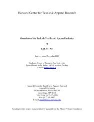
host CD4 helper T cells (Fig. 1). <strong>The</strong> recognition<br />
of antigens through this indirect pathway<br />
may lead to the activation of T helper cells,<br />
which then secrete cytokines <strong>and</strong> regulate cellmediated<br />
immune response <strong>and</strong> inflammation.<br />
69,70 Small molecules, such as reactive oxy-
972<br />
FIG. 1. Schematic representation of antigen recognition<br />
via (A) the direct pathway <strong>and</strong> (B) the indirect pathway.<br />
APC, antigen-presenting cells; TCR, T cell receptor.<br />
gen species <strong>and</strong> cytokines, may also pass<br />
through the polymer membrane <strong>and</strong> damage<br />
the transplanted tissue.<br />
Owing to the complexity of the immune response<br />
mechanism, it is important to underst<strong>and</strong><br />
the mechanism of immune protection,<br />
<strong>and</strong> considerable efforts have been made to investigate<br />
this issue. Chen et al. 71 developed cell<br />
lines that have resistance to cytokines <strong>and</strong> oxygen<br />
radical-induced damage. Recently, macrophage<br />
depletion has also been used as a tool to<br />
protect xenografts from immune destruction. It<br />
has been found that depletion of peritoneal<br />
macrophages with clodronate liposomes improved<br />
the survival of macroencapsulated<br />
porcine neonatal pancreatic cell clusters. 35<br />
Safley et al. 72 evaluated the cellular immune response<br />
in non-obese diabetic (NOD) mice after<br />
two different pathways of T cell co-stimulation<br />
were blocked. Alginate–poly-L-lysine-encapsulated<br />
porcine islet xenografts were transplanted<br />
intraperitoneally in NOD mice treated<br />
with CTLA4-immunoglobulin to block CD28/<br />
B7 <strong>and</strong> with anti-CD154 monoclonal antibody<br />
to inhibit CD40/CD40–lig<strong>and</strong> interactions. It<br />
was concluded that blocking two different<br />
pathways of T cell co-stimulation inhibited T<br />
cell-dependent inflammatory responses, <strong>and</strong><br />
KIZILEL ET AL.<br />
significantly prolonged the survival of encapsulated<br />
islet xenografts.<br />
Another approach to improving the survival<br />
of microencapsulated islets can be to use local<br />
immunosuppression. Studies have shown that<br />
preimplantation of an immunoisolating device<br />
improved the survival of an encapsulated islet<br />
graft <strong>and</strong> reduced fibroblast outgrowth. 73 Functional<br />
capsules may also be designed to release<br />
the immunosuppressive agent in a controlled<br />
manner. Local immunosuppression induced using<br />
this approach may prevent the occurrence<br />
of an undesirable reaction around the implant.<br />
Inflammatory reaction in response to the implantation<br />
of cell-free capsules involves a sequence<br />
of events similar to a foreign body reaction,<br />
starting with an acute inflammatory<br />
response <strong>and</strong> leading to a chronic inflammatory<br />
response or granulation tissue development<br />
<strong>and</strong> fibrous capsule development. <strong>The</strong> intensity<br />
<strong>and</strong> duration of each of these responses<br />
are dependent upon several factors, such as the<br />
extent of injury created in the implantation,<br />
biomaterial chemical composition, surface free<br />
energy, surface charge, porosity, surface<br />
roughness, <strong>and</strong> implant size <strong>and</strong> shape. 74,75 <strong>The</strong><br />
biocompatibility of the material is determined<br />
by the extent <strong>and</strong> deviation from the optimal<br />
wound healing conditions. 76<br />
Sources of donor islets<br />
Despite all of the advances in islet transplantation<br />
<strong>and</strong> in encapsulation technology, some<br />
challenges must be still overcome before the<br />
bioartificial pancreas can be broadly applied.<br />
<strong>The</strong> most significant of these challenges is the<br />
shortage of donor islets. This shortage is the<br />
motivation for researchers to find alternative<br />
sources of insulin-producing cells. Promising<br />
approaches for resolving this problem are differentiation<br />
of stem cells into cells with -celllike<br />
characteristics77–80 <strong>and</strong> genetic engineering<br />
of adult cells for secretion of recombinant insulin.<br />
81–86 Based on similarities between mechanisms<br />
that control the development of both the<br />
adult pancreas <strong>and</strong> the central nervous system,<br />
methods promoting neural differentiation of<br />
embryonic stem cells have been adapted by researchers<br />
to derive insulin-producing cells. 87–91<br />
Hori et al. 92 investigated the possibility of directing<br />
neural progenitors to develop into glu-
THE BIOARTIFICIAL PANCREAS 973<br />
cose-responsive insulin-producing cells using<br />
inductive signals involved in normal pathways<br />
of islet development. Compared with other<br />
methods for insulin-producing cell development<br />
from human stem cells, 91,93 the method<br />
developed by this group produced insulin at<br />
the highest levels yet achieved from an exp<strong>and</strong>able,<br />
human stem cell-derived tissue.<br />
However, the method developed in the study<br />
still needs to be improved, perhaps through the<br />
addition of glucagon-like peptide-1, transforming<br />
growth factor- lig<strong>and</strong>s, or other factors<br />
to potentiate -cell maturation, growth,<br />
<strong>and</strong> insulin secretion. 94–96<br />
Gene therapy has also been suggested as a<br />
treatment of insulin-dependent diabetes mellitus.<br />
This strategy can be applied by preventing<br />
the autoimmune destruction of -cells, 97,98 by<br />
regenerating -cells, 99,100 or by engineering insulin-secreting<br />
surrogate -cells. 99,101–103 Sapir<br />
et al. 104 recently suggested the potential use of<br />
adult human liver as an alternate tissue for autologous<br />
-cell-replacement therapy. Using<br />
pancreatic <strong>and</strong> duodenal homeobox gene 1<br />
(PDX-1) <strong>and</strong> soluble factors, they managed to<br />
induce a comprehensive developmental shift of<br />
adult human liver cells into functional insulinproducing<br />
cells. According to the findings of<br />
that study, PDX-1-treated human liver cells expressed<br />
insulin, stored it in defined granules,<br />
<strong>and</strong> secreted the hormone in a glucose-regulated<br />
manner. When these cells were implanted<br />
under the renal capsule of immunodeficient<br />
mice with diabetes, the cells ameliorated hyperglycemia<br />
for prolonged periods of time. <strong>The</strong><br />
technique offers both the potential of a cell-replacement<br />
therapy for patients with diabetes<br />
<strong>and</strong> allows the patient to be the donor of his or<br />
her own insulin-producing tissue.<br />
Intestinal K cells have also been engineered<br />
to express <strong>and</strong> secrete insulin in a glucose dependent<br />
manner, generating functional cells.<br />
105,106 With the development of robust <strong>and</strong><br />
safe gene therapy, this strategy may lead to effective<br />
therapy in the future.<br />
Preservation of encapsulated cell systems<br />
<strong>The</strong> last challenge for a bioartificial pancreas<br />
to become a routine clinical application is the<br />
long-term cryopreservation of encapsulated<br />
cells. Validated technologies are required for<br />
long-term preservation of encapsulated cell systems<br />
to maintain a product inventory, <strong>and</strong> in order<br />
to meet end-user dem<strong>and</strong>s. One of the current<br />
strategies for overcoming the problem of<br />
cryopreservation of tissue is the reduction of cryoprotectant<br />
[dimethyl sulfoxide (DMSO)] concentration<br />
or complete replacement by equally<br />
powerful substances. Recent work in Dr. Opara’s<br />
laboratory has demonstrated that cryopreservation<br />
of islet cells cultured overnight in the presence<br />
of 10% polyvinylpyrrolidone yielded higher<br />
intact islet recovery compared with islets frozen<br />
in the presence of DMSO <strong>and</strong> glycerol. 107 <strong>The</strong><br />
lower islet cell integrity <strong>and</strong> function were explained<br />
on the basis of hypothesis that low-molecular-weight<br />
compounds, such as DMSO <strong>and</strong><br />
glycerol, permeate the cell <strong>and</strong> interact hydrophobically<br />
with intracellular proteins, which<br />
results in perturbed cytoskeletal architecture of<br />
the frozen cells <strong>and</strong> diminished islet cell integrity<br />
<strong>and</strong> function.<br />
<strong>The</strong> other promising technical improvement<br />
of the cryopreservation technology is the reduction<br />
of total sample volume. Miniaturization<br />
of the cryosubstrates from 1 mL to microliters<br />
reduces temperature gradients in cryosubstrates,<br />
which makes it possible to achieve<br />
higher freezing rates <strong>and</strong> more homogeneous<br />
freezing of the sample. 108 Islets cryopreserved<br />
with this strategy were highly functional even<br />
with the lower DMSO concentrations. 109<br />
Misler et al. 110 studied stimulus–secretion<br />
coupling in whole islets as well as single cells<br />
from carefully selected cryopreserved <strong>and</strong><br />
thawed human islets of Langerhans. <strong>The</strong>y found<br />
that without using any of the recent advances in<br />
cryopreservation technique, cryopreserved <strong>and</strong><br />
thawed human islets, which were selected based<br />
on their smooth surface <strong>and</strong> diameter, responded<br />
to glucose in a calcium- <strong>and</strong> metabolicdependent<br />
fashion. In another study by von<br />
Mach et al., 111 viability of pancreatic islets after<br />
cryopreservation was correlated negatively with<br />
their size, <strong>and</strong> suppression of insulin release was<br />
not observed for islets 300 m.<br />
DIFFERENT FORMS OF BIOARTIFICIAL<br />
PANCREAS<br />
Various configurations have been studied for<br />
the purpose of immunoisolation of islets. <strong>The</strong>se
974<br />
include biohybrid vascular devices, extravascular<br />
chambers, <strong>and</strong> encapsulation. 112,113 Encapsulation<br />
is the technique of islet immunoisolation<br />
using biopolymeric spheres of<br />
different sizes. Currently, results in rodents<br />
<strong>and</strong> dogs with diabetes indicate that spherical<br />
micro- <strong>and</strong> macrocapsules appear to have the<br />
highest therapeutic potential. 60,65,114 A microcapsule<br />
typically contains only a few hundred<br />
cells or a single islet, <strong>and</strong> to provide a therapeutic<br />
dosage, tens of hundreds of thous<strong>and</strong>s<br />
of cells are required. In contrast, macrocapsules<br />
employ much larger depots to contain the full<br />
therapeutic dosage in one or a few implants.<br />
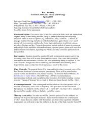
Macrocapsules are shaped as cylinders or<br />
planes, <strong>and</strong> typically one dimension is in the<br />
range of 2–6 mm (Fig. 2a). 115,116 One advantage<br />
of the implantation of macrocapsules is the ease<br />
of retrieval in case of complications. However,<br />
despite reports of reduced risks in the implantation<br />
of macrocapsules, it has been shown that<br />
macroencapsulated islets have significantly impaired<br />
insulin secretion because of necrosis in<br />
the center of aggregated islet clumps as a result<br />
of diffusion limitations of nutrients <strong>and</strong><br />
oxygen. 116,117<br />
Encouraging results have been obtained by<br />
immobilization of the islets in a matrix before<br />
final macroencapsulation. 115,118 Limited diffusion<br />
of nutrients, as well as slow exchange of<br />
glucose <strong>and</strong> insulin, occurs as a result of relative<br />
large surface-to-volume ratio of these devices.<br />
To ensure sufficient nutrient <strong>and</strong> oxygen<br />
diffusion to the cells, islet cell density within<br />
the macrocapsules is kept to around 5–10% of<br />
the volume fraction. This approach, however,<br />
requires the implantation of large devices in order<br />
to provide adequate masses of insulin-producing<br />
cells, <strong>and</strong> large grafts are not practical<br />
<strong>and</strong> cannot be transplanted in conventional<br />
sites. 119<br />
In contrast, microencapsulation offers an optimal<br />
volume-to-surface ratio for fast exchange<br />
of hormones <strong>and</strong> nutrients without a large device.<br />
<strong>The</strong> technique involves enclosure of one<br />
or two islets within small spheres, which have<br />
diameters of less than 1 mm (Fig. 2b). Also, because<br />
of reduced volume of the islet transplants,<br />
<strong>and</strong> in contrast to macroencapsulated islets, microcapsules<br />
can theoretically be transplanted in<br />
delicate sites, such as the portal vein. <strong>The</strong> trans-<br />
(a) Macroencapsulation<br />
(b) Microencapsulation<br />
plantation of microcapsules into the peritoneal<br />
cavity, an outpatient-based procedure, 33,120–122<br />
makes it particularly attractive to patients <strong>and</strong><br />
doctors. Interestingly, intraperitoneally implanted<br />
microencapsulated islets have shown<br />
promising results in large animal 122–124 models<br />
as well as in limited clinical trials. 33<br />
TECHNIQUES IN<br />
MICROENCAPSULATION<br />
KIZILEL ET AL.<br />
FIG. 2. Immunoisolated islets within (a) membrane diffusion<br />
chambers (macrocapsules) <strong>and</strong> (b) microcapsules.<br />
Materials used for microencapsulation<br />
Various materials have been used as biopolymeric<br />
coats in islet microencapsulation. <strong>The</strong>se<br />
have included alginate, agarose, tissue-engineered<br />
chondrocytes, polyacrylates, 125,126 <strong>and</strong><br />
poly(ethylene glycol) (PEG).<br />
Alginate–polylysine-based microencapsulation<br />
of islets, first described by Lim <strong>and</strong> Sun, 32<br />
has been found not to interfere with cellular function,<br />
<strong>and</strong> these microcapsules have been shown<br />
to be stable for years in small <strong>and</strong> large animals<br />
as well as in human beings. 127–132 Alginate is a<br />
linear polysaccharide extracted from seaweed. 133<br />
It is composed of three types of 1 4-linked<br />
polyuronic acid blocks containing poly-L-guluronic<br />
acid segments (G blocks), poly-D-manuronic<br />
acid segments (M blocks), <strong>and</strong> segments<br />
of alternating L-guluronic <strong>and</strong> D-mannuronic<br />
acid residues, <strong>and</strong> it has gel-forming properties<br />
in the presence of most polyvalent cations. 133,134<br />
Impurities, such as monomers, catalysts, <strong>and</strong> initiators<br />
present in synthetic polymers, may contribute<br />
to the failure of the encapsulated islet im-
THE BIOARTIFICIAL PANCREAS 975<br />
plants. 135 Other impurities, such as pyrogens <strong>and</strong><br />
mitogens, can be found in polymers derived<br />
from natural substances, such as seaweed. 135 Up<br />
to 90% of the impurities can be removed by electrophoresis<br />
or by the Klock extraction purification<br />
procedure. 130,135 Conflicting reports exist on<br />
whether alginate with high M blocks or high G<br />
blocks results in increased fibrosis. 53 It has been<br />
shown, however, that the inflammatory response<br />
to alginate becomes less with purification,<br />
irrespective of the composition used. 36,136<br />
<strong>The</strong> immunoisolating permselectivity of alginate<br />
microcapsules can be achieved by soaking<br />
the initial alginate beads in 0.05–0.1%<br />
polylysine dissolved in normal saline for 6–20<br />
min. This occurs as a result of the binding of<br />
negatively charged carboxyl groups on the alginate<br />
to positively charged -amino groups on<br />
the amino acid polymer. <strong>The</strong> droplets are again<br />
rinsed with normal saline, followed by the addition<br />
of an outer alginate layer by soaking in<br />
a lower concentration of 0.06–0.25% sodium alginate<br />
for 4 min. It is extremely important that<br />
the outer coating of alginate on top of the<br />
polylysine membrane is complete. In the case<br />
of incomplete cover of the polylysine coating<br />
<strong>and</strong> when an impure alginate is used for the<br />
outer coating, microcapsules may become completely<br />
covered by cell overgrowths within 1<br />
week of transplantation because of the inflammatory<br />
reactions attributable to polylysine <strong>and</strong><br />
the impurities in the alginate. 137 To dissolve the<br />
intracapsular sodium alginate by chelation of<br />
the cross-linking calcium, the resulting microcapsules<br />
are treated with 55 mM sodium citrate<br />
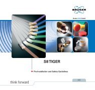
for 7 min. <strong>The</strong> hollow microcapsules containing<br />
a few islets are then washed with saline<br />
(Fig. 3). It appears that liquefaction of the inner<br />
alginate core of microcapsules is required<br />
for optimal function of the enclosed islets. 138<br />
One successful transplantation of encapsulated<br />
islets performed with this technique was reported<br />
by a team of investigators at Duke University.<br />
<strong>The</strong> encapsulated islets were made<br />
with an outer alginate coating <strong>and</strong> inner poly-<br />
L-ornithine layer for permselectivity. To liquefy<br />
the inner alginate core of each bead a salt treatment<br />
was used. After transplantation, encapsulated<br />
islet cells were shown to keep a baboon<br />
with diabetes from requiring exogenous insulin<br />
for more than 9 months. 122<br />
FIG. 3. Microencapsulation of islets with alginate–<br />
polylysine.<br />
For a capsule to be stable in vivo it has to be<br />
water insoluble, as capsule dissolution will<br />
elicit a continuing inflammatory response <strong>and</strong><br />
will ultimately expose the transplanted cells to<br />
the host. Both alginate <strong>and</strong> polylysine are water-soluble<br />
polymers <strong>and</strong> are both expected to<br />
elicit an inflammatory or foreign body response.<br />
However, findings indicate that the<br />
polyelectrolyte complex formed during encapsulation<br />
is biocompatible <strong>and</strong> less soluble in<br />
water than the individual components. 62,139,140<br />
Differences in biocompatibility were also noted<br />
between empty <strong>and</strong> cell-containing capsules in<br />
NOD mice, 141,142 which could be due to the difficulty<br />
of producing reproducible cell-containing<br />
capsules. For better biocompatibility, one<br />
group has suggested the use of high guluronic<br />
acid-containing alginates, 53 while another<br />
group modified alginate–polylysine capsules<br />
with PEG. 132 An alternative approach has been<br />
chosen to obtain stronger <strong>and</strong> more biocompatible<br />
microcapsules using synthetic polyacrylates<br />
such as the water-insoluble synthetic<br />
copolymer hydroxyethyl methacrylate-methyl<br />
methacrylate. 143 This research group has selected<br />
the acrylate monomers because of their<br />
diversity <strong>and</strong> has shown that mammalian cells
976<br />
may be microencapsulated in uncharged <strong>and</strong><br />
polyelectrolyte polymer, in polyelectrolyte<br />
complexes, <strong>and</strong> inside a cohesive precipitate<br />
from a destabilized emulsion, without loss of<br />
viability.<br />
Polymerization of acrylamide monomer on<br />
islet cells encapsulated in agarose microspheres<br />
has also been reported. 144 However, when<br />
polymerization is in direct contact with tissue,<br />
this can generate excessive local heating <strong>and</strong><br />
cytotoxicity. 145 Poly(styrene sulfonic acid), one<br />
of the most potent complement-interacting<br />
polymers, was selected for the purpose of consuming<br />
cytolytic complement activity <strong>and</strong><br />
mixed with agarose for the preparation of microbeads<br />
to protect xenogeneic islets in mice<br />
with diabetes from the humoral immune reaction.<br />
63,146 Antibodies <strong>and</strong> complement proteins<br />
play a major role in the rejection of xenografts.<br />
Binding of antibodies to the antigens on the<br />
xenogeneic cell surface cannot alone damage<br />
the islet cell. For destruction of the xenogeneic<br />
cells to occur, complement activation by antigen–antibody<br />
complexes present on the cell<br />
surface is required. 147 Various polymers bearing<br />
hydroxyl groups such as regenerated cellulose,<br />
cellulose acetate, <strong>and</strong> poly(ethylene-covinyl<br />
alcohol) activate the complement system<br />
through the alternative pathway, 148 whereas<br />
other polymers bearing sulfonic acid or sulfate<br />
groups strongly interact with complement proteins<br />
<strong>and</strong> decrease the cytolytic complement activity.<br />
149<br />
As previously noted, a fibrotic reaction after<br />
implantation would limit the diffusion of essential<br />
nutrients or metabolites, leading to reduced<br />
viability or impaired functional activity<br />
of the encapsulated cells. Several studies have<br />
reported the usefulness of coating membrane<br />
surfaces with poly(ethylene oxide) (PEO) in order<br />
to reduce protein adsorption <strong>and</strong> associated<br />
fibrotic reaction. 150 This was achieved by<br />
exposing the poly(acrylonitrile-vinyl chloride)<br />
membrane surface to amine-terminated PEO<br />
after acid hydrolysis of the nitrile group to a<br />
carboxylic acid. Membrane surfaces were also<br />
exposed to PEO-succinimide after base reduction<br />
to an amine. 151 <strong>The</strong> in vivo response<br />
proved that a smaller number of macrophages<br />
<strong>and</strong> foreign body giant cells were present on<br />
the PEO-grafted membrane surface without<br />
KIZILEL ET AL.<br />
any change in transport properties. <strong>The</strong> other<br />
application to modify the membrane surface<br />
was achieved by synthesizing a graft copolymer<br />
having poly-L-lysine as the backbone <strong>and</strong><br />
the monomethoxy PEG as pendant chains. Microcapsules<br />
with sodium alginate were formed<br />
using this polycationic copolymer, which demonstrated<br />
reduced protein adsorption, complement<br />
binding, <strong>and</strong> cell adhesion in vitro compared<br />
with materials with unmodified poly-<br />
L-lysine. 132<br />
That artificial materials used for immunoisolation<br />
purposes are not completely inert <strong>and</strong><br />
can induce foreign body <strong>and</strong> inflammatory reactions<br />
have led researchers to study tissue-engineered<br />
capsules of rat chondrocytes. Pollok<br />
et al. 126 proposed the encapsulation of islets<br />
with a layer of chondrocytes <strong>and</strong> their matrix<br />
to prevent immunorecognition <strong>and</strong> destruction<br />
of transplanted allogeneic or xenogeneic islets.<br />
This investigation demonstrated the membrane’s<br />
ability as an immunoisolation barrier<br />
by utilizing the immunoprivileged properties<br />
of the chondrocyte matrix. This approach<br />
might offer a therapeutic advantage for patients<br />
with diabetes, using the patient’s own<br />
chondrocytes from a cartilage biopsy specimen<br />
as the encapsulation material.<br />
PEG is another polymer that has been utilized<br />
for the purpose of encapsulating islets. In<br />
order to obtain stable <strong>and</strong> biocompatible gels<br />
without any cytotoxicity generation, Pathak et<br />
al. 152 <strong>and</strong> Cruise et al. 153 reported rapid photopolymerization<br />
of water-soluble PEG-based<br />
macromers in direct contact with islet cells.<br />
PEG is biocompatible, nontoxic, non-immunogenic,<br />
<strong>and</strong> hydrophilic <strong>and</strong> can be chemically<br />
cross-linked into hydrogels for a variety of applications.<br />
154,155 Diffusion profiles of proteins<br />
through PEG diacrylate hydrogels showed that<br />
gels formed with the proper formulation were<br />
capable of being immunoprotective. <strong>The</strong> semipermeable<br />
properties of cross-linked PEG hydrogels<br />
made by interfacial photopolymerization<br />
of PEG diacrylate were also studied by<br />
Cruise et al. 156 by forming hydrogels upon<br />
poly(vinylidiene fluoride) microporous filters.<br />
This approach allowed them to study the effect<br />
of molecular weight <strong>and</strong> concentration of the<br />
PEG diacrylate on the diffusivity of biological<br />
molecules.
THE BIOARTIFICIAL PANCREAS 977<br />
Techniques used for droplet generation<br />
<strong>The</strong> other major consideration in microencapsulation<br />
technology is how to get the cells<br />
into the individual polymer capsules. Techniques<br />
to form such a physical barrier include<br />
air-jet syringe pump droplet generator, interfacial<br />
photopolymerization, <strong>and</strong> selective withdrawal.<br />
One of the main devices used for microencapsulation<br />
of islets with alginate–polylysine is<br />
the air-jet syringe pump droplet generator. 157<br />
<strong>The</strong> device consists of an air jacket surrounding<br />
an alginate nozzle through which alginate<br />
solution is injected. 158,159 Islets are suspended<br />
in a solution of 1.4–3% sodium alginate, <strong>and</strong><br />
spherical droplets of this suspension are<br />
formed by an air-jet syringe pump generator.<br />
As alginate droplets are forced out of the end<br />
of the needle by the syringe pump, the droplets<br />
are pulled off by the shear forces set up by the<br />
flowing air stream. <strong>The</strong> size of the spherical<br />
droplets is controlled by adjusting the flow rate<br />
of the air jacket. <strong>The</strong> spherical droplets are collected<br />
in a funnel containing HEPES acid buffer<br />
supplemented with 1.1% CaCl2 solution, which<br />
transformed alginate droplets into rigid beads<br />
by gelation. 110 Excess fluid is drained with the<br />
aid of a filtering device consisting of a nylon<br />
mesh <strong>and</strong> a stopcock at the exit end of the funnel,<br />
<strong>and</strong> the droplets are washed in normal<br />
saline.<br />
As noted earlier, an air jet-syringe pump extrusion<br />
method generates gel droplets containing<br />
entrapped islets from the suspension of the<br />
islets in aqueous sodium alginate solution.<br />
However, there are various constraints concerning<br />
the use of this procedure to produce<br />
microcapsule diameters of less than 700 m. To<br />
form perfectly spherical capsules, the viscosity<br />
of the gel-forming liquid must be greater than<br />
30 cp. Also, to prevent the blockage of the needle<br />
by the islets, the minimum internal diameter<br />
of the needle must be greater than 300 m.<br />
Finally, the volumetric airflow rate must remain<br />
below 2,000 mL/min in order to produce<br />
capsules of uniform diameter. This procedure<br />
was improved by employing an electrostatic<br />
droplet generator to form uniform, smooth,<br />
<strong>and</strong> perfectly spherical microcapsules having a<br />
diameter about 150–500 m. 160 A droplet,<br />
which is charged with high static voltage, is<br />
suspended from a needle <strong>and</strong> attracted to a collecting<br />
vessel with opposing polarity. Once the<br />
voltage threshold potential is passed, the<br />
droplet moves from the needle to the collecting<br />
vessel. Droplets with predetermined sizes<br />
may be repeatedly generated as the voltage<br />
pulse height, pulse frequency <strong>and</strong> length, <strong>and</strong><br />
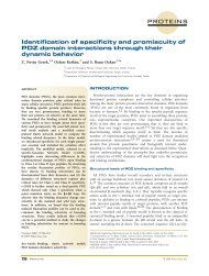
extrusion rate of the droplets are adjustable. As<br />
shown in Figure 4, droplets are formed as the<br />
plunger is driven by the syringe pump <strong>and</strong> expelled<br />
towards a collecting vessel containing a<br />
hardening solution, which may be aqueous calcium<br />
chloride in the case of an aqueous droplet<br />
forming liquid containing sodium alginate.<br />
Negative polarity was attached to the needle,<br />
while positive polarity was attached to the<br />
metal ring. As the voltage applied during<br />
droplet formation is a static voltage, the viability<br />
of the encapsulated islets is not compromised.<br />
Biomolecules such as proteins <strong>and</strong> cells can<br />
also be deposited as patterned droplets onto<br />
surfaces for the development of cell-based<br />
biosensing devices for applications such as<br />
high-throughput drug screening. 161 <strong>The</strong>re are<br />
numerous techniques for microfabrication of<br />
patterned polymer surfaces for protein delivery.<br />
Lithographic techniques <strong>and</strong> microcontact<br />
printing have been most widely used to generate<br />
patterned droplets of cells. 162–168 PEG hy-<br />
FIG. 4. Schematic representation of an electrostatic<br />
droplet generator.
978<br />
drogel is the most commonly used material for<br />
cell patterning purposes. In the case of surfaceinitiated<br />
photopolymerization of PEG, the technique<br />
involves immobilization of initiator onto<br />
a substrate surface using a stamp such as<br />
poly(dimethyl siloxane), followed by polymerization<br />
of precursor solution. 169 One recent<br />
study focused on the cellular delivery based on<br />
micro- <strong>and</strong> nanotechnology. 170 To provide an<br />
immunoisolating microenvironment for islet<br />
cells, nanoporous biocapsules were bulk <strong>and</strong><br />
surface micromachined to present uniform <strong>and</strong><br />
well-controlled pore sizes. <strong>The</strong> designs of the<br />
membranes with defined nanopores were fabricated<br />
using a thermally grown silicon oxide<br />
s<strong>and</strong>wiched between two structural layers of<br />
silicon.<br />
Another technique used to generate droplets<br />
of encapsulated islet cells was proposed by<br />
Cruise et al. 153 using interfacial photopolymerization.<br />
<strong>The</strong> technique involves physical adsorption<br />
of the initiator eosin Y on the islet cell<br />
surface, which then allows polymerization to<br />
occur on the islet–prepolymer interface. <strong>The</strong> interfacial<br />
photoinitiation process employed with<br />
this technique results in conformal coating of<br />
cross-linked PEG-based hydrogel on the islet<br />
cell surface.<br />
<strong>The</strong> process of selective withdrawal is another<br />
novel method reported recently for the<br />
purpose of coating cell clusters, such as islet<br />
cells. 171 Briefly, the process involves the insertion<br />
of a tube into a container such that its tip<br />
is suspended at a specific height above an interface<br />
separating two immiscible fluids (Fig.<br />
5). In the case of a low fluid withdrawal rate,<br />
only the upper fluid (oil) is withdrawn through<br />
the tube. Increasing the fluid flow rate or de-<br />
Oil<br />
Water<br />
To pump<br />
FIG. 5. Diagram of the selective withdrawal apparatus.<br />
creasing the height of the tip above the interface<br />
results in a transition where the lower liquid<br />
(water) is incorporated in a thin spout<br />
along with the oil. <strong>The</strong> technique was demonstrated<br />
through encapsulation of a poppy seed<br />
in a poly(styrenesulfonic acid). <strong>The</strong> particles<br />
were coated with the prepolymer solution,<br />
which has styrene sulfonic acid sodium salt<br />
<strong>and</strong> triethylene glycol diacrylate, <strong>and</strong> mixed<br />
with the initiator eosin Y <strong>and</strong> co-initiator triethanolamine<br />
used for the purpose of initiating<br />
polymerization. 171 After the selective withdrawal<br />
step, the coated particles were collected<br />
<strong>and</strong> photopolymerized with a halogen lamp.<br />
<strong>The</strong> technique could also be used to encapsulate<br />
cells through surface initiated photopolymerization<br />
of PEG. <strong>The</strong> photoinitiator eosin Y<br />
could be immobilized on the islet cell surface<br />
through covalent attachment, 169 <strong>and</strong> after the<br />
selective withdrawal process, cells could be collected<br />
<strong>and</strong> illuminated for 2–3 min with an argon<br />
ion laser in order to cross-link the coat<br />
around the cells.<br />
CONCLUSIONS<br />
KIZILEL ET AL.<br />
<strong>The</strong> successful development of a bioartificial<br />
pancreas involves several considerations. Two<br />
major obstacles in islet transplantation are the<br />
limited supply of islet cells <strong>and</strong> the use of immunosuppressive<br />
drugs to prevent transplant<br />
rejection. It is hoped that the use of encapsulated<br />
islets as a form of bioartificial pancreas<br />
would overcome these obstacles. Three potential<br />
sources of islet cell tissue, including human<br />
or allogeneic cells, porcine or xenogenic cells,<br />
<strong>and</strong> engineered cells, are currently under investigation.<br />
Among these sources, human islet<br />
cells would be the least immunogenic; however,<br />
there is a shortage of pancreata retrieved<br />
from human cadaveric donors, <strong>and</strong> these cells<br />
have a limited secretory capacity <strong>and</strong> life span.<br />
<strong>The</strong> use of porcine islet cells could be a better<br />
option, because these are readily available, <strong>and</strong><br />
there is an unlimited supply of donors. However,<br />
one major concern about the use of islets<br />
harvested from pigs has been the possibility of<br />
transmittance of porcine endogenous retroviruses<br />
(PERVs) to humans during transplantation.<br />
This concept arose from studies on the
THE BIOARTIFICIAL PANCREAS 979<br />
infection of humans <strong>and</strong> immunodeficient mice<br />
after pig cell xenotransplantation. 172,173 <strong>The</strong>re<br />
are studies, however, showing no possibility of<br />
transmission of PERVs from pigs to human recipients<br />
of islet xenografts. 120,174,175 Indeed, one<br />
recent study showed that the porcine genome<br />
harbors a limited number of infectious PERV<br />
sequences, which suggests that many breeds of<br />
pig fail to produce PERVs capable of infecting<br />
human cells even in laboratory testing. 176 This<br />
raises the possibility of using special breeds of<br />
pig for an unlimited supply of islet cells for encapsulation<br />
<strong>and</strong> use as a bioartificial pancreas<br />
in clinical xenotransplantation.<br />
<strong>The</strong> other attractive approach to generate unlimited<br />
supply of islets is the use of embryonic<br />
stem cells, which have the ability to differentiate<br />
into a variety of cell lineages in vitro. Recent<br />
studies in small animals showing successful<br />
transplantation of pancreatic stem cells<br />
suggest that this approach could provide an<br />
unlimited source of allogeneic insulin-producing<br />
tissue in the future. 177–179<br />
<strong>The</strong> maintenance of cell viability is another<br />
important concern in the development of a successful<br />
bioartificial pancreas. When cells are encapsulated<br />
<strong>and</strong> implanted in an environment<br />
without a natural circulation, lack of an adequate<br />
supply of oxygen <strong>and</strong> nutrients results in<br />
cell necrosis, making implantation unsuccessful.<br />
To improve long-term islet survival <strong>and</strong><br />
function, vascularization of transplanted islets<br />
has been proposed. 180–182 An ideal architecture<br />
for implantation would be to promote vascularization<br />
around encapsulated cells using a<br />
novel approach. Recently, a technique to form<br />
covalently bonded multilayers of PEG hydrogels<br />
has been developed. This approach may<br />
be used to encapsulate cells with bilayers such<br />
that outer layer would promote vascularization<br />
around the inner layer. 183,184<br />
<strong>The</strong>re are other areas of urgent need for the<br />
successful development of a bioartificial pancreas<br />
through islet cell microencapsulation. All<br />
current techniques for cell microencapsulation<br />
are slow in producing desirable microcapsules,<br />
requiring days to generate enough encapsulated<br />
islets for one transplantation. 185 An ideal<br />
procedure for routine use of encapsulated islets<br />
would require the development of massively<br />
parallel concepts in microencapsulation, which<br />
would generate sufficient quantities of microcapsules<br />
for multiple transplantations in a matter<br />
of minutes. In the absence of a technique<br />
based on such a principle, there is a need for<br />
an adequate procedure for long-term storage of<br />
encapsulated cells to enhance their use for<br />
transplantation. It is also highly desirable to develop<br />
rapid in vitro techniques for determination<br />
of the functional viability of encapsulated<br />
islets prior to transplantation.<br />
In summary, a potential cure for Type 1 diabetes<br />
could be the use of bioartificial pancreatic<br />
constructs based on insulin-secreting cells<br />
(allogeneic or xenogeneic islet cells) that are immunoisolated<br />
with the microencapsulation<br />
technique. Additionally, enhanced survival of<br />
the graft might be supported with a novel approach<br />
to induce neovascularization, which<br />
could make this technology a clinical reality in<br />
the near future. This will in turn serve as an<br />
important progress in cell therapy for the treatment<br />
of diabetes as well as other diseases, such<br />
as cancer, hemophilia, liver failure, <strong>and</strong> Parkinson’s<br />
disease.<br />
REFERENCES<br />
1. Kleinman JC, Donahue RP, Harris MI, Finucane FF,<br />
Madans JH, Brock DB: Mortality among diabetics in<br />
a national sample. Am J Epidemiol 1988;128:389–401.<br />
2. Drury TF: Disability among adult diabetics. In: National<br />
Diabetes Group, eds. Diabetes in America: Diabetes<br />
Data Compiled. Publication 85-1468. Bethesda,<br />
MD: National Diabetes Data Group, 1985.<br />
3. World Health Organization: Definition, Diagnosis<br />
<strong>and</strong> Classification of Diabetes Mellitus <strong>and</strong> Its Complications.<br />
Report of a WHO Consultation. Geneva:<br />
World Health Organization, 1999.<br />
4. Nathan DM: Long term complications of diabetes<br />
mellitus. N Engl J Med 1993;328:1676–1685.<br />
5. Barrett-Connor E, Orchard T: Insulin-dependent diabetes<br />
mellitus <strong>and</strong> ischemic heart disease. Diabetes<br />
Care 1985;8:65–70.<br />
6. Barret-Conor E, Khaw KT: Diabetes mellitus <strong>and</strong> independent<br />
risk factor for stroke. Am J Epidemiol<br />
1988;128:116–123.<br />
7. Bild DE, Selby JV, Sinnock P, Browner WS, Braveman<br />
P, Showstack JA: Lower extremity amputation<br />
in people with diabetes: epidemiology <strong>and</strong> prevention.<br />
Diabetes Care 1989;12:24–29.<br />
8. Geiss LS, Herman WH, Teutsch SM: Diabetes <strong>and</strong><br />
renal mortality in the United States. Am J Public<br />
Health 1985;75:1325–1326.
980<br />
9. Moss SE, Klein R, Klein BE: <strong>The</strong> incidence of vision<br />
loss in a diabetic population. Ophthalmology 1988;<br />
95:1340–1348.<br />
10. Rubin RJ, Altman WM, Mendelson DN: Health care<br />
expenditures for people with diabetes mellitus. J<br />
Clin Endocrinol Metab 1994;78:809A–809F.<br />
11. Diabetes Control <strong>and</strong> Complications Trial Research<br />
Group: <strong>The</strong> effect of intensive treatment of diabetes<br />
on the development <strong>and</strong> progression of long-term<br />
complications in insulin-dependent diabetes mellitus.<br />
N Engl J Med 1993;329:977–986.<br />
12. Ido Y, Vindignia A, Chang K, Stramm L, Chance R,<br />
Heath WF, DiMarchi RD, Di Cera E, Williamson JR:<br />
Prevention of vascular <strong>and</strong> neural dysfunction in diabetic<br />
rats by C-peptide. Science 1997;277:563–566.<br />
13. Johansson BL, Borg K, Fernqvist-Forbes E, Kernell<br />
A, Odergren T, Wahren J: Beneficial effects of C-peptide<br />
on incipient nephropathy <strong>and</strong> neuropathy in patients<br />
with Type 1 diabetes mellitus. Diabet Med<br />
2000;17:181–189.<br />
14. Hansen A, Johansson BL, Wahren J, Bibra HV: Cpeptide<br />
exerts beneficial effects on myocardial blood<br />
flow <strong>and</strong> function in patients with Type 1 diabetes.<br />
Diabetes 2002;51:3077–3082.<br />
15. Ekberg K, Brismar T, Johansson BL, Jonsson B, Lindström<br />
P, Wahren J: Amelioration of sensory nerve<br />
dysfunction by C-peptide in patients with Type 1 diabetes.<br />
Diabetes 2003;52:536–541.<br />
16. Sutherl<strong>and</strong> DE, Gruessner RW, Gruessner AC: <strong>Pancreas</strong><br />
transplantation for treatment of diabetes mellitus.<br />
World J Surg 2001;25:487–496.<br />
17. Venstrom JM, McBride MA, Rother KI, Hirshberg B,<br />
Orchard TJ, Harlan DM: Survival after pancreas<br />
transplantation in patients with diabetes <strong>and</strong> preserved<br />
kidney function. JAMA 2003;290:2817–2823.<br />
18. Sutherl<strong>and</strong> DE, Stratta RJ, Gruessner AC: <strong>Pancreas</strong><br />
transplant outcome by recipient category: single<br />
pancreas versus combined kidney-pancreas. Curr<br />
Opin Organ Transplant 1998;3:231–241.<br />
19. de Groot M, Schuurs TA, van Schilfgaarde R: Causes<br />
of limited survival of microencapsulated pancreatic<br />
islet grafts. J Surg Res 2004;121:141–150.<br />
20. Kendall WFJ, Opara EC: Immunoisolation techniques<br />
for islet cell transplantation. Expert Opin Biol<br />
<strong>The</strong>r 2002;2:503–511.<br />
21. Alej<strong>and</strong>ro R, Feldman EC, Bloom AD, Kenyon NS:<br />
Effects of cyclosporin on insulin <strong>and</strong> C-peptide secretion<br />
in healthy beagles. Diabetes 1989;38:698–703.<br />
22. Gunnarsson R, Klintmalm G, Lundgren G, Wilczek<br />
H, Ostman J, Groth CG: Deterioration in glucose metabolism<br />
in pancreatic transplant recipients given cyclosporine.<br />
Lancet 1983;2:571–572.<br />
23. Schlumpf R, Largiader F, Uhlschmid GK, Baumgartner<br />
D: Is cyclosporine toxic for transplanted pancreatic<br />
islets? Transplant Proc 1986;28:1169–1170.<br />
24. Van Schilfgaarde R, van der Burg MPM, van<br />
Suylichem HG, Goosen HG, Frolich M: Does cyclosporine<br />
influence beta cell function? Transplant<br />
Proc 1986;28:1175–1176.<br />
KIZILEL ET AL.<br />
25. Shapiro AM, Lakey JR, Ryan EA, Korbutt GS, Toth E,<br />
Warnock GL, Kneteman NM, Rajotte RV: Islet transplantation<br />
in seven patients with Type I diabetes<br />
mellitus using a glucocorticoid-free immunosuppressive<br />
regimen. N Engl J Med 2000;343:230–238.<br />
26. Ryan EA, Lakey JRT, Paty BW, Imes S, Korbutt GS,<br />
Kneteman, NM, Bigam D, Rajotte RV, Shapiro AM:<br />
Successful islet transplantation: continued insulin reverse<br />
provides long-term glycemic control. Diabetes<br />
2002;51:2148–2157.<br />
27. Brunicardi FC, Mullen Y: Issues in clinical islet transplantation.<br />
<strong>Pancreas</strong> 1994;9:281–290.<br />
28. De Vos P, Van Schilfgaarde R: Biocompatibility issues.<br />
In: Chick WL, ed. Cell Encapsulation Technology<br />
<strong>and</strong> <strong>The</strong>rapeutics. Boston: Birkhauser, 1999:<br />
63–79.<br />
29. De Vos P, Van Straaten JF, Nieuwenhuizen AG, de<br />
Groot M, Ploeg RJ, De Haan BJ, Van Schlifgaarde R:<br />
Why do microencapsulated islet grafts fail in the<br />
presence of fibrotic overgrowth? Diabetes 1999;48:<br />
1381–1388.<br />
30. Ortiz C, Zhang D, Xie Y, Davisson VJ, Ben-Amotz<br />
D: Identification of insulin variants using Raman<br />
spectroscopy. Anal Biochem 2004;332:245–252.<br />
31. Lanza RP, Ecker DM, Kuhtreiber WM, Marsh JP,<br />
Ringeling J, Chick WL: Transplantation of islets using<br />
microencapsulation: studies in diabetic rodents<br />
<strong>and</strong> dogs. J Mol Med 1999;77:206–210.<br />
32. Lim F, Sun AM: Microencapsulated islets as bioartificial<br />
endocrine pancreas. Science 1980;210:908–910.<br />
33. Soon-Shiong P, Heintz RE, Merideth N, Yao QX, Yao<br />
ZW, Zheng TL, Murphy M, Moloney MK, Schmehl<br />
M, Harris M, Mendez R, Mendez R, S<strong>and</strong>ford PA:<br />
Insulin independence in a type I diabetic patient after<br />
encapsulated islet transplantation. Lancet<br />
1994;343:950–951.<br />
34. Zekorn TD, Horcher A, Siebers U, Federlin K, Bretzel<br />
RG: Synergistic effect of microencapsulation <strong>and</strong><br />
immunoalteration on islet allograft survival in bioartificial<br />
pancreas. J Mol Med 1999;77:193–199.<br />
35. Omer A, Keegan M, Czismadia E, De Vos P, Van<br />
Rooijen N, Bonner-Weir S, Weir GC: Macrophage depletion<br />
improves survival of porcine neonatal pancreatic<br />
cell clusters contained in alginate macrocapsules<br />
transplanted into rats. Xenotransplantation<br />
2003;10:240–251.<br />
36. de Vos P, Hamel AF, Tatarkiewicz K: Considerations<br />
for successful transplantation of encapsulated pancreatic<br />
islets. Diabetologia 2002;45:159–173.<br />
37. De Vos P, Vegter D, De Haan BJ, Strubbe JH, Bruggink<br />
JE, Van Schilfgaarde R: Kinetics of intraperitoneally<br />
infused insulin in rats. Functional implications<br />
for the bioartificial pancreas. Diabetes 1996;<br />
45:1102–1107.<br />
38. Kessler L, Jesser C, Lombard Y, Karsten V, Belcourt<br />
A, Pinget M, Poindron P: Cytotoxicity of peritoneal<br />
murine macrophages against encapsulated pancreatic<br />
rat islets: in vivo <strong>and</strong> in vitro studies. J Leuk Biol<br />
1996;60:729–736.
THE BIOARTIFICIAL PANCREAS 981<br />
39. Toso C, Oberholzer J, Ceausoglu I, Ris F, Rochat B,<br />
Rehor A, Bucher P, W<strong>and</strong>rey C, Schuldt U, Belenger<br />
J, Bosco D, Morel P, Hunkeler D: Intra-portal injection<br />
of 400 microcapsules in a large animal model.<br />
Transplant Int 2003;16:405–410.<br />
40. Qian JH, Hashimoto T, Fujiwara H, Hamaoka T:<br />
Studies on the induction of tolerance of alloantigens.<br />
I. <strong>The</strong> abrogation of potentials for delayed-type-hypersensitivity<br />
responses to alloantigens by portal venous<br />
inoculations with allogeneic cells. J Immunol<br />
1985;134:3656–3661.<br />
41. Alej<strong>and</strong>ro R, Mintz DH, Noel J, Latif Z, Koh N, Russell<br />
E, Miller J: Islet cell transplantation in Type I<br />
diabetes mellitus. Transplant Proc 1987;19:2359–<br />
2361.<br />
42. Brendel MD, Hering B, Schultz AO, Bretzel RG:<br />
International Islet Transplant Registry. Newsletter<br />
8. Giessen, Germany: Justus-Liebig-University of<br />
Giessen, 1999.<br />
43. Pfeiffer EF: Artificial pancreas, glucose sensors <strong>and</strong><br />
the impact upon diabetology. Int J Artif Organs<br />
1993;16:636–644.<br />
44. Pfeiffer EF, Kerner W, Beischer W: Substitution of<br />
islet cell function by mechanical device: extracorporeal<br />
artificial pancreas. Adv Exp Med Biol 1979;<br />
119:501–508.<br />
45. Kruse-Jarres JD, Braun G, Naegele R, Bresch M,<br />
Lehmann U: Blood glucose monitoring <strong>and</strong> computer<br />
regulation by means of an artificial endocrine<br />
pancreas. Horm Metab Res Suppl 1979;8:42–45.<br />
46. Kerner W, Beischer W, Herfarth C, Pfeiffer EF: Application<br />
of an artificial endocrine pancreas in the<br />
management of the diabetic surgical patient. Horm<br />
Metab Res Suppl 1979;8:159–161.<br />
47. Hunkeler D: Allo transplants xeno: as bioartificial organs<br />
move to the clinic. Ann N Y Acad Sci 2001;944:<br />
1–6.<br />
48. Encapsulation & immunoprotective strategies of<br />
islet cells. Diabetes Technol <strong>The</strong>r 2002;4:361.<br />
49. Prokop A: <strong>Bioartificial</strong> pancreas: materials, devices,<br />
function <strong>and</strong> limitations. Diabetes Technol <strong>The</strong>r<br />
2001;3:431–449.<br />
50. Chang TMS: Semipermeable microcapsules. Science<br />
1964;146:524–525.<br />
51. Sun AM, O’Shea GM, Goosen MF: Injectable microencapsulated<br />
islet cells as bioartificial pancreas.<br />
Appl Biochem Biotechnol 1984;10:87–99.<br />
52. Wieg<strong>and</strong> F, Kroncke KD, Kolb-Bachofen V: Macrophage<br />
generated nitric-oxide as cytptoxic factor in<br />
destruction of alginate-encapsulated islets. Transplant<br />
Proc 1993;56:1206–1212.<br />
53. Soon-Shiong P, Otterlie M, Skjak-Braek G, Smidsrod<br />
O, Heintz R, Lanza RP, Espevik T: An immunologic<br />
basis for the fibrotic reaction to implanted microcapsules.<br />
Transplant Proc 1991;23:758–759.<br />
54. Str<strong>and</strong> BL, Ryan TL, In’t Veld P, Kulseng B, Rokstad<br />
AM, Skjak-Brek G, Espevik T: Poly-L-lysine induces<br />
fibrosis on alginate microcapsules via the induction<br />
of cytokines. Cell Transplant 2001;10:263–275.<br />
55. De Vos P, Wolters GHJ, Fritschy WM, Van Schilfgaarde<br />
R: Obstacles in the application of microencapsulation<br />
in islet transplantation. Int J Artif Organs<br />
1993;16:205–212.<br />
56. Goosen MFA: In: Lanza RR, Chick WL, eds. Immunoisolation<br />
of Pancreatic Islets. Austin, TX: R.G.<br />
L<strong>and</strong>es Co., 1994:21–44.<br />
57. Soon-Shiong P, Feldman E, Nelson R, Heintz R, Yao<br />
Q, Yao Z, Zheng T, Merideth N, Skjak-Braek G, Espevik<br />
T, Smidsrod O, S<strong>and</strong>ford P: Long-term reversal<br />
of diabetes by the injection of immunoprotected<br />
islets. Proc Natl Acad Sci U S A 1993;90:5843–5847.<br />
58. Lanza R, Langer R, Chick WL: Principles of Tissue<br />
Engineering, 2 nd ed. Orl<strong>and</strong>o, FL: Academic Press,<br />
2000.<br />
59. Colton CK, Avgoustiniatos ES: Bioengineering in development<br />
of the hybrid artificial pancreas. J Biomech<br />
Eng 1991;113:152–170.<br />
60. Brissova M, Lacik I, Powers AC, Anilkumar AV,<br />
Wang TG: Control <strong>and</strong> measurement of permeability<br />
for design of microcapsule cell delivery system.<br />
J Biomed Mater Res 1998;39:61–70.<br />
61. Cole DR, Waterfall M, McIntyre M, Baird JD: Microencapsulated<br />
islet grafts in the BB/E rat: a possible<br />
role for cytokines in graft failure. Diabetologia<br />
1992;35:231–237.<br />
62. Fan MY, Lum Z, Levesque L, Tai IT, Sun AM: Reversal<br />
of diabetes in BB rats by transplantation of encapsulated<br />
rat islets. Diabetes 1990;39:519–522.<br />
63. Iwata H, Takagi T, Kobayashi K, Oka T, Tsuki T, Ito<br />
F: Strategy for developing microbeads applicable to<br />
islet xenotransplantation into a spontaneous diabetic<br />
NOD mice. J Biomed Mater Res 1994;28:1201–1207.<br />
64. Lanza RR, Chick WL, eds.: Immunoisolation of Pancreatic<br />
Islets. Austin, TX: R.G. L<strong>and</strong>es Co., 1994.<br />
65. Lacik I, Brissova M, Anilkumar AV, Powers AC,<br />
Wang TG: New capsule with tailored properties for<br />
the encapsulation of living cells. J Biomed Mater Res<br />
1998;39:52–60.<br />
66. Lum ZP, Tai P, Krestow I, Tai T, Sun AM: Xenografts<br />
of microencapsulated rat islets into diabetic mice.<br />
Transplant Proc 1992;53:1180–1183.<br />
67. Iwata H, Murakami Y, Ikada Y: Control of complement<br />
activities for immunoisolation. Ann N Y Acad<br />
Sci 1999;875:7–23.<br />
68. Paul LC: Mechanism of humoral xenograft rejection.<br />
In: Cooper DKC, Kemp E, Reemtsma K, White D,<br />
eds. Xenotransplantation. Berlin: Springer-Verlag,<br />
1991:47–67.<br />
69. Auchincloss HJ, Moses R, Conti D, Sundt T, Smith<br />
C, Sachs DH, Winn HJ: Rejection of transgenic skin<br />
expressing a xeno-class I antigen is CD4-dependent<br />
<strong>and</strong> CD8-independent. Transplant Proc 1990;22:<br />
1059–1060.<br />
70. Sayegh MH, Watschinger B, Carpenter CB: Mechanisms<br />
of T cell recognition of alloantigen. <strong>The</strong> role<br />
of peptides. Transplantation 1994;57:1295–1302.<br />
71. Chen G, Hohmeier HE, Gasa R, Tran VV, Newgard<br />
CB: Selection of insulinoma cell lines with resistance
982<br />
to interleukin-1beta- <strong>and</strong> gamma-interferon-induced<br />
cytotoxicity. Diabetes 2000;49:562–570.<br />
72. Safley SA, Kapp LM, Tucker-Burden C, Hering B,<br />
Kapp JA, Weber CJ: Inhibition of cellular immune<br />
responses to encapsulated porcine islet xenografts<br />
by simultaneous blockade of two different costimulatory<br />
pathways. Transplantation 2005;79:409–418.<br />
73. Rafael E, Wu GS, Hultenby K, Tibell A, Wernerson<br />
A: Improved survival of macroencapsulated islets of<br />
Langerhans by preimplantation of the immunoisolating<br />
device: a morphometric study. Cell Transplant<br />
2003;12:407–412.<br />
74. Anderson JM: Inflammatory response to implants.<br />
ASAIO Trans 1988;34:101–107.<br />
75. Anderson JM: Mechanisms of inflammation <strong>and</strong> infection<br />
with implanted devices. Cardiovasc Pathol<br />
1993;2(Suppl):33S–41S.<br />
76. Anderson JM: Biocompatibility of tissue engineered<br />
implants. In: CW Patrick Jr, AG Mikos, LV McIntire,<br />
eds. Frontiers in Tissue Engineering. Oxford: Elsevier<br />
Science, 1998:152–165.<br />
77. Bonner-Weir S, Sharma A: Pancreatic stem cells. J<br />
Pathol 2002;197:519–526.<br />
78. Lumelsky N, Blondel O, Laeng P, Velasco I, Ravin<br />
R, McKay R: Differentiation of embryonic stem cells<br />
to insulin-secreting structures similar to pancreatic<br />
islets. Science 2001;292:1389–1394.<br />
79. Peck AB, Cornelius JG, Schatz D, Ramiya VK: Generation<br />
of islets of Langerhans from adult pancreatic<br />
stem cells. J Hepatobil Pancreat Surg 2002;9:704–709.<br />
80. Ramiya VK, Maraist M, Arfors KE, Schatz DA, Peck<br />
AB, Cornelius JG: Reversal of insulin-dependent diabetes<br />
using islets generated in vitro from pancreatic<br />
stem cells. Nat Med 2000;6:278–282.<br />
81. Chen R, Meseck ML, Woo SL: Auto-regulated hepatic<br />
insulin gene expression in type I diabetic rats.<br />
Mol <strong>The</strong>r 2001;3:584–590.<br />
82. Lee HC, Kim SJ, Kim KS, Shin HC, Yoon JW: Remission<br />
in models of Type I diabetes by gene therapy<br />
using a single-chain insulin analogue. Nature<br />
2000;408:483–488.<br />
83. Tang SC, Sambanis A: Development of genetically<br />
engineered human intestinal cells for regulated insulin<br />
secretion using rAAV-mediated gene transfer.<br />
Biochem Biophys Res Commun 2003;303:645–652.<br />
84. Tang SC, Sambanis A: Preproinsulin mRNA engineering<br />
<strong>and</strong> its application to the regulation of insulin<br />
secretion from human hepatomas. FEBS Lett<br />
2003;537:193–197.<br />
85. Thule PM, Liu JM: Regulated hepatic insulin gene<br />
therapy of STZ-diabetic rats. Gene <strong>The</strong>r 2000;<br />
7:1744–1752.<br />
86. Thule PM, Liu J, Philips LS: Glucose regulated production<br />
of human insulin in rat hepatocytes. Gene<br />
<strong>The</strong>r 2000;7:205–214.<br />
87. Lumelsky N, Blondel O, Laeng P, Velasco I, Ravin<br />
R, McKay R: Differentiation of embryonic stem cells<br />
to insulin secreting structures similar to pancreaticislets.<br />
Science 2001;292:1389–1394.<br />
KIZILEL ET AL.<br />
88. Hori Y, Rulifson IC, Tsai BC, Heit JJ, Cahoy JD, Kim<br />
SK: Growth inhibitors promote differentiation of insulin-producing<br />
tissue from embryonic stem cells.<br />
Proc Natl Acad Sci U S A 2002;99:16105–16110.<br />
89. Blyszczuk P, Czyz J, Kania G, Wagner M, Roll U, St-<br />
Onge L, Wobus AM: Expression of Pax4 in embryonic<br />
stem cells promotes differentiation of nestinpositive<br />
progenitor <strong>and</strong> insulin-producing cells. Proc<br />
Natl Acad Sci U S A 2003;100:998–1003.<br />
90. Moritoh Y, Yamato E, Yasui Y, Miyazaki S, Miyazaki<br />
J: Analysis of insulin-producing cells during in vitro<br />
differentiation from feeder-free embryonic stem<br />
cells. Diabetes 2003;52:1163–1168.<br />
91. Segev H, Fishman B, Ziskind A, Shulman M, Itskovitz-Eldor<br />
J: Differentiation of human embryonic<br />
stem cells into insulin-producing clusters. Stem Cells<br />
2004;22:265–274.<br />
92. Hori Y, Gu X, Xie X, Kim SK: Differentiation of insulin-producing<br />
cells from human neural progenitor<br />
cells. PLoS Med 2005;2:e103.<br />
93. Assady S, Maor G, Amit M, Itskovitz-Eldor J, Skorecki<br />
KL, Tzukerman M: Insulin production by human<br />
embryonic stem cells. Diabetes 2001;50:1691–<br />
1697.<br />
94. Xu G, Stoffers DA, Habener JF, Bonner-Weir S: Exendin-4<br />
stimulates both beta-cell replication <strong>and</strong><br />
neogenesis, resulting in increased beta-cell mass <strong>and</strong><br />
improved glucose tolerance in diabetic rats. Diabetes<br />
1999;48:2270–2276.<br />
95. Gromada J, Brock B, Schmitz O, Rorsman P:<br />
Glucagon-like peptide-1: regulation of insulin secretion<br />
<strong>and</strong> therapeutic potential. Basic Clin Pharmacol<br />
Toxicol 2004;95:252–262.<br />
96. Harmon EB, Apelqvist AA, Smart NG, Gu X, Osborne<br />
DH, Kim SK: GDF11 modulates NGN3 islet<br />
progenitor cell number <strong>and</strong> promotes beta-cell differentiation<br />
in pancreas development. Development<br />
2004;131:6163–6174.<br />
97. Bach JF: Immunotherapy of insulin-dependent diabetes<br />
mellitus. Curr Opin Immunol 2001;13:601–605.<br />
98. Giannoukakis N, Rudert WA, Robbins PD, Trucco<br />
M: Targeting autoimmune diabetes with gene therapy.<br />
Diabetes 1999;48:2107–2121.<br />
99. Docherty K: Gene therapy for diabetes mellitus. Clin<br />
Sci (Lond) 1997;92:321–330.<br />
100. Soria B, Skoudy A, Martin F: From stem cells to beta<br />
cells: new strategies in cell therapy of diabetes mellitus.<br />
Diabetologia 2001;44:407–415.<br />
101. Selden RF, Skoskiewicz MJ, Russell PS, Goodman<br />
HM: Regulation of insulin-gene expression. Implications<br />
for gene therapy. N Engl J Med 1987;<br />
317:1067–1076.<br />
102. Newgard CB: Cellular engineering <strong>and</strong> gene therapy<br />
strategies for insulin replacement in diabetes. Diabetes<br />
1994;43:341–350.<br />
103. Halban PA, Kahn SE, Lernmark A, Rhodes CJ: Gene<br />
<strong>and</strong> cell-replacement therapy in the treatment of<br />
type 1 diabetes: how high must the st<strong>and</strong>ards be set?<br />
Diabetes 2001;50:2181–2191.
THE BIOARTIFICIAL PANCREAS 983<br />
104. Sapir T, Shternhall K, Meivar-Levy I, Blumenfeld T,<br />
Cohen H, Skutelsky E, Eventov-Friedman S, Barshack<br />
I, Goldberg I, Pri-Chen S, Ben-Dor L, Polak-<br />
Charcon S, Karasik A, Shimon I, Mor E, Ferber S:<br />
Cell-replacement therapy for diabetes: generating<br />
functional insulin-producing tissue from adult<br />
human liver cells. Proc Natl Acad Sci U S A<br />
2005;102:7964–7969.<br />
105. Fujita Y, Cheung AT, Kieffer TJ: Harnessing the gut<br />
to treat diabetes. Pediatr Diabetes 2004;5:57–69.<br />
106. Cheung AT, Dayan<strong>and</strong>an B, Lewis JT, Korbutt GS,<br />
Rajotte RV, Bryer-Ash M, Boylan MO, Wolfe MM,<br />
Kieffer TJ: Glucose-dependent insulin release from<br />
genetically engineered K cells. Science 2000;290:<br />
1959–1962.<br />
107. El-Shewy HM, Kendall WFJ, Darrabie M, Collins BH,<br />
Opara EC: Polyvinyl pyrrolidone: a novel cryoprotectant<br />
in islet cell cryopreservation. Cell Transplant<br />
2004;13:237–243.<br />
108. Zimmermann H, Katsen AD, Ihmig FR, Durst CHP,<br />
Shirley SG, Fuhr GR: First steps of an interdisciplinary<br />
approach towards miniaturised cryopreservation<br />
for cellular nanobiotechnology. IEE Proc Nanobiotechnol<br />
2004;151:134–138.<br />
109. Zimmermann H, Zimmermann D, Reuss R, Feilen<br />
PJ, Manz B, Katsen A, Weber M, Ihmig FR, Ehrhart<br />
F, Gessner P, Behringer M, Steinbach A, Wegner LH,<br />
Sukhorukov VL, Vasquez JA, Schneider S, Weber<br />
MM, Volke F, Wolf R, Zimmermann U: Towards a<br />
medically approved technology for alginate-based<br />
microcapsules allowing long-term immunoisolated<br />
transplantation. J Mater Sci Mater Med 2005;16:<br />
491–501.<br />
110. Misler S, Dickey A, Barnett DW: Maintenance of<br />
stimulus-secretion coupling <strong>and</strong> single beta-cell<br />
function in cryopreserved-thawed human islets of<br />
Langerhans. Pflugers Arch 2005;450:395–404.<br />
111. von Mach MA, Schlosser J, Weil<strong>and</strong> M, Feilen PJ,<br />
Ringel M, Hengstler JG, Weilemann LS, Beyer J,<br />
Kann P, Schneider S: Size of pancreatic islets of<br />
Langerhans: a key parameter for viability after cryopreservation.<br />
Acta Diabetol 2003;40:123–129.<br />
112. Lanza RP, Chick WL: Transplantation of encapsulated<br />
cells <strong>and</strong> tissues. Surgery 1997;121:1–9.<br />
113. Lanza RP, Chick WL: Immunoisolation strategies for<br />
the transplantation of pancreatic islets. Ann N Y<br />
Acad Sci 1997;831:323–331.<br />
114. Wang TG: <strong>Progress</strong> in bioartificial pancreas. J Regen<br />
Med 2000;1:93–98.<br />
115. Jain K, Yang H, Cai BR, Haque B, Hurvitz AI, Diehl<br />
C, Miyata T, Smith BH, Stenzel K, Suthanthiran M: Retrievable,<br />
replaceable, macroencapsulated pancreatic<br />
islet xenografts. Long-term engraftment without immunosuppression.<br />
Transplantation 1995;59:319–324.<br />
116. Trivedi N, Keegan M, Steil GM, Hollister-Lock J,<br />
Hasenkamp WM, Colton CK, Bonner-Weir S, Weir<br />
GC: Islets in alginate macrobeads reverse diabetes<br />
despite minimal acute insulin secretory responses.<br />
Transplantation 2001;71:203–211.<br />
117. Lacy PE, Hegre OD: Maintenance of normoglycemia<br />
in diabetic mice by subcutaneous xenografts of encapsulated<br />
islets. Science 1991;254:1782–1784.<br />
118. Scharp DW, Swanson CJ, Olack BJ, Latta PP, Hegre<br />
OD, Doherty EJ, Gentile FT, Flavin KS, Ansara MF,<br />
Lacy P: Protection of encapsulated human islets implanted<br />
without immunosuppression in patients<br />
with type I or type II diabetes <strong>and</strong> in nondiabetic<br />
control subjects. Diabetes 1994;43:1167–1170.<br />
119. van Suylichem PT, Strubbe JH, Houwing H, Wolters<br />
GH, van Schilfgaarde R: Insulin secretion by rat islet<br />
isografts of a defined endocrine volume after transplantation<br />
to three different sites. Diabetologia<br />
1992;35:917–923.<br />
120. Elliott RB, Escobar L, Garkavenko O, Croxson MC,<br />
Schroeder BA, McGregor M, Ferguson G, Beckman<br />
N, Ferguson S: No evidence of infection with porcine<br />
endogenous retrovirus in recipients of encapsulated<br />
porcine islet xenografts. Cell Transplant 2000;9:<br />
895–901.<br />
121. Sun YL, Ma X, Zhou D, Vacek I, Sun AM: Normalization<br />
of diabetes in spontaneously diabetic<br />
cynomologus monkeys by xenografts of microencapsulated<br />
porcine islets without immunosuppression.<br />
J Clin Invest 1996;98:1417–1422.<br />
122. Kendall WF, Collins BH, Hobbs HA, Bollinger RR,<br />
Opara EC: Long-term normoglycemia induced by<br />
microencapsulated porcine islet xenotransplantation<br />
in a diabetic baboon [abstract]. Acta Chir Austriaca<br />
2001;33(Suppl 174):56.<br />
123. Soon-Shiong P, Feldman E, Nelson R, Komtebedde<br />
J, Smidsrod O, Skjak-Braek G, Espevik T, Heintz R,<br />
Lee M: Successful reversal of spontaneous diabetes<br />
in dogs by intraperitoneal microencapsulated islets.<br />
Transplantation 1992;54:769–774.<br />
124. Sun Y, Ma X, Zhou D, Vacek I, Sun AM: Normalization<br />
of diabetes in spontaneously diabetic cynomologous<br />
monkeys by xenografts of microencapsulated<br />
porcine islets without immunosuppression. J Clin Invest<br />
1996;98:1417–1422.<br />
125. Uludag H, De Vos P, Tresco PA: Technology of<br />
mammalian cell encapsulation. Adv Drug Deliv Rev<br />
2000;42:29–64.<br />
126. Pollok JM, Kolin PA, Lorenzen M, Torok E, Kaufmann<br />
PM, Kluth D, Bohuslavizki KH, Gundlach M,<br />
Rogiers X: Islets of Langerhans encapsulated with a<br />
tissue engineered membrane of rat chondrocytes<br />
maintain insulin secretion <strong>and</strong> glucose insulin feedback<br />
for at least 30 days in culture. Transplant Proc<br />
2001;33:1713–1714.<br />
127. S<strong>and</strong>ler S, Andersson A, Eizirik DL, Hellerstrom C,<br />
Espevik T, Kulseng B, Thu B, Pipeleers DG, Skjak-<br />
Braek G: Assessment of insulin secretion in vitro<br />
from microencapsulated fetal porcine islet like cell<br />
clusters <strong>and</strong> rat, mouse, <strong>and</strong> human pancreatic islets.<br />
Transplantation 1997;63:1712–1718.<br />
128. Halle JP, Leblond FA, Pariseau JF, Jutras P, Brabant<br />
MJ, Lepage Y: Studies on small (300 microns) microcapsules:<br />
II—Parameters governing the produc-
984<br />
tion of alginate beads by high voltage electrostatic<br />
pulses. Cell Transplant 1994;3:365–372.<br />
129. Duvivier-Kali VF, Omer A, Parent RJ, O’Neil JJ, Weir<br />
GC: Complete protection of islets against allorejection<br />
<strong>and</strong> autoimmunity by a simple barium alginate<br />
membrane. Diabetes 2001;50:1698–1705.<br />
130. Klock G, Frank H, Houben R, Zekorn T, Horcher A,<br />
Siebers U, Wöhrle M, Federlin K, Zimmermann U:<br />
Production of purified alginates suitable for use in<br />
immunoisolated transplantation. Appl Microbiol<br />
Biotechnol 1994;40:638–643.<br />
131. Kulseng B, Thu T, Espevik T, Skjk-Brk G: Alginate<br />
poly-lysine microcapsules as immune barrier: permeability<br />
of cytokines <strong>and</strong> immunoglobins over the<br />
capsule membrane. Cell Transplant 1997;6:387–394.<br />
132. Sawhney AS, Hubbell JA: Poly(ethylene oxide)graft-poly(L-lysine)<br />
copolymers to enhance the biocompatibility<br />
of poly(L-lysine)-alginate microcapsule<br />
membranes. Biomaterials 1992;13:863–870.<br />
133. Lim F: Microencapsulation of living mammalian<br />
cells. Adv Biotechnol Processes 1988;7:185–197.<br />
134. Smidsrod O, Skjak-Braek G: Alginate as immobilization<br />
matrix for cells. Trends Biotechnol 1990;8:<br />
71–78.<br />
135. Prokop A, Wang TG: Purification of polymers used<br />
for fabrication of an immunoisolation barrier. Ann<br />
N Y Acad Sci 1997;831:223–231.<br />
136. Zekorn T, Klock G, Horcher A, Siebers U, Wohrle M,<br />
Kowalski M, Arnold M, Federlin K, Bretzel R, Zimmermann<br />
U: Lymphoid activation by different crude<br />
alginates <strong>and</strong> the effect of purification. Transplant<br />
Proc 1992;24:2952–2953.<br />
137. Ricker A, Stockberger S: Hyperimmune response to<br />
microencapsulated xenogeneic tissue in NOD mice<br />
[abstract]. Diabetes 1986;35:61A.<br />
138. Garfinkel MR, Harl<strong>and</strong> RC, Opara EC: Optimization<br />
of the microencapsulated islet for transplantation. J<br />
Surg Res 1998;76:7–10.<br />
139. Ar’Rajab A, Bengmark S, Ahren B: Insulin secretion<br />
in streptozotocin-diabetic rats transplanted with immunoisolated<br />
islets. Transplantation 1991;51:571–<br />
574.<br />
140. Calafiore R, Calcinaro F, Basta G, Pietropoalo M,<br />
Falorni A, Piermattei M, Brunetti P: A method for<br />
the massive separation of highly purified, adult<br />
porcine islets of Langerhans. Metabolism 1990;39:<br />
175–181.<br />
141. Carani BS, Edwin G, eds.: Annals of the New York<br />
Academy of Sciences, Vol. 1037: Immunology of Diabetes<br />
III. New York: New York Academy of Sciences,<br />
2005.<br />
142. Mazaheri R, Atkison P, Stiller C, Dupre J, Vose J,<br />
O’Shea G: Transplantation of encapsulated allogeneic<br />
islets into diabetic BB/W rats. Effects of immunosuppression.<br />
Transplantation 1991;51:750–754.<br />
143. Sefton MV, Stevenson WTK: Microencapsulation of<br />
live animal cells using polyacrylates. Adv Polym Sci<br />
1993;107:145–198.<br />
144. Dupuy B, Gin H, Baquey C, Ducassou D: In situ<br />
polymerization of a microencapsulating medium<br />
KIZILEL ET AL.<br />
round living cells. J Biomed Mater Res 1988;22:<br />
1061–1070.<br />
145. Gin H, Dupuy B, Baquey C, Ducassou D, Aubertin<br />
J: Agarose encapsulation of islets of Langerhans: reduced<br />
toxicity in vitro. J Microencapsul 1987;4:<br />
239–242.<br />
146. Takagi T, Iwata H, Tashiro H, Tsuji T, Ito F: Development<br />
of a novel microbead applicable to xenogeneic<br />
islet transplantation. J Control Rel 1994;31:<br />
283–291.<br />
147. Iwata H, Murakami Y, Ikada Y: Control of complement<br />
activities for immunoisolation. Ann N Y Acad<br />
Sci 1999;875:7–23.<br />
148. Deppsich R, Goehl H, Ritz E, Haensch GM: Complement<br />
activation on artificial surfaces in biomedical<br />
therapies. In: Rother K, Till GO, Hänsch GM, eds.<br />
<strong>The</strong> Complement System, 2 nd ed. Berlin: Springer-<br />
Verlag, 1998, pp. 487–504.<br />
149. Pangburn MK: Alternative pathway: activation <strong>and</strong><br />
regulation. In: Rother K, Till GO, Hänsch GM, eds.<br />
<strong>The</strong> Complement System, 2 nd ed. Berlin: Springer-<br />
Verlag, 1998:93–115.<br />
150. Chaikof EL: Engineering <strong>and</strong> material considerations<br />
in islet cell transplantation. Annu Rev Biomed<br />
Eng 1999;1:103–127.<br />
151. Shoichet MS, Winn SR, Gentile FT, Athavale S, Harris<br />
JM: Poly(ethylene oxide) grafted thermoplastic<br />
membranes for use as cellular hybrid bioartificial organs<br />
in the central nervous system. Biotechnol Bioengin<br />
1994;43:563–572.<br />
152. Pathak CP, Sawhney AS, Hubbell JA: Rapid photopolymerization<br />
of immunoprotective gels in contact<br />
with cells <strong>and</strong> tissue. J Am Chem Soc 1992;<br />
114:8311–8312.<br />
153. Cruise GM, Hegre OD, Scharp DS, Hubbell JA: A<br />
sensitivity study of the key parameters in the interfacial<br />
photopolymerization of poly(ethylene glycol)<br />
diacrylate upon porcine islets. Biotechnol Bioengin<br />
1998;57:655–665.<br />
154. Harris JM: Poly(ethylene Glycol) Chemistry: Biotechnical<br />
<strong>and</strong> Biomedical Applications. New York:<br />
Plenum Press, 1992.<br />
155. Sawhney AS, Pathak CP, Hubbell JA: Bioerodible<br />
hydrogels based on photopolymerized poly(ethylene<br />
glycol)-co-poly(-hydroxy acid) diacrylate<br />
macromers. Macromolecules 1993;26:581–587.<br />
156. Cruise GM, Scharp DS, Hubbell JA: Characterization<br />
of permeability <strong>and</strong> network structure of interfacially<br />
photopolymerized poly(ethylene glycol)<br />
diacrylate hydrogels. Biomaterials 1998;19:1287–<br />
1294.<br />
157. Wolters GH, Fritschy WM, Gerrits D, Van Schilfgaarde<br />
R: A versatile alginate droplet generator applicable<br />
for microencapsulation of pancreatic islets.<br />
J Appl Biomater 1992;3:281–286.<br />
158. Opara EC: <strong>The</strong> therapeutic potential of islet cell<br />
transplants in the treatment of diabetes. Expert Opin<br />
Invest Drugs 1998;7:1–11.<br />
159. Hobbs H, Kendall W, Darrabie M, Collins B, Bridges<br />
S, Opara EC: Substitution of polyornithine for
THE BIOARTIFICIAL PANCREAS 985<br />
polylysine in alginate microcapsules [abstract]. Diabetes<br />
2000;49:A111.<br />
160. Hommel M, Sun AM, Goosen MFA, inventors; Connaught<br />
Laboratories Limited, assignee: Droplet generation.<br />
US Patent 4,956,128. September 11, 1990.<br />
161. Sundberg C: High-throughput <strong>and</strong> ultra-highthroughput<br />
screening: solution- <strong>and</strong> cell-based approaches.<br />
Curr Opin Biotechnol 2000;11:47–53.<br />
162. Kane RS, Takayama S, Ostuni E, Ingber DE, Whitesides<br />
GM: Patterning proteins <strong>and</strong> cells using soft<br />
lithography. Biomaterials 1999;20:2363–2376.<br />
163. Singhvi R, Kumer A, Lopez GP, Stephanopoulos GN,<br />
Wang DIC, Whitesides GM, Ingber DE: Engineering<br />
cell shape <strong>and</strong> function. Science 1994;264:696–698.<br />
164. Amirpour ML, Ghosh P, Lackowski WM, Crooks<br />
RM, Pishko MV: Mammalian cell cultures on micropatterned<br />
surfaces of weak-acid, polyelectrolyte<br />
hyperbranched thin films on gold. Anal Chem<br />
2001;73:1560–1566.<br />
165. Chen CS, Mrksich M, Huang S, Whitesides GM, Ingber<br />
DE: Micropatterned surfaces for control of cell<br />
shape, position, <strong>and</strong> function. Biotechnol Prog<br />
1998;14:356–363.<br />
166. Ito Y: Surface micropatterning to regulate cell functions.<br />
Biomaterials 1999;20:2333–2342.<br />
167. Koh W, Revzin A, Simonian A, Reeves T, Pishko M:<br />
Control of mammalian cell <strong>and</strong> bacteria adhesion on<br />
substrates micropatterned with poly(ethylene glycol)<br />
hydrogels. Biomed Microdev 2003;5:11–19.<br />
168. Matsuda T, Sugawara T: Development of surface<br />
photochemical modification method for micropatterning<br />
of cultured cells. J Biomed Mater Res<br />
1995;29:749–756.<br />
169. Kizilel S, Perez-Luna VH, Teymour F: Photopolymerization<br />
of poly(ethylene glycol) diacrylate on<br />
eosin-functionalized surfaces. Langmuir 2004;20:<br />
8652–8658.<br />
170. Desai TA, West T, Cohen M, Boiarski T, Rampersaud<br />
A: Nanoporous microsystems for islet cell replacement.<br />
Adv Drug Deliv Rev 2004;56:1661–1673.<br />
171. Cohen I, Li H, Hougl<strong>and</strong> JL, Mrksich M, Nagel SR:<br />
Using selective withdrawal to coat microparticles.<br />
Science 2001;292:265–267.<br />
172. Patience C, Takeuchi Y, Weiss RA: Infection of human<br />
cells by an endogeneous retrovirus of pigs. Nat<br />
Med 1997;3:282–286.<br />
173. van der Laan LJW, Lockey C, Griffeth BC, Frasier FS,<br />
Wilson CA, Onions DE, Hering BJ, Long Z, Otto E,<br />
Torbett BE, Salomon DR: Infection by porcine endogeneous<br />
retrovirus after islet xenotransplantation in<br />
SCID mice. Nature 2000;407:90–94.<br />
174. Heneine W, Tibell A, Switzer WM, S<strong>and</strong>strom P,<br />
Rosales GV, Mathews A, Korsgren O, Chapman LE,<br />
Folks TM, Groth CG: No evidence of infection with<br />
porcine endogeneous retrovirus in recipients of<br />
porcine islet-cell xenografts. Lancet 1998;352:695–<br />
699.<br />
175. Paradis K, Langford G, Long Z, Heneine W, S<strong>and</strong>strom<br />
P, Switzer WM, Chapman LE, Lockey C,<br />
Onions D, Otto E: Search for cross species transmis-<br />
sion of porcine endogeneous retrovirus in patients<br />
treated with living pig tissue. <strong>The</strong> XEN 111 Study<br />
Group. Science 1999;285:1236–1241.<br />
176. Niebert M, Rogel-Gaillard C, Chardon P, Tonjes RR:<br />
Characterization of chromosomally assigned replication-component<br />
gamma porcine endogenous<br />
retroviruses derived from a large white pig <strong>and</strong> expression<br />
in human cells. J Virol 2002;76:2714–2720.<br />
177. Soria B, Roche E, Berna G, Leon-Quinto T, Reig JA,<br />
Martin F: Insulin secreting cells derived from embryonic<br />
stem cells normalize glycemia in streptozotocin<br />
induced diabetic mice. Diabetes 2000;49:157–162.<br />
178. Soria B, Andreu E, Berna G, Fuentes E, Gil A, Leon-<br />
Quinto T, Martin F, Montanya E, Nadal A, Reig JA,<br />
Ripoll C, Roche E, Sanchez-Andres JV, Segura J: Engineering<br />
pancreatic islets. Pflugers Arch 2000;<br />
440:1–18.<br />
179. Ramiya VK, Maraist M, Arfors KE, Schatz DA, Peck<br />
AB, Cornelius JG: Reversal of insulin dependent diabetes<br />
using islets generated in vitro from pancreatic<br />
stem cells. Nat Med 2000;6:278–282.<br />
180. Zhang N, Richter A, Suriawinata J, Harbaran S, Altomonte<br />
J, Cong L, Zhang H, Song K, Meseck M,<br />
Bromberg J, Dong H: Elevated vascular endothelial<br />
growth factor production in islets improves islet<br />
graft vascularization. Diabetes 2004;53:963–970.<br />
181. Sigrist S, Mechine-Neuville A, M<strong>and</strong>es K, Calenda<br />
V, Braun S, Legeay G, Bellocq JP, Pinget M, Kessler<br />
L: Influence of VEGF on the viability of encapsulated<br />
pancreatic rat islets after transplantation in diabetic<br />
mice. Cell Transplant 2003;12:627–635.<br />
182. Narang AS, Cheng K, Henry J, Zhang C, Sabek O,<br />
Fraga D, Kotb M, Gaber AO, Mahato RI: Vascular<br />
endothelial growth factor gene delivery for revascularization<br />
in transplanted human islets. Pharm Res<br />
2004;21:15–25.<br />
183. Kizilel S: <strong>The</strong>oretical <strong>and</strong> experimental investigation<br />
for interfacial photopolymerization of poly(ethylene<br />
glycol) diacrylate [Ph.D. thesis]. Chicago: Biomedical<br />
Engineering, Illinois Institute of Technology, 2004.<br />
184. Kizilel S, Sawardecker E, Teymour F, Perez-Luna<br />
VH: Sequential formation of covalently bonded hydrogel<br />
multilayers through surface initiated photopolymerization.<br />
Biomaterials 2005 Sept 10 [Epub<br />
ahead of print].<br />
185. De Vos P, De Haan BJ, Van Schilfgaarde R: Upscaling<br />
the production of microencapsulated pancreatic<br />
islets. Biomaterials 1997;18:1085–1090.<br />
Address reprint requests to:<br />
Emmanuel C. Opara, Ph.D.<br />
Pritzker Institute of Biomedical Science &<br />
Engineering<br />
Illinois Institute of Technology<br />
10 West 32 nd Street, E1-116<br />
Chicago, IL 60616<br />
E-mail: opara@iit.edu


![]_[O](https://img.yumpu.com/10363126/1/190x138/-o.jpg?quality=85)