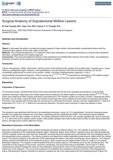
Surgical Anatomy of Supratentorial Midline Lesions
Surgical Anatomy of Supratentorial Midline Lesions
Surgical Anatomy of Supratentorial Midline Lesions
Create successful ePaper yourself
Turn your PDF publications into a flip-book with our unique Google optimized e-Paper software.
To Print: Click your browser's PRINT button.<br />
NOTE: To view the article with Web enhancements, go to:<br />
http://www.medscape.com/viewarticle/507334<br />
<strong>Surgical</strong> <strong>Anatomy</strong> <strong>of</strong> <strong>Supratentorial</strong> <strong>Midline</strong> <strong>Lesions</strong><br />
M. Gazi Yasargil, M.D.; Ugur Ture, M.D.; Dianne C. H. Yasargil, R.N.<br />
Neurosurg Focus. 2005;18(6) ©2005 American Association <strong>of</strong> Neurological Surgeons<br />
Posted 07/27/2005<br />
Abstract and Introduction<br />
Abstract<br />
Object: In this paper the authors correlate the surgical aspects <strong>of</strong> deep median and paramedian supratentorial lesions with the<br />
connective fiber systems <strong>of</strong> the white matter <strong>of</strong> the brain.<br />
Methods: The cerebral hemispheres <strong>of</strong> 10 cadaveric brains were dissected in a mediolateral direction by using the fiber dissection<br />
technique, corresponding to the surgical approach.<br />
Conclusions: This study illuminates the delicacy <strong>of</strong> the intertwined and stratified fiber laminae <strong>of</strong> the white matter, and establishes<br />
that these structures can be preserved at surgical exploration in patients.<br />
Introduction<br />
Falcine meningiomas; AVMs; cavernomas; intrinsic tumors <strong>of</strong> the medial frontal, parietal, and occipital lobes, cingulate gyrus, corpus<br />
callosum, parasplenial region, thalamus, and hypothalamus; and also lesions in the third and lateral ventricles can now be<br />
successfully explored and re moved via an anterior, middle, or posterior interhemispheric approach, or via a<br />
supracerebellar-transtentorial approach, without causing injury. [1-7,9,11-17,20-27] Comprehensive descriptions <strong>of</strong> the related surgical<br />
anatomy and the specific surgical strategy, tactics, and techniques can be found in earlier publications. [22-25]<br />
Dissection<br />
Preparation <strong>of</strong> Specimens<br />
Ten previously frozen, formalin-fixed human brains were dissected with the aid <strong>of</strong> an operating microscope by using the fiber<br />
dissection technique, beginning at the medial aspect and proceeding to the lateral aspect in a step-by-step fashion. [8,10,18,19] The<br />
brains were obtained as fresh autopsy specimens and preserved in 10% formalin solution for at least 2 months. Following the period<br />
<strong>of</strong> fixation, the pia mater, arachnoid mater, and vessels <strong>of</strong> each specimen were carefully removed under the operating microscope.<br />
The brains were washed with running water for several hours to eliminate the formalin, drained, and then refrigerated for 1 week at a<br />
temperature <strong>of</strong> 2 10 to 2 15° C. Before we commenced dissection, the brains were immersed in water and allowed to thaw.<br />
<strong>Surgical</strong> Tools<br />
The dissection was performed with the aid <strong>of</strong> an operating microscope, using 4 to 10 3 magnification. During the freezing process,<br />
formalin ice crystals form between the nerve fibers, expanding and thus separating the fibers. This freezing process facilitates<br />
dissection <strong>of</strong> the fine fiber bundles in particular. Our primary dissection tools were thin, s<strong>of</strong>t, wooden spatulas with various sized tips<br />
(1, 2, 4, and 6 mm) and a surgical suction system. S<strong>of</strong>t wooden spatulas were used to peel away the fiber bundles along anatomical<br />
planes, and high-power suction aided the process.<br />
Mediolateral Dissection <strong>of</strong> the White Matter<br />
Dissection <strong>of</strong> the medial aspect <strong>of</strong> the cerebral hemisphere proceeds as follows (Figure 1A). The midbrain is severed to achieve<br />
adequate visualization <strong>of</strong> the mediobasal temporal region. Dissection begins at the anterior cingulate sulcus and extends posteriorly<br />
until the cingulate cortex has been removed. Dissection proceeds along the callosal sulcus and extends from the subcallosal area to<br />
the hippocampal sulcus posteriorly. The indusium griseum and lateral and medial longitudinal striae become visible within the callosal<br />
sulcus. The indusium griseum is an extension <strong>of</strong> the hippocampal formation, a fine layer covering the corpus callosum. The indusium<br />
griseum extends anteroinferiorly to the paraterminal gyrus, which merges into the diagonal band <strong>of</strong> Broca, located in the anterior<br />
perforated substance. The cingulum is demonstrated around the corpus callosum, and extends as far as the subcallosal area. The<br />
connections <strong>of</strong> the cingulum to the frontal, precentral, postcentral, and precuneal areas are illustrated. The arcuate or U fibers<br />
underlying the medial frontoparietal cortex are also displayed. Removing the cortex <strong>of</strong> the parahippocampal gyrus exposes the<br />
inferior arm <strong>of</strong> the cingulum. The uncus and uncalsulcus are identified, the uncalsulcus being an extension <strong>of</strong> the hippocampal<br />
sulcus. Retracting the cingulum beneath the splenium exposes the tail <strong>of</strong> the hippocampus and the subsplenial gyrus (Figure 1B).
Figure 1.<br />
Photographs <strong>of</strong> cadaveric brains showing serial dissections <strong>of</strong> the medial aspect <strong>of</strong> the left cerebral hemisphere. A:<br />
Medial aspect <strong>of</strong> the right cerebral hemisphere. Connective arms (arrows) link the cingulate gyrus to gyrus rectus, to<br />
anterior, middle, and posterior portions <strong>of</strong> the medial frontal gyrus, and to the precuneus, and the structure continues<br />
as a parahippocampal gyrus. B: The midbrain is removed to achieve sufficient visualization <strong>of</strong> the mediobasal temporal<br />
region. Dissecting the cortex <strong>of</strong> the cingulate and parahippocampal gyrus demonstrates the whole length <strong>of</strong> the<br />
cingulum and its connections. C: After cutting the medial portion <strong>of</strong> the corpus callosum, the caudate nucleus in the<br />
lateral wall <strong>of</strong> the lateral ventricle is demonstrated. D: Removal <strong>of</strong> the choroid plexus, cingulum, and mediobasal<br />
temporal region, dissection <strong>of</strong> the radiating fibers <strong>of</strong> the corpus callosum, and partial removal <strong>of</strong> the fornix and<br />
amygdala further reveals the stria terminalis, the thalamus, and the hypothalamus, which are covered by the<br />
transparent ependyma. E: Partial removal <strong>of</strong> the ependyma and the caudate nucleus in the lateral ventricle, and<br />
dissecting away the subcallosal stratum and the anterior portion <strong>of</strong> the radiation <strong>of</strong> the corpus callosum demonstrates<br />
cortical extensions <strong>of</strong> the anterior and superior thalamic peduncles as well as the corona radiata and the intersection <strong>of</strong><br />
the corpus callosum with the corona radiata. F: Following total removal <strong>of</strong> the ependyma and caudate nucleus, the<br />
tapetum <strong>of</strong> the corpus callosum and the posterior and inferior thalamic peduncles are demonstrated. G: The tapetum,<br />
the stria terminalis, and the amygdala have been dissected away. The thalamus and fibers <strong>of</strong> the anterior, superior,<br />
posterior, and inferior thalamic peduncles as well as the optic radiation on the ro<strong>of</strong> <strong>of</strong> the temporal horn are<br />
demonstrated. Dissecting away the hypothalamus and thalamus demonstrated the column <strong>of</strong> the fornix and the<br />
mammillothalamic tract. H: Removal <strong>of</strong> the hypothalamus and thalamus with anterior, superior, posterior, and inferior<br />
peduncles (the mammillary body and column <strong>of</strong> the fornix are preserved) demonstrates the lateral portion <strong>of</strong> the corona<br />
radiata and internal capsule. A = amygdala; ac = anterior commissure; acs = anterior calcarine sulcus; af = arcuate<br />
fibers; atp = anterior thalamic peduncle (internal capsule); b = body <strong>of</strong> corpus callosum; bf = body <strong>of</strong> fornix; cc = corpus<br />
callosum; ces = central sulcus; cf = column <strong>of</strong> fornix; cg = cingulate gyrus; chp = choroid plexus; cin = cingulum; cis =<br />
cingulate sulcus; cn = caudate nucleus; cols = collateral sulcus; cp = cerebral peduncle; cr = corona radiata; cs =<br />
callosal sulcus; cu = cuneus; e = ependyma; fg = fusiform gyrus; fm = forceps major (radiation <strong>of</strong> corpus callosum); fmi<br />
= forceps minor (radiation <strong>of</strong> corpus callosum); fo = fornix; g = genu <strong>of</strong> corpus callosum; gr = gyrus rectus; h =<br />
hypothalamus; ic = internal capsule; icc = intersection <strong>of</strong> corpus callosum with corona radiata; ic1 = frontopontine tract<br />
(internal capsule); ic2 = pyramidal tract (internal capsule); ic3 = occipitopontine tract (internal capsule); ic4 =<br />
temporopontine tract (internal capsule); ig = indusium griseum; ist = isthmus cinguli; itp = inferior thalamic peduncle<br />
(internal capsule); lg = lingual gyrus; m = midbrain; mb = mammillary body; mr = marginal ramus <strong>of</strong> cingulate sulcus; mt<br />
= mammillothalamic tract; oc = optic chiasm; or = optic radiation; ot = optic tract; pb = pineal body; pc = precuneus; pcl<br />
= paracentral lobule; pcs = posterior calcarine sulcus; pg = paraterminal gyrus; pos = parietooccipital sulcus; ppc =<br />
prepiriform cortex (tip <strong>of</strong> the parahippocampal gyrus); prcu = precuneus; pt = pulvinar thalami; ptp = posterior thalamic<br />
peduncle (internal capsule); r = rostrum <strong>of</strong> corpus callosum; rcc = radiations <strong>of</strong> corpus callosum; sa = subcallosal area;<br />
sas = sagittal stratum; sm = stria medullaris thalami; sn = substantia nigra; sp = splenium <strong>of</strong> corpus callosum; ss =<br />
subcallosal stratum; st = stria terminalis; stp = superior thalamic peduncle (internal capsule); t = thalamus; ta = tapetum<br />
<strong>of</strong> corpus callosum; tp = temporal pole; u = uncus.
After cutting through the medial portion <strong>of</strong> the corpus callosum, the crus <strong>of</strong> the fornix and the hippocampal commissure are exposed.<br />
The fimbria can be traced to the crus, body, and column <strong>of</strong> the fornix, and terminates in the mammillary body. The frontal horn, body,<br />
and atrial portions <strong>of</strong> the lateral ventricle with the choroid plexus, as well as the head and body portions <strong>of</strong> the caudate nucleus are<br />
demonstrated (Figure 1C).<br />
The hippocampus, choroid plexus, fimbria, crus, and body portions <strong>of</strong> the fornix compose the entire anatomy <strong>of</strong> the lateral ventricle.<br />
After further removal <strong>of</strong> the medial frontoparietal cortex, removal <strong>of</strong> the cingulum and further dissection <strong>of</strong> the corpus callosum<br />
reveals the radiating fibers <strong>of</strong> the corpus callosum. The callosal fibers form a major portion <strong>of</strong> the commissural system and serve to<br />
interconnect the hemispheres. The genu portion <strong>of</strong> these fibers is known as the forceps minor, and the splenial portion is called the<br />
forceps major. The uncus is deflected to separate the amygdala from its complex connections, revealing the temporal horn <strong>of</strong> the<br />
lateral ventricle. The hippocampus is dissected free <strong>of</strong> the collateral eminence to gain entrance in to the collateral sulcus. The tail <strong>of</strong><br />
the hippocampus and the fornix are separated from the choroid plexus along the choroidal fissure. The fornix is incised at the junction<br />
<strong>of</strong> the body and column, and the choroid plexus is removed along the choroidal fissure. The stria terminalis, located between the<br />
caudate nucleus and thalamus, connects the bed nucleus <strong>of</strong> the stria terminalis and parts <strong>of</strong> the hypothalamus to the amygdala<br />
(Figure 1D).<br />
The removal <strong>of</strong> the frontal horn ependyma (which is a single layer <strong>of</strong> specialized epithelium lining the ventricles) and the body <strong>of</strong> the<br />
lateral ventricle allows exposure <strong>of</strong> the head and body <strong>of</strong> the caudate nucleus and the subcallosal stratum. The subcallosal stratum is<br />
a subependymal structure located between the caudate nucleus and the radiations <strong>of</strong> the corpus callosum. The caudate nucleus is<br />
observed to ex tend along the wall <strong>of</strong> the lateral ventricle and the tail <strong>of</strong> the caudate reaches forward to the level <strong>of</strong> the amygdala. The<br />
caudate nucleus has the same s<strong>of</strong>t consistency as the put amen. Removal <strong>of</strong> the head and body portions <strong>of</strong> the caudate nucleus<br />
reveals the anterior and superior thalamic peduncles. The next step involves dissecting away the anterior portions <strong>of</strong> the subcallosal<br />
stratum and the radiations <strong>of</strong> the corpus callosum to allow identification <strong>of</strong> the extensions <strong>of</strong> the anterior and superior thalamic<br />
peduncles to the cortex. These peduncles are the anteromedial portion <strong>of</strong> the internal capsule, and they connect the frontoparietal<br />
regions <strong>of</strong> the cortex with the thalamus (Figure 1E).<br />
After total removal <strong>of</strong> the ependyma lining the lateral wall and ro<strong>of</strong> <strong>of</strong> the lateral ventricle, the posterior portion <strong>of</strong> the subcallosal<br />
stratum and the tapetum <strong>of</strong> the corpus callosum are demonstrated, both <strong>of</strong> which were found to be subependymal structures. The<br />
tapetum, a subgroup <strong>of</strong> callosal fibers in the splenial region, forms the ro<strong>of</strong> and lateral wall <strong>of</strong> the atrial portion <strong>of</strong> the lateral ventricle<br />
and sweeps around the temporal horn, thereby separating the fibers <strong>of</strong> the optic radiation from the temporal horn. Further removal <strong>of</strong><br />
the caudate nucleus exposes the posterior and inferior thalamic peduncles. During our fiber dissection, identification <strong>of</strong> the precise<br />
location <strong>of</strong> the border separating the tapetum and the subcallosal stratum eluded us. Nevertheless, we did note a distinct difference<br />
between these two structures, and we suspect that the border lies be tween the body and the atrial portions <strong>of</strong> the lateral ventricle. In<br />
the subcallosal stratum, we were unable to identify a definite fiber system. There was, however, a fiber system clearly present in the<br />
tapetum. In addition, we made a significant observation; that the subcallosal stratum has fine, microscopic connections with the<br />
superior margin <strong>of</strong> the caudate nucleus (Figure 1F).<br />
The tapetum extends from the splenium, forming the lateral wall <strong>of</strong> the atrium and the ro<strong>of</strong> <strong>of</strong> the temporal horn beneath the<br />
ependyma. After removing the remaining posterior layer <strong>of</strong> the subcallosal stratum, we dissected away the tapetum, the stria<br />
terminalis, and the amygdala, exposing the entire anatomy <strong>of</strong> the anterior, superior, posterior, and inferior thalamic peduncles as well<br />
as the optic radiation over the ro<strong>of</strong> <strong>of</strong> the temporal horn. Resection <strong>of</strong> the body <strong>of</strong> the corpus callosum further exposed the superior<br />
thalamic peduncle. The thalamic peduncles as a whole form the medial portion <strong>of</strong> the internal capsule and connect the cerebral<br />
cortex to the thalamus. The inferior thalamic peduncle connects the temporal lobe to the thalamus. Following the column <strong>of</strong> the fornix<br />
into the hypothalamus demonstrates its connection with the mammillary body. The anterior commissure is located anterior to the<br />
column <strong>of</strong> the fornix. Further dissection into the hypothalamus and thalamus reveals the column <strong>of</strong> the fornix, optic tract, mammillary<br />
body, and mammillothalamic tract, which is also known as the tract <strong>of</strong> Vicq d'Azyr. Extensive dissection into the thalamus reveals its<br />
connections with the thalamic peduncles (Figure 1G).<br />
Removal <strong>of</strong> the anterior, superior, posterior (which includes the optic radiation), and inferior thalamic peduncles together with the<br />
thalamus and the lateral geniculate body concludes the dissection and demonstrates the whole corona radiata and the lateral portion<br />
<strong>of</strong> the internal capsule from a medial view, and the cerebral peduncle. These structures are composed <strong>of</strong> frontopontine fibers,<br />
pyramidal tract, and occipitopontine and temporopontine fibers (Figure 1H).<br />
<strong>Surgical</strong> Considerations<br />
The striking advances in neurovisualization technology confirm the observations <strong>of</strong> neuropathologists, neurologists, and<br />
neurosurgeons that each type <strong>of</strong> CNS lesion has a predilection to present in distinct sites in osseous, meningeal, cisternal,<br />
parenchymal, ventricular, or vascular compartments. In each <strong>of</strong> these locations, the lesions may <strong>of</strong>ten reach a considerable size<br />
without causing any or only discreet signs and symptoms. It can be assumed that a lesion may compress and displace normal brain<br />
structures to a greater degree, but lack the capacity to transgress and destroy the unique architecture <strong>of</strong> the gray and white matter <strong>of</strong><br />
the CNS. This fact affords us the opportunity to devise and initiate adequate treatment plans.<br />
The main principle <strong>of</strong> a neurosurgical procedure is always to perform a pure lesionectomy, using tactics to avoid compromising<br />
normal homeostasis <strong>of</strong> the CNS. This surgical principle becomes a challenge to uphold when considering deep, localized, so-called<br />
"midline lesions," which may originate from the medial part <strong>of</strong> the frontal, parietal, or occipital lobe; from the anterior, middle, or<br />
posterior parts <strong>of</strong> the cingulate gyrus; from the parasplenial region (posterior cingulate gyrus, inferior precuneus, and posterior<br />
parahippocampal gyrus); corpus callosum; thalamus; hypothalamus; or third and lateral ventricles. All lesions in these locations<br />
(tumors, AVMs, and cavernomas) can be explored and removed through an anterior, middle, or posterior interhemispheric, and<br />
supracerebellar-infratentorial approach (Figure 2). The specific method <strong>of</strong> these approaches prevents infliction <strong>of</strong> harm to the dorsal<br />
neopallial areas and to the connective fiber systems <strong>of</strong> the white matter.
Figure 2.<br />
Schematic drawing showing surgical approaches to supratentorial midline lesions. 1 = anterior frontal approach; 2 =<br />
posterior frontal approach; 3a = parietooccipital approach; 3b = parietooccipital transtentorial approach. (Reprinted with<br />
permission from Yasargil MG: Microneurosurgery IVB: Microneurosurgery <strong>of</strong> CNS Tumors. Stuttgart: Georg Thieme<br />
Verlag, 1996, p 315.)<br />
It is an indisputable fact that falcine and callosal lesions, and saccular aneurysms <strong>of</strong> A2 and A3 segments are explored and removed<br />
(or occluded with clips) via an exploration in to the interhemispheric fissure. This is considered a routine approach for the majority <strong>of</strong><br />
midline lesions. Endovascular and gamma knife surgeries have proven effective for some types <strong>of</strong> tumors and for AVMs <strong>of</strong> smaller<br />
dimensions. Computer-assisted stereotactic, endoscopic, or microsurgical procedures are certainly accurate in targeting these<br />
lesions, but transcerebral trajectories to approach a lesion surgically are accompanied by unavoidable injuries to the neopallial<br />
cortices and connective fiber systems <strong>of</strong> the white matter.<br />
Illustrative Cases<br />
A few patients with typical deep midline lesions chosen from several hundred cases treated by the senior author (M.G.Y.) at the<br />
University <strong>of</strong> Arkansas for Medical Sciences in Little Rock illustrate the effectiveness <strong>of</strong> inter hem i spheric approaches (Figs. 3-12).
Figure 3.<br />
Case 1. A-C: Preoperative MR images demonstrating a large tumor occupying the anterior and subcallosal areas <strong>of</strong> the<br />
right cingulate gyrus. D-F: Postoperative MR images obtained after removal <strong>of</strong> the tumor.
Figure 4.<br />
Case 2. A-C: Preoperative MR images demonstrating occlusive hydrocephalus due to a lesion in the third ventricle.<br />
D-F: Postoperative MR images obtained after complete removal <strong>of</strong> the lesion.
Figure 5.<br />
Case 3. A-C: Preoperative MR images revealing a well-defined lesion in the right anterior lateral thalamic region. D-F:<br />
Postoperative MR images obtained after the lesion was re moved.<br />
Figure 6.<br />
Case 4. A and B: Preoperative MR images revealing a large tumor originating in the region <strong>of</strong> the septum pellucidum<br />
and extending through the middle <strong>of</strong> the corpus callosum and interhemispheric fissure to the surface <strong>of</strong> the left pre- and<br />
postcentral gyri. C and D: Postoperative MR images obtained after exploration and complete removal <strong>of</strong> the tumor.
Figure 7.<br />
Case 5. A-C: Preoperative MR images demonstrating a well-circumscribed lesion within the right precuneus area, with<br />
extension into the posterior part <strong>of</strong> the right parahippocampal gyrus. D-F: Postoperative MR images obtained after<br />
exploration and total resection.
Figure 8.<br />
Case 6. A-C: Preoperative MR images revealing a large lesion within the left precuneus and posterior parahippocampal<br />
gyrus. D-F: Postoperative MR images obtained after exploration and complete resection <strong>of</strong> the lesion.
Figure 9.<br />
Case 7. A-C: Preoperative MR images demonstrating a compact lesion in the left atrium. D-F: Postoperative MR<br />
images obtained after complete removal <strong>of</strong> the tumor.
Figure 10.<br />
Case 8. A-C: Preoperative MR images revealing a large, left-sided mediobasal tumor extending into the dorsolateral<br />
region <strong>of</strong> the mesencephalon. D-F: Postoperative MR images obtained after tumor removal.
Figure 11.<br />
Case 9. A-C: Preoperative MR images revealing a well-circumscribed intrinsic tumor in the left posterior thalamic<br />
region. D-F: Postoperative MR images obtained after radical tumor removal.
Case 1<br />
Figure 12.<br />
Case 10. A-C: Preoperative MR images demonstrating a cavernoma in the left posterior thalamic region. D-F:<br />
Postoperative MR images obtained after complete removal <strong>of</strong> the lesion.<br />
This 26-year-old woman suffered from simple and complex partial seizures and impairment <strong>of</strong> her short-term memory. Admission MR<br />
images (Figure 3A-C) demonstrated a large tumor occupying the anterior and subcallosal areas <strong>of</strong> the right cingulate gyrus. The<br />
tumor was removed via a frontal interhemispheric approach (Figure 3D-F, postoperative MR images). Histological studies revealed a<br />
pilocytic as trocytoma. She had no neurological deficits pre- or postoperatively. Her postoperative course was uneventful. There was<br />
a remarkable improvement in her short-term memory difficulties, and she regained her full working capacity.<br />
Case 2<br />
This 10-year-old girl with chronic headaches underwent CT scanning, which demonstrated occlusive hydrocephalus due to a lesion in<br />
the third ventricle. After a ventriculoatrial shunt was placed, the lesion was explored and completely removed through a right frontal<br />
parasagittal osteoplastic craniotomy, via an anterior interhemispheric transcallosal-transforaminal approach. Histological studies<br />
revealed a pilocytic astrocytoma. The patient had no neurological, mental, or endocrine deficits pre- or postoperatively. Nine years <strong>of</strong><br />
follow up revealed no recurrence <strong>of</strong> the tumor (Figure 4A-C, preoperative and Figure 4D-F, postoperative MR images).<br />
Case 3<br />
This 12-year-old boy presented with headache after hitting a soccer ball with his head. The admission CT and MR imaging studies<br />
revealed a well-defined lesion in the right anterior lateral thalamic region, which was removed via an interhemispheric transcallosal<br />
approach. Histological studies revealed pilocytic astrocytoma. The patient's pre- and postoperative neurological and mental status<br />
was normal (Figure 5A-C, preoperative and Figure 5D-F, postoperative MR images).<br />
Case 4<br />
This 7-year-old girl experienced progressive weakness distally in her right leg. The admission CT and MR imaging studies revealed a<br />
large tumor originating in the region <strong>of</strong> the septum pellucidum, extending through the middle <strong>of</strong> the corpus callosum and
interhemispheric fissure to the surface <strong>of</strong> the left pre- and postcentral gyri. The lesion was explored and completely removed through<br />
a left posterior frontal parasagittal craniotomy, and further exploration was performed along the interhemispheric fissure in a<br />
supero-inferior dissection (Figure 6A and B, preoperative and Figure 6C and D, postoperative MR images). The postoperative course<br />
was uneventful, and the foot process recovered fully within a few months. Histological findings remained inconclusive; the tumor was<br />
thought to be either a neurocytoma or an oligodendroglioma.<br />
Case 5<br />
This 42-year-old woman suffered single and complex partial seizures. The admission CT and MR imaging studies demonstrated a<br />
well-circumscribed lesion within the right precuneus area, with extension into the posterior part <strong>of</strong> the right parahippocampal gyrus.<br />
With the patient in a sit ting position, the lesion was explored and completely re moved through a right parietooccipital osteoplastic<br />
craniotomy and via a posterior interhemispheric approach (Figure 7A-C, preoperative and Figure 7D-F, postoperative MR images).<br />
The histological studies revealed low-grade oligodendroglioma. The patient was free <strong>of</strong> neurological and mental deficits pre- and<br />
postoperatively, and her visual field is normal. She has regained her full working capacity and has had no seizures.<br />
Case 6<br />
This 36-year-old woman reported difficulty with reading and memory problems. She had right hemianopia and papilledema. The<br />
admission MR images (Figure 8A-C) revealed a large lesion within the left precuneus and posterior parahippocampal gyrus. The<br />
dorsal part <strong>of</strong> the tumor had been removed in another hospital. The tumor was completely removed after a second exploration; this<br />
was done through a left parietooccipital craniotomy and via an interhemispheric approach with the patient in a sitting position (Figure<br />
8D-F, postoperative MR images). Histological studies revealed a low-grade oligodendroglioma. There were no neurological and<br />
mental deficits and no visual field deficits pre- or postoperatively. The patient regained her full working capacity, and has had no<br />
seizures.<br />
Case 7<br />
This 25-year-old woman suffered from a headache that increased in intensity over 5 months, fatigue, and short-term memory<br />
problems. The admission CT and MR imaging studies demonstrated a compact lesion in the left atrium. With the patient in a sitting<br />
position, the tumor was explored through a left parietooccipital osteoplastic craniotomy, via a posterior interhemispheric approach.<br />
The dorsomedial extension <strong>of</strong> the tumor into the posterior part <strong>of</strong> cingulate gyrus was identified. Through a 10-mm-long incision, the<br />
well-encapsulated, very vascularized lesion was completely removed. Histological studies revealed an atypical meningioma. The<br />
patient's pre- and postoperative neurological and mental status were found to be normal; in particular no visual field deficit was<br />
detected. She regained her full working capacity (Figure 9A-C, preoperative and Figure 9D-F, postoperative MR images).<br />
Case 8<br />
In this 42-year-old woman, who had suffered from typical temporal seizures for a couple <strong>of</strong> years, admission MR imaging studies<br />
(Figure 10A-C) revealed a large, left-sided mediobasal tumor extending into the dorsolateral region <strong>of</strong> the mesencephalon. The tumor<br />
was explored and removed (except for the amygdala area) via a supracerebellar-transtentorial approach (Figure 10D-F,<br />
postoperative MR images). The patient's postoperative course was uneventful. Preoperatively and postoperatively she had no<br />
neurological or mental deficits, and her visual field was intact. The histological studies revealed a low-grade oligodendroglioma. After<br />
surgery, the seizures did not recur and the patient could continue to work at her full capacity.<br />
Case 9<br />
In this 12-year-old girl suffering from a progressive right hemisyndrome but no visual field defect, the admission MR imaging studies<br />
revealed a well-circumscribed intrinsic tumor in the left posterior thalamic region (Figure 11A-C). With the patient in a sitting position,<br />
the lesion was explored via a left supracerebellar-transtentorial approach, and an anaplastic astrocytoma was radically removed with<br />
the aid <strong>of</strong> an operating microscope (Figure 11D-F, postoperative MR images). Postoperatively there was rapid improvement <strong>of</strong> the<br />
hemisyndrome, and there was no visual field deficit. The adjuvant radio- and chemotherapy could not change the course <strong>of</strong> her<br />
disease, and she died 2 years post surgery.<br />
Case 10<br />
This 45-year-old woman suffered an onset <strong>of</strong> a right hemisyndrome and homonymous hemianopia. The admission MR images<br />
(Figure 12A-C) demonstrated a cavernoma in the left posterior thalamic region. With the patient in a sit ting position, this lesion was<br />
explored via a supracerebellar-transtentorial approach, and we were able to re move the lesion completely (Figure 12D-F). The<br />
histological findings confirmed a cavernoma. Postoperatively the moderate hemiparesis improved remarkably, whereas the visual<br />
field deficit remained unchanged.<br />
Discussion<br />
The planning <strong>of</strong> a neurosurgical procedure incorporates a study <strong>of</strong> the parenchymal, vascular, cisternal, and ventricular architecture<br />
<strong>of</strong> the brain on MR imaging, MR angiography, MR venography, and serial cerebral angiograms, which are usually analyzed in the<br />
axial (base-up > top-down), coronal (anteroposterior > posteroanterior), and sagittal view (lateromedial > mediolateral). Because the<br />
lesions are explored with the patient either supine or in a sitting position, but are approached via the interhemispheric fissure in a<br />
mediolateral direction, the related fiber system <strong>of</strong> the white matter is also shown in a mediolateral direction in the dissected cadaveric<br />
brains in this study.<br />
Inferiorly extending tumors <strong>of</strong> the hypothalamus and third ventricle can be explored and resected via a pterional-transsylvian and<br />
translamina-terminalis approach. Tumors in the posterior part <strong>of</strong> the third ventricle and the thalamus are approached via the posterior
interhemispheric fissure or via a suboccipital-supracerebellar route.<br />
<strong>Lesions</strong> in the third ventricle and the anterior two thirds <strong>of</strong> the lateral ventricle are explored through the anterior or middle part <strong>of</strong> the<br />
interhemispheric fissure, and lesions in the trigonum (atrium) are explored via a posterior interhemispheric approach with the patient<br />
in the sitting position. Exploration <strong>of</strong> the ventricle requires a small incision (10-15 cm) in the commissural fiber system <strong>of</strong> the corpus<br />
callosum. [22-25] One exception to this recommendation is when a tumor expands to the surface <strong>of</strong> the frontal or parietal lobe. This,<br />
however, is an extremely rare occurrence.<br />
Dorsal transcerebral approaches traverse neocortical areas, and injuries to cortices and to the complex stratification <strong>of</strong> associative,<br />
commissural, and projection fiber systems are impossible to avoid. Interhemispheric transcallosal approaches are definitely the<br />
preferred surgical strategies, <strong>of</strong>fering good access to the lesion, permitting conservation <strong>of</strong> normal tissue and structures, and<br />
resulting in a positive outcome for the patient.<br />
References<br />
1. Apuzzo MLJ, Lit<strong>of</strong>sky NS: Surgery in and around the anterior third ventricle, in Apuzzo MLJ (ed): Brain Surgery: Com<br />
plication Avoidance and Management. New York: Church ill-Livingstone, 1993, pp 541-580<br />
2. Bellotti C, Pappada G, Sani R, et al: The transcallosal approach for lesions affecting the lateral and third ventricles. <strong>Surgical</strong><br />
considerations and results in a series <strong>of</strong> 42 cases. Acta Neurochir 111:103-107, 1991<br />
3. D'Angelo VA, Galarza M, Catapano D, et al: Lateral ventricle tumors: surgical strategies according to tumor origin and<br />
development -- a series <strong>of</strong> 72 cases. Neurosurgery 56 (Suppl 1): 36-45, 2005<br />
4. Dandy WE: Benign Tumors in the Third Ventricle <strong>of</strong> the Brain: Diagnosis and Treatment. Springfield, IL: Charles C Thomas,<br />
1933<br />
5. Geffen G, Walsh A, Simpson D, et al: Comparison <strong>of</strong> the effects <strong>of</strong> transcortical and transcallosal removal <strong>of</strong> intraventricular<br />
tumours. Brain 103:773-788, 1980<br />
6. Hutter BO, Spetzger U, Bertalanffy H, et al: Cognition and quality <strong>of</strong> life in patients after transcallosal microsurgery for midline<br />
tumors. J Neurosurg Sci 41:123-129, 1997<br />
7. Jeeves MA, Simpson DA, Geffen G: Functional consequences <strong>of</strong> the transcallosal removal <strong>of</strong> intraventricular tumours. J<br />
Neurol Neurosurg Psychiatry 42:134-142, 1979<br />
8. Klingler J: Erleichterung der makroskopischen Praeparation des Gehirns durch den Gefrierprozess. Schweiz Arch Neurol<br />
Psychiat 36:247-256, 1935<br />
9. Konovalov AN, Gorelyshev SK, Khuhlaeva EA: Surgery <strong>of</strong> diencephalic and brainstem tumors, in Schmidek HH, Sweet WH<br />
(eds): Operative Neurosurgical Technique. Indications, Methods and Results, ed 3. Philadelphia: Saunders, Vol 1, 1995, pp<br />
765-782<br />
10. Ludwig E, Klingler J: Atlas cerebri humani der innere Bau des Gehirns dargestellt auf Grund makroskopischer Preparate.<br />
Basel: Karger S, 1956<br />
11. Misra BK, Rout D, Padamadan J, et al: Transcallosal approach to anterior and mid-third ventricular tumors -- a review <strong>of</strong> 62<br />
cases. Ann Acad Med Singapore 22 (Suppl 3):435-440, 1993<br />
12. Pendl G: Pineal and Midbrain <strong>Lesions</strong>. Wien: Springer, 1985<br />
13. Rhoton AL Jr: The lateral and third ventricles. Neurosurgery 51 (Suppl 4):S207-S271, 2002<br />
14. Rosenfeld JV, Harvey AS, Wrennall J, et al: Transcallosal resection <strong>of</strong> hypothalamic hamartomas, with control <strong>of</strong> seizures, in<br />
children with gelastic epilepsy. Neurosurgery 48:108-118, 2001<br />
15. Standefer M, Bay JW, Trusso R: The sitting position in neurosurgery: a retrospective analysis <strong>of</strong> 488 cases. Neurosurgery<br />
14:649-658, 1984<br />
16. Timurkaynak E, Rhoton AL Jr, Barry M: Microsurgical anatomy and operative approaches to the lateral ventricles.<br />
Neurosurgery 19:685-723, 1986<br />
17. Ture U, Yasargil MG, Al-Mefty O: The transcallosal-transforaminal approach to the third ventricle with regard to the venous<br />
variations in this region. J Neurosurg 87:706-715, 1997<br />
18. Ture U, Yasargil MG, Friedman AH, et al: Fiber dissection technique: lateral aspect <strong>of</strong> the brain. Neurosurgery 47:417-427,<br />
2000<br />
19. Ture U, Yasargil MG, Pait TG: Is there a superior occipit<strong>of</strong>rontal fasciculus? A microsurgical anatomic study. Neurosurgery<br />
40:1226-1232, 1997<br />
20. Voigt K, Yasargil MG: Cerebral cavernous hemangioma or cavernomas. Incidence, pathology, localization, diagnosis, clinical<br />
features and treatment. Review <strong>of</strong> the literature and report <strong>of</strong> an unusual case. Neurochir 19:59-68, 1976<br />
21. Woiciechowsky C, Vogel S, Lehmann R, et al: Transcallosal removal <strong>of</strong> lesions affecting the third ventricle: an anatomic and<br />
clinical study. Neurosurgery 36:117-123, 1995<br />
22. Yasargil MG: Microneurosurgery I: Microsurgical <strong>Anatomy</strong> <strong>of</strong> the Basal Cisterns and Vessels <strong>of</strong> the Brain, Diagnostic Studies,<br />
General Operative Techniques and Pathological Considerations <strong>of</strong> the Intracranial Aneurysms. Stuttgart: Georg Thieme<br />
Verlag, 1984, pp 5-168<br />
23. Yasargil MG: Microneurosurgery IIIB: Arm <strong>of</strong> the Brain, Clinical Considerations, General and Special Operative Techniques,<br />
<strong>Surgical</strong> Results, Nonoperated Cases, Cavernous and Venous Angiomas, Neuroanesthesia. Stuttgart: Georg Thieme Verlag,<br />
1988<br />
24. Yasargil MG: Microneurosurgery IVB: Microneurosurgery <strong>of</strong> CNS Tumors. Stuttgart: Georg Thieme Verlag, 1996, pp 237-342<br />
25. Yasargil MG, Ture U, Roth P: Combined approaches, in Apuzzo MLJ (ed): Surgery <strong>of</strong> the Third Ventricle, ed 2. Baltimore:<br />
Williams & Wilkins, 1998, pp 541-552<br />
26. Yamamoto I, Rhoton AL Jr, Peace DA: Microsurgery <strong>of</strong> the third ventricle: Part I. Microsurgical anatomy. Neurosurgery<br />
8:334-356, 1981<br />
27. Yonekawa Y, Imh<strong>of</strong> HG, Taub E, Curcic M, Kaku Y, Roth P, Wieser HG, Groscurth P. Supracerebellar transtentorial<br />
approach to posterior temporomedial structure. J Neurosurg 94:339-345, 2001<br />
Abbreviation Notes<br />
AVM = arteriovenous malformation; CNS = central nervous system; CT = computerized tomography; MR = magnetic resonance.
Reprint Address<br />
M. Gazi Yasargil, M.D., Department <strong>of</strong> Neurosurgery, University <strong>of</strong> Arkansas for Medical Sciences, 4301 West Markham, #507, Little<br />
Rock, Arkansas 72205.<br />
M. Gazi Yasargil, M.D., Ugur Ture, M.D., Dianne C. H. Yasargil, R.N., Departments <strong>of</strong> Neurosurgery, University <strong>of</strong> Arkansas for<br />
Medical Sciences, Little Rock, Arkansas; and Ondokuz Mayis University School <strong>of</strong> Medicine, Samsun, Turkey
Copyright © by the Congress <strong>of</strong> Neurological Surgeons Volume 40(6), June 1997, pp 1226-1232<br />
Is There a Superior Occipit<strong>of</strong>rontal Fasciculus? A Microsurgical Anatomic Study<br />
[Anatomic Report]<br />
Türe, Ugur MD[modifier hacek]; Yasargil, M. Gazi MD; Pait, T. Glenn MD<br />
Department <strong>of</strong> Neurosurgery, University <strong>of</strong> Arkansas for Medical Sciences, Little Rock,<br />
Arkansas<br />
Received, May 1, 1996. Accepted, January 7, 1997.<br />
Reprint requests: Ug[modifier hacek]ur Türe, M.D., Department <strong>of</strong> Neurosurgery,<br />
University <strong>of</strong> Arkansas for Medical Sciences, 4301 West Markham, Slot 507, Little Rock, AR<br />
72205.<br />
Abstract<br />
OBJECTIVE: Using a fiber-dissection technique, our aim was to expose and study the myelinated fiber bundles <strong>of</strong> the brain to achieve a<br />
clearer conception <strong>of</strong> their configurations and locations. During the course <strong>of</strong> our study, the superior occipit<strong>of</strong>rontal fasciculus became the focus<br />
<strong>of</strong> our interest. Many publications have defined this as a bundle <strong>of</strong> association fibers, located between the corpus callosum and the caudate<br />
nucleus, that connects the frontal and occipital lobes. By examining this area using fiber dissection, we realized that the descriptions <strong>of</strong> the<br />
anatomy are inadequate; thus, we focused on the elucidation <strong>of</strong> the anatomic structures <strong>of</strong> this region and, in particular, that known as the<br />
superior occipit<strong>of</strong>rontal fasciculus.<br />
METHODS: Twenty previously frozen, formalin-fixed human brains were dissected under the operating microscope using the<br />
fiber-dissection technique.<br />
RESULTS: On coronal sections <strong>of</strong> the brain, a structure on the superolateral aspect <strong>of</strong> the caudate nucleus usually has been identified as the<br />
superior occipit<strong>of</strong>rontal fasciculus. However, our fiber dissections revealed that this structure is the superior thalamic peduncle, that it is<br />
composed <strong>of</strong> projection fibers rather than association fibers, and that it does not interconnect the occipital and frontal lobes.<br />
CONCLUSION: The structures <strong>of</strong> the brain are better understood when the fiber-dissection technique is used to explore their configurations<br />
and locations. The resulting information is especially beneficial for planning strategies and tactics <strong>of</strong> neurosurgical procedures.<br />
The white matter <strong>of</strong> the cerebral hemispheres consists <strong>of</strong> myelinated fiber bundles, called fasciculi, which are divided into three groups: 1)<br />
association, 2) commissural, and 3) projection. Association fibers interconnect cortical regions within the same hemisphere. The main<br />
association fasciculi are the arcuate fibers, the cingulum, the uncinate fasciculus, the superior and inferior longitudinal fasciculi, and the superior<br />
and inferior occipit<strong>of</strong>rontal fasciculi. The commissural fibers cross the midline and interconnect the two hemispheres. These fiber tracts include<br />
the corpus callosum, the anterior commissure, and the hippocampal commissure. Projection fibers connect the cerebral cortex with subcortical<br />
regions. These radiating projection fibers form the corona radiata, and, near the rostral part <strong>of</strong> the brain stem, they form a compact band <strong>of</strong> fibers<br />
known as the internal capsule (3, 11, 23, 33).<br />
We used the fiber-dissection technique to reveal the association, commissural, and projection fibers <strong>of</strong> the brain. This technique, which<br />
involves peeling away the white matter tracts <strong>of</strong> the brain to display its internal anatomic organization, was the first to allow a true<br />
three-dimensional appreciation <strong>of</strong> the brain. As early as the 17th century, this technique was used to demonstrate many tracts and fasciculi <strong>of</strong> the<br />
brain (4, 6, 9, 17, 21, 22, 29, 31). Since the development <strong>of</strong> the microtome and histological techniques, fiber dissection has not been extensively<br />
used. Klinger (16) cultivated an interest in the fiber-dissection technique and developed an improved method <strong>of</strong> brain fixation and fiber<br />
dissection that now bears his name, Klinger's technique (16, 17, 21). He maintained that dissecting fiber tracts <strong>of</strong> the white matter was the best<br />
method for acquiring an accurate knowledge and understanding <strong>of</strong> the internal structures <strong>of</strong> the brain.<br />
During our anatomic study, we were unable to identify the superior occipit<strong>of</strong>rontal fasciculus. Our analysis <strong>of</strong> numerous publications<br />
revealed inconsistencies in the definitions, locations, and patterns <strong>of</strong> this fasciculus (1-5, 7, 8, 11, 18, 23, 24, 26, 27, 30, 32). On coronal sections<br />
<strong>of</strong> the brain, a structure 2 to 3 mm in width and situated on the superolateral aspect <strong>of</strong> the caudate nucleus and lateral to the subcallosal stratum<br />
exists. Studies <strong>of</strong> histological cross sections suggested the possibility that this structure is a collection <strong>of</strong> association fibers interconnecting the
frontal and occipital lobes, forming the superior occipit<strong>of</strong>rontal fasciculus (Fig. 1, A and B) (7, 8, 18, 23, 24, 30, 32). Thus, we focused on the<br />
clarification <strong>of</strong> the anatomic structures <strong>of</strong> this region.<br />
FIGURE 1. A, coronal section through the center <strong>of</strong> the third ventricle, the mamillary body, and the hippocampus. In this panel, number 22 is<br />
identified as the superior occipit<strong>of</strong>rontal fasciculus (arrow) (our dissection identified the structure marked 22 as the superior thalamic<br />
peduncle).23, stria terminalis; 32, optic tract;33, cerebral peduncle; 35, hippocampus (from, Nieuwenhuys R, Voogd J, van Huijzen C: The<br />
Human Central Nervous System. Berlin, Springer-Verlag, 1988, p 101, with permission [23]). B, long association bundles <strong>of</strong> the right<br />
hemisphere in a lateral view. In this schematic figure, number 1 is identified as the superior occipit<strong>of</strong>rontal fasciculus (arrow) (in our dissection,<br />
we observed no fasciculus composed <strong>of</strong> association fibers following the pattern as shown in 1).2, site <strong>of</strong> corona radiata; 3, superior longitudinal<br />
fasciculus; 6, outline <strong>of</strong> insula;7, inferior occipit<strong>of</strong>rontal fasciculus; 8, inferior longitudinal fasciculus; 9, site <strong>of</strong> anterior commissure;10, uncinate<br />
fasciculus (from, Nieuwenhuys R, Voogd J, van Huijzen C: The Human Central Nervous System. Berlin, Springer-Verlag, 1988, p 367, with<br />
permission [23]).<br />
MATERIALS AND METHODS
We dissected 20 previously frozen, formalin-fixed human brains under the operating microscope using the fiber-dissection technique <strong>of</strong><br />
Klingler (16, 17, 21). The brains were removed from the craniums no later than 10 to 12 hours postmortem and were fixed in a 10% formalin<br />
solution for at least 2 months. To maintain the normal contours <strong>of</strong> the brain, the basilar artery was ligated and used to suspend the brain in the<br />
formalin solution. The specimens were then washed under running water for several hours to remove the formalin and were refrigerated at<br />
temperatures ranging from -10 to -15°C for 1 week. Afterwards, they were immersed in water and allowed to thaw. The specimens were then<br />
dissected using the operating microscope with 6× to 40× magnification. The primary dissection tools were handmade, thin, wooden spatulas with<br />
various tip sizes.<br />
RESULTS<br />
In 16 <strong>of</strong> the 20 specimens, we dissected the medial aspect <strong>of</strong> the cerebral hemispheres. After removing the cortex, the hippocampus, the<br />
medial portion <strong>of</strong> the corpus callosum, and the fornix, we demonstrated the entire anatomy <strong>of</strong> the lateral ventricle (Fig. 2A). The removal <strong>of</strong> the<br />
ependyma (which is a single layer <strong>of</strong> specialized epithelium lining the ventricles) <strong>of</strong> the frontal horn and the body <strong>of</strong> the lateral ventricle allowed<br />
the exposure <strong>of</strong> the subcallosal stratum. The subcallosal stratum is a subependymal structure located between the caudate nucleus and the<br />
radiation <strong>of</strong> the corpus callosum. The head and body <strong>of</strong> the caudate nucleus were removed to demonstrate the fibers <strong>of</strong> the anterior and superior<br />
thalamic peduncles (Fig. 2B). After total removal <strong>of</strong> the ependyma <strong>of</strong> the lateral wall and the ro<strong>of</strong> <strong>of</strong> the lateral ventricle, we demonstrated the<br />
posterior portion <strong>of</strong> the subcallosal stratum and the tapetum <strong>of</strong> the corpus callosum, both <strong>of</strong> which were found to be subependymal structures.<br />
The tapetum, a subgroup <strong>of</strong> callosal fibers in the splenial region, forms the ro<strong>of</strong> and lateral wall <strong>of</strong> the atrial portion <strong>of</strong> the lateral ventricle and<br />
sweeps around the temporal horn, thereby separating the fibers <strong>of</strong> the optic radiation from the temporal horn. During our fiber dissection, we<br />
could not identify the precise location <strong>of</strong> the border separating the tapetum and the subcallosal stratum. However, we noted a distinct difference<br />
between these two structures, and we suspect that the border lies between the body and atrial portions <strong>of</strong> the lateral ventricle. In the subcallosal<br />
stratum, we could not identify a definite fiber system. For this reason, we prefer to use the nomenclature "stratum" (a layered, sheetlike mass <strong>of</strong><br />
substance <strong>of</strong> nearly uniform thickness), to describe this structure rather than "fasciculus," as some authors do (1, 3, 23, 28, 32). There was,<br />
however, a fiber system clearly present in the tapetum. Also, we observed that the subcallosal stratum had microscopic connections with the<br />
superior margin <strong>of</strong> the caudate nucleus. We next dissected away the anterior portions <strong>of</strong> the subcallosal stratum and the radiation <strong>of</strong> the corpus<br />
callosum to allow identification <strong>of</strong> the extensions <strong>of</strong> the anterior and superior thalamic peduncles to the cortex (Fig. 2C). After removing the<br />
remaining portions <strong>of</strong> the subcallosal stratum and the caudate nucleus, we dissected away the tapetum, the stria terminalis, and the amygdala,<br />
exposing the entire anatomy <strong>of</strong> the anterior, superior, posterior, and inferior thalamic peduncles as well as the optic radiation on the ro<strong>of</strong> <strong>of</strong> the<br />
temporal horn (Figs. 2D and 3).<br />
FIGURE 2. Serial dissections <strong>of</strong> the medial aspect <strong>of</strong> the left cerebral hemisphere. A, dissecting the cortex and the corpus callosum and partially<br />
removing the fornix (f) and amygdala(a) further reveals the caudate nucleus (cn) in the lateral wall <strong>of</strong> the lateral ventricle, as well as the stria<br />
terminalis(st), the thalamus (t), and the hypothalamus(h), which are covered by the transparent ependyma (e). The radiation <strong>of</strong> the corpus<br />
callosum (rcc), the anterior commissure (ac), the midbrain (m), the mamillary body(mb), the optic chiasm (oc), and the pineal body(pb) are also
labeled. B, after partial removal <strong>of</strong> the ependyma (e) and the caudate nucleus (cn) in the frontal horn and body <strong>of</strong> the lateral ventricle, we<br />
demonstrated the subcalcallosum; stratum (ss), the anterior thalamic peduncle(atp), and the superior thalamic peduncle (stp).rcc, radiation <strong>of</strong> the<br />
corpus callosum; st, stria terminalis; f, fornix; t, thalamus.C, after totally removing the ependyma <strong>of</strong> the lateral wall and ro<strong>of</strong> <strong>of</strong> the lateral<br />
ventricle, dissecting away the anterior portions <strong>of</strong> the subcallosal stratum (ss) and the radiation <strong>of</strong> the corpus callosum(rcc), we demonstrated<br />
cortical extensions <strong>of</strong> the anterior thalamic peduncle (atp) and superior thalamic peduncle(stp), as well as the corona radiata (cr), the intersection<br />
<strong>of</strong> the corpus callosum with the corona radiata (icc), the tapetum <strong>of</strong> the corpus callosum (ta), and the inferior thalamic peduncle (itp). cn, caudate<br />
nucleus;st, stria terminalis; f, fornix;t, thalamus; a, amygdala. D, tapetum, subcallosal stratum, caudate nucleus, stria terminalis, and amygdala<br />
have been dissected away. The thalamus (t) and fibers <strong>of</strong> the anterior thalamic peduncle (atp), superior thalamic peduncle (stp), posterior<br />
thalamic peduncle (ptp), and inferior thalamic peduncle(itp), as well as the optic radiation (or) on the ro<strong>of</strong> <strong>of</strong> the temporal horn, are<br />
demonstrated. The fibers <strong>of</strong> the superior thalamic peduncle form an angle (arrow) inferiorly and continue to the thalamus. The change in<br />
direction <strong>of</strong> the superior thalamic peduncle inferiorly is not demonstrated on histological coronal sections. It is clearly shown that there is no<br />
fasciculus on the superolateral aspect <strong>of</strong> the caudate nucleus and lateral to the subcallosal stratum connecting the frontal lobe to the occipital<br />
lobe, which has been described in previous publications as being the superior occipit<strong>of</strong>rontal fasciculus. According to our observations, this<br />
structure is the superior thalamic peduncle. cr, corona radiata;icc, intersection <strong>of</strong> the corpus callosum with the corona radiata;rcc, radiation <strong>of</strong> the<br />
corpus callosum; f, fornix.<br />
FIGURE 3. The medial aspect <strong>of</strong> the left hemisphere. The anterior and middle portions <strong>of</strong> the corpus callosum, the ependyma <strong>of</strong> the lateral<br />
ventricle, the subcallosal stratum, the caudate nucleus, the stria terminalis, the fornix (f), and the thalamus(t) have been dissected away. The<br />
angle (arrow) <strong>of</strong> the fibers belonging to the superior thalamic peduncle (stp) are shown. ac, anterior commissure; atp, anterior thalamic peduncle;<br />
cr, corona radiata; icc, intersection <strong>of</strong> corpus callosum with corona radiata; on, optic nerve;pg, parahippocampal gyrus; rcc, radiation <strong>of</strong> corpus<br />
callosum; s, splenium <strong>of</strong> corpus callosum; sn, substantia nigra.<br />
In the four remaining specimens, we performed coronal sections through the center <strong>of</strong> the third ventricle. We identified a structure 2 to 3<br />
mm in width, on the superolateral aspect <strong>of</strong> the caudate nucleus and lateral to the subcallosal stratum. This structure was previously thought to<br />
be the superior occipit<strong>of</strong>rontal fasciculus. Continuing further the dissection <strong>of</strong> the ependyma <strong>of</strong> the lateral ventricle, the caudate nucleus, the stria<br />
terminalis, and the thalamus, we observed that the fibers constituting this structure formed an angle inferiorly, extending to the thalamus and,<br />
therefore, belonging to the superior thalamic peduncle (Fig. 4, A and B).
FIGURE 4. A, anterior view coronal section <strong>of</strong> the left hemisphere through the center <strong>of</strong> the thalamus(t). The superolateral aspect <strong>of</strong> the caudate<br />
nucleus (cn) and, lateral to the subcallosal stratum (ss), the superior thalamic peduncle (stp), described in previous publications as the superior<br />
occipit<strong>of</strong>rontal fasciculus, are clearly shown. The ependyma (e) is a single layer <strong>of</strong> specialized epithelium lining the ventricle. cg, cingulate<br />
gyrus; cc, corpus callosum; cp, choroid plexus;st, stria terminalix; f, fornix; ic, internal capsule; p, putamen. B, slightly anteromedial view. The<br />
ependyma, the caudate nucleus, and the stria terminalis have been dissected away. The arrows indicate the course taken inferiorly by the<br />
superior thalamic peduncle. cc, corpus callosum;cg, cingulate gyrus; f, fornix;gp, globus pallidus; i, insula;ic, internal capsule; p, putamen;t,<br />
thalamus.<br />
Studies <strong>of</strong> histological coronal cross sections led to the identification <strong>of</strong> a structure on the superolateral aspect <strong>of</strong> the caudate nucleus,<br />
which was speculated to be formed by association fibers connecting the occipital and frontal lobes. It was referred to as the "superior<br />
occipit<strong>of</strong>rontal fasciculus" (7, 8, 18, 23, 24, 30, 32). Our serial dissections <strong>of</strong> 20 brain specimens clearly demonstrated, however, that these fibers<br />
are projection fibers(rather than association fibers) belonging to the superior thalamic peduncle, which radiates from the posterior limb <strong>of</strong> the<br />
internal capsule, and also that its fibers form a connection between the ventral thalamic nuclei and posterior frontal and parietal lobes.<br />
DISCUSSION<br />
Little is known concerning the relations, courses, and connections <strong>of</strong> the fibers <strong>of</strong> the white matter. These fibers are difficult to follow by
histological techniques, and descriptions are largely based on experimental studies, which provide a fairly complete account <strong>of</strong> these connections<br />
in subhuman primates (23, 33). In comparison to histological sections, dissection following the fiber tracts <strong>of</strong> the white matter <strong>of</strong> the brain is an<br />
older method. The fiber-dissection technique was one <strong>of</strong> the earliest methods used to demonstrate the internal structures <strong>of</strong> the brain. In 1685,<br />
Vieussens (31) completed the first successful study using the fiber-dissection technique. He demonstrated the corona radiata, the internal<br />
capsule, the cerebral peduncle, and the pyramidal tracts <strong>of</strong> the pons and medulla oblongata. In 1810, Gall and Spurzheim (9) dissected the<br />
corona radiata, the internal capsule, and the medullary decussation <strong>of</strong> the pyramids. Mayo (22), in 1827, published pictures <strong>of</strong> several dissected<br />
brains, which are considered to be the best to date. Other early anatomists also demonstrated many tracts and fasciculi <strong>of</strong> the brain using this<br />
technique (4, 6, 17, 21, 29).<br />
Because performing the fiber-dissection technique is relatively difficult and time-consuming, its neglect became almost inevitable after the<br />
development <strong>of</strong> the microtome and histological techniques. During the early part <strong>of</strong> the 20th century, a few anatomists, such as Johnston (15),<br />
Jamieson (14), Hoeve (12), and Curran (4), still preferred the fiber-dissection technique for studying brain anatomy. In 1909, Curran (4)<br />
described the inferior occipit<strong>of</strong>rontal fasciculus using this technique. He stated that one <strong>of</strong> the limitations <strong>of</strong> cross-section studies is the inability<br />
<strong>of</strong> these sections to clearly demonstrate acute vertical changes in the direction <strong>of</strong> the fibers. In 1929, Hultkrantz (13) published an atlas with<br />
illustrations <strong>of</strong> fiber-dissected brains. In 1935, Klingler (16) developed an improved method <strong>of</strong> brain fixation and fiber dissection that now bears<br />
his name. His atlas on fiber dissection, published with Ludwig in 1956, contains detailed anatomic studies <strong>of</strong> the brain (21). Although his studies<br />
were impressive, this technique never became widely used. Illustrations <strong>of</strong> the internal structure <strong>of</strong> the brain in current textbooks are usually<br />
pictures <strong>of</strong> sections or schematic drawings. Only a few fiber dissections from earlier textbooks are still reproduced (2, 8, 11, 26, 27, 33).<br />
The superior occipit<strong>of</strong>rontal fasciculus was described at the end <strong>of</strong> the 19th century, but its location and pattern have never been clearly<br />
defined (1-8, 11, 13, 18, 23, 24, 26, 27, 30, 32). The prevailing consensus is that this fasciculus interconnects the frontal and occipital lobes and<br />
passes over the superolateral aspect <strong>of</strong> the caudate nucleus as association fibers. On coronal sections <strong>of</strong> the brain, it is identified on the<br />
superolateral aspect <strong>of</strong> the caudate nucleus and lateral to the subcallosal stratum as a structure 2 to 3 mm in width (7, 8, 11, 23, 24, 30, 32).<br />
However, Platzer (27) and De Armond et al. (5), in separate atlases, identified this structure on the coronal sections <strong>of</strong> the brain as the "superior<br />
longitudinal fasciculus." Fix and Punte (8) used the nomenclatures"occipit<strong>of</strong>rontal fasciculus" and "subcallosal fasciculus" interchangeably in<br />
their book. Curran (4) was unable to identify the superior occipit<strong>of</strong>rontal fasciculus using the fiber-dissection technique. Instead, he discovered<br />
and named the "inferior occipit<strong>of</strong>rontal fasciculus" that interconnects the frontal and occipital lobes in the inferior part <strong>of</strong> the extreme capsule.<br />
Ludwig and Klingler (21), and then Gluhbegovic and Williams (10), demonstrated and referred to the "inferior occipit<strong>of</strong>rontal fasciculus" but did<br />
not mention the"superior occipit<strong>of</strong>rontal fasciculus." They exposed the superior thalamic peduncle; however, they did not discuss that these<br />
fibers appear to compose what others refer to as the "superior occipit<strong>of</strong>rontal fasciculus" on coronal sections. Ludwig and Klingler (21) also<br />
identified what we call the "subcallosal stratum" as the "subcallosal fasciculus." Hultkrantz (13) identified the "subcallosal stratum" as the<br />
"subcallosal fasciculus" or the "occipit<strong>of</strong>rontal fasciculus <strong>of</strong> Forel." In Dorland's Medical Dictionary(1), the "superior occipit<strong>of</strong>rontal fasciculus"<br />
and the"subcallosal fasciculus" are both defined as "a collection <strong>of</strong> association fibers lying just internal to the intersection <strong>of</strong> the internal capsule<br />
and corpus callosum, interconnecting the cortex <strong>of</strong> the occipital and temporal lobes with that <strong>of</strong> the insula and frontal lobe, and probably<br />
comprising a significant part <strong>of</strong> the tapetum." Hoeve (12) also mentioned a relationship between the "occipit<strong>of</strong>rontal fasciculus <strong>of</strong> Forel" and the<br />
tapetum. Most likely, he was describing what we call the "subcallosal stratum." Crosby et al. (3) identified what we call the"subcallosal stratum"<br />
as the superior occipit<strong>of</strong>rontal fasciculus or subcallosal fasciculus. Both Parent (26) and Carpenter (2) identified the "inferior occipit<strong>of</strong>rontal<br />
fasciculus" but made no mention <strong>of</strong> the "superior occipit<strong>of</strong>rontal fasciculus" in their books. The superior occipit<strong>of</strong>rontal fasciculus was not<br />
mentioned in the current Nomina Anatomica (25), perhaps because <strong>of</strong> the brevity <strong>of</strong> the list (the inferior occipit<strong>of</strong>rontal fasciculus, clearly<br />
demonstrated by Curran [4], also did not appear there). Williams et al. (33) did not specifically mention either the superior or inferior<br />
occipit<strong>of</strong>rontal fasciculus. They referred to only the "occipit<strong>of</strong>rontal fasciculus," but its anatomic description corresponds to that <strong>of</strong> the "superior<br />
occipit<strong>of</strong>rontal fasciculus." These confusing nomenclatures and descriptions are an indication that this structure is not clearly understood.<br />
Riley (28), in his atlas based on myelin-stained material, used the terms "superior occipit<strong>of</strong>rontal fasciculus" and "stratum reticulatum<br />
coronae radiatae" interchangeably. He stated that this structure is thought by Marburg to represent a thalamocortical radiation. He also<br />
mentioned the subcallosal stratum in the same definition with the subcallosal fasciculus, adding that its constituents are not clear. Krieg (18), in<br />
1942, described the superior and inferior occipit<strong>of</strong>rontal fasciculi as association fibers; however, in 1966, he preferred the term "medio-frontal<br />
bundle" instead <strong>of</strong> "superior occipit<strong>of</strong>rontal fasciculus" (19). His definitions regarding the medi<strong>of</strong>rontal bundle and the subcallosal fasciculus<br />
(subcallosal stratum) are revealing.<br />
In the angle between capsule and callosum are two bundles not generally understood. The lateral blends with the internal capsule, but its<br />
fibers are more nearly horizontal than adjacent capsular ones. This proves to be the projection from the medial thalamic nucleus to the frontal<br />
areas. The other tract, subcallosal fasciculus, is not understood at all. It is coextensive with the lateral ventricle, but seems to arise from nowhere
and to end nowhere, and is composed <strong>of</strong> little more than a feltwork <strong>of</strong> poorly myelinated fibers. Some <strong>of</strong> them seem to end in the caudate (19).<br />
In 1973, he published his studies on the cerebral fiber systems based on chimpanzee brains (with degeneration-stained preparations) and<br />
human newborn, infant, and adult brains (with myelin-stained sections)(20). He concedes the difficulty <strong>of</strong> interpreting histological techniques,<br />
because the axonal pathways "appear to be an inextricable feltwork in myelin stained sections." He recommended experimental studies with<br />
monkeys using a degeneration-stained technique to better understand the human brain; however, he also added that the human brain does not<br />
follow the same pattern as the monkey's brain. In this study, he renamed the medi<strong>of</strong>rontal bundle the "juxtacaudate system" because it is<br />
complex and difficult to unravel and added that this system must belong to the thalamic radiations.<br />
Our study using the fiber-dissection technique clearly shows that when referring to the coronal section <strong>of</strong> the brain, the structure on the<br />
superolateral aspect <strong>of</strong> the caudate nucleus and lateral to the subcallosal stratum is not the "superior occipit<strong>of</strong>rontal fasciculus"; rather, it is the<br />
"superior thalamic peduncle" and is, therefore, composed <strong>of</strong> projection fibers. In our opinion, it has been incorrectly identified and described as<br />
the "superior occipit<strong>of</strong>rontal fasciculus" because <strong>of</strong> the limitations <strong>of</strong> cross-section studies, which fail to elucidate the angle taken by these fibers.<br />
The superior thalamic peduncle diverges from the posterior limb <strong>of</strong> the internal capsule, and its fibers form a two-way connection between the<br />
ventral thalamic nuclei and rolandic area and adjacent portions <strong>of</strong> the frontal and parietal lobes (2, 3, 23). Fibers, carrying general somatic<br />
sensory signals from the body and head, form part <strong>of</strong> this radiation and terminate in the postcentral gyrus.<br />
Also, according to some authors, what we call the "subcallosal stratum" is the superior occipit<strong>of</strong>rontal fasciculus or occipit<strong>of</strong>rontal<br />
fasciculus <strong>of</strong> Forel (3, 12, 13). We do not agree with those authors. The subcallolsal stratum is a subependymal structure that is located in the<br />
superolateral wall <strong>of</strong> the frontal horn and body <strong>of</strong> the lateral ventricle, and, during our dissection, we could not identify a definite fiber system.<br />
The subcallosal stratum disappears near the atrial portion <strong>of</strong> the lateral ventricle and the tapetum, which is the subgroup <strong>of</strong> callosal fibers that<br />
belong to the commissural system, and appears in the superolateral wall <strong>of</strong> the lateral ventricle (Fig. 2C). Therefore, what we call the<br />
"subcallosal stratum" does not connect the frontal and occipital lobes.<br />
CONCLUSION<br />
Previous anatomic studies relied on histological cross sections and incorrectly indicated that the structure located on the superolateral<br />
aspect <strong>of</strong> the caudate nucleus is composed <strong>of</strong> association fibers, forming the"superior occipit<strong>of</strong>rontal fasciculus." Our fiber dissections revealed<br />
this structure to be the "superior thalamic peduncle," which is composed <strong>of</strong> projection fibers.<br />
The fiber-dissection technique confirmed that the "inferior occipit<strong>of</strong>rontal fasciculus," which Curran (4) described in detail, connects the<br />
occipital lobes to the frontal lobes and is, therefore, composed <strong>of</strong> association fibers. Considering the results <strong>of</strong> our study that a "superior<br />
occipit<strong>of</strong>rontal fasciculus," as such, does not exist, a more apt nomenclature for the "inferior occipit<strong>of</strong>rontal fasciculus" would be the<br />
"occipit<strong>of</strong>rontal fasciculus."<br />
Because other anatomic techniques do not consistently provide an accurate perspective <strong>of</strong> the brain's complex structures, a revival <strong>of</strong> the<br />
fiber-dissection technique <strong>of</strong> the white matter is strongly advocated. This technique is time-consuming and intricate to perform, but it is<br />
beneficial to increasing our knowledge <strong>of</strong> brain anatomy, which is essential for neurosurgical procedures.<br />
ACKNOWLEDGMENTS<br />
We thank Dianne C.H. Yasargil, R.N., and B. Lee Ligon, Ph.D., for editing the text, Ching Hearnsberger, R.N., for helping to prepare the<br />
manuscript, and Grant Sinson, M.D., for recommendations. We are grateful to Pr<strong>of</strong>essor Ossama Al-Mefty for support and guidance during the<br />
completion <strong>of</strong> this study at the Microsurgical <strong>Anatomy</strong> Laboratory <strong>of</strong> the Department <strong>of</strong> Neurosurgery at the University <strong>of</strong> Arkansas for Medical<br />
Sciences in Little Rock.<br />
This study was presented in part at the 45th Annual Meeting <strong>of</strong> Congress <strong>of</strong> Neurological Surgeons, San Francisco, CA, October 1995.<br />
REFERENCES<br />
1. Anderson DM: Dorland's Illustrated Medical Dictionary. Philadelphia, W.B. Saunders Co., 1994, ed 28, pp 611, 1586, 1587. [Context Link]<br />
2. Carpenter MB: Core Text <strong>of</strong> Neuroanatomy. Baltimore, Williams & Wilkins, 1991, ed 4, pp 279-283. [Context Link]<br />
3. Crosby EC, Humphrey T, Lauer EW: Correlative <strong>Anatomy</strong> <strong>of</strong> the Nervous System. New York, MacMillan, 1962, pp 394-409. [Context Link]<br />
4. Curran EJ: A new association fiber tract in the cerebrum. J Comp Neurol 19:645-656, 1909. [Context Link]
5. De Armond SJ, Fusco MM, Dewey MM: Structure <strong>of</strong> the Human Brain. New York, Oxford University Press, 1989, pp 42-45. [Context Link]<br />
6. Dejerine J: Anatomie des Centres Nerveux. Paris, J Rueff et Cie, 1895, vol 1. [Context Link]<br />
7. Duvernoy H: The Human Brain. Wien, Springer-Verlag, 1991, pp 218-231. [Context Link]<br />
8. Fix JD, Punte CS: Atlas <strong>of</strong> the Human Brain Stem and Spinal Cord. Baltimore, University Park Press, 1981. [Context Link]<br />
9. Gall FJ, Spurzheim JC: Anatomie et Physiologie du Système Nerveux en Genéréal et du Cerveau en Particulier. Paris, F Schoell, 1810.<br />
[Context Link]<br />
10. Gluhbegovic N, Williams TH: The Human Brain: A Photographic Guide. Hagerstown, Harper & Row, 1980, pp 116-135. [Context Link]<br />
11. Heimer L: The Human Brain and Spinal Cord. New York, Springer-Verlag, 1995, ed 2, pp 83-92. [Context Link]<br />
12. Hoeve HJH: A modern method <strong>of</strong> teaching the anatomy <strong>of</strong> the brain. Anat Rec 3:247-257, 1909. [Context Link]<br />
13. Hultkrantz JW: Gehirnpräparation mittels Zerfaserung: Anleitung zum makroskopischen Studium des Gehirns. Berlin, J Springer, 1929.<br />
[Context Link]<br />
14. Jamieson EB: The means <strong>of</strong> displaying, by ordinary dissection, the larger tracts <strong>of</strong> white matter <strong>of</strong> the brain in their continuity. J Anat Physiol<br />
18:225-234, 1909. [Context Link]<br />
15. Johnston JB: A new method <strong>of</strong> brain dissection. Anat Rec 2:345-358, 1908. [Context Link]<br />
16. Klingler J: Erleichterung der makroskopischen Praeparation des Gehirns durch den Gefrierprozess. Schweiz Arch Neurol Psychiatry<br />
36:247-256, 1935. [Context Link]<br />
17. Klingler J, Gloor P: The connections <strong>of</strong> the amygdala and <strong>of</strong> the anterior temporal cortex in the human brain. J Comp Neurol 115:333-369,<br />
1960. Bibliographic Links Document Delivery Library Holdings [Context Link]<br />
18. Krieg WJS: Functional Neuroanatomy. Philadelphia, The Blakiston Company, 1942, pp 342-346. [Context Link]<br />
19. Krieg WJS: Functional Neuroanatomy. Evanston, Brain Books, 1966, ed 3, pp 327, 423, 732-746. [Context Link]<br />
20. Krieg WJS: Architectonics <strong>of</strong> Human Cerebral Fiber Systems. Evanston, Brain Books, 1973, pp VII and 41-44 and plates 68, 70, 75, 149.<br />
[Context Link]<br />
21. Ludwig E, Klingler J: Atlas Cerebri Humani. Basel, Karger, 1956, pp 15-17, and Tabulae 29-31, 33-35, 39, and 45-47. [Context Link]<br />
22. Mayo HM: A Series <strong>of</strong> Engravings Intended to Illustrate the Structure <strong>of</strong> the Brain and Spinal Cord in Man. London, Burgess Hill, 1827.<br />
[Context Link]<br />
23. Nieuwenhuys R, Voogd J, van Huijzen C: The Human Central Nervous System. Berlin, Springer-Verlag, 1988, pp 70, 90-101, 237, 245,<br />
365-375. [Context Link]<br />
24. Noback CR, Demarest RJ: The Human Nervous System: Basic Principles <strong>of</strong> Neurobiology. Singapore, McGraw-Hill, 1984, ed 3, p 493.<br />
[Context Link]<br />
25. Nomina Anatomica: Authorized by the Twelfth International Congress <strong>of</strong> Anatomists in London, 1985, ed 6, p A78. [Context Link]<br />
26. Parent A: Carpenter's Human Neuroanatomy. Baltimore, Williams & Wilkins, 1996, ed 9, pp 38-41. [Context Link]<br />
27. Platzer W: Pernkopf Anatomie. Munich, Urban and Schwarzenberg, 1987, vol 1, pp 212-217. [Context Link]<br />
28. Riley HA: An Atlas <strong>of</strong> the Basal Ganglia, Brain Stem and Spinal Cord. New York, Hafner, 1960, pp 576, 671, 672. [Context Link]<br />
29. Rolando L: Della Struttura degli Emisferi Cerebrali. Turin, Memorie della Regia Accademia delle Scienze di Torino, 1829, vol 35, pp<br />
103-145. [Context Link]<br />
30. Talairach J, Tournoux P: Co-Planar Stereotaxic Atlas <strong>of</strong> the Human Brain. Stuttgart, Georg Thieme Verlag, 1988, pp 20-119. [Context Link]<br />
31. Vieussens R: Neurographia Universalis. Lyons, Lugduni, Apud Joannem Certe, 1685. [Context Link]<br />
32. Villiger E, Ludwig E: Atlas <strong>of</strong> Cross Section <strong>Anatomy</strong> <strong>of</strong> the Brain. New York, McGraw-Hill, 1951. [Context Link]<br />
33. Williams PL, Bannister LH, Berry MM, Collins P, Dyson M, Dussek JE, Ferguson MVJ: Gray's <strong>Anatomy</strong>. New York, Churchill Livingstone,<br />
1995, ed 38, pp 1176-1180, 1189. [Context Link]<br />
COMMENTS
Türe et al. present an anatomic report <strong>of</strong> their experience with brain dissection. They emphasize the complexity and confusion <strong>of</strong> the<br />
neuroanatomic nomenclature applied to the human brain and the complexity and confusion <strong>of</strong> the anatomy itself. Further, they demonstrate the<br />
necessity <strong>of</strong> supplementing gross dissection with a variety <strong>of</strong> microscopic anatomy-staining techniques to clarify the several origins and<br />
terminations <strong>of</strong> both the association bundles and projection fibers <strong>of</strong> the cerebral white matter.<br />
The incorporation <strong>of</strong> a dissection microscope may well stimulate a resurgence <strong>of</strong> brain dissection using the Klingler (1) technique, at least<br />
among neurosurgeons. The reference list provides a compilation <strong>of</strong> atlases that have artistic renderings <strong>of</strong> gross dissections <strong>of</strong> the brain<br />
extending over approximately 400 years.<br />
On accepting the findings presented by Türe et al., one is tempted to agree with them that there is not a superior occipit<strong>of</strong>rontal fasciculus.<br />
The structure heret<strong>of</strong>ore given that name is actually the superior thalamic peduncle. However, I think that the authors provide additional<br />
evidence for an older view <strong>of</strong>, and name for, this bundle. The structure that they dissected free <strong>of</strong> adjacent gray and white matter is or has been<br />
known by the name <strong>of</strong> the occipit<strong>of</strong>rontal (frontooccipital) fasciculus (2), as well as by several other names, including the"stratum reticulatum<br />
coronae radiatae <strong>of</strong> Sachs" (2). Further, this same bundle has already been described as representing a subdivision <strong>of</strong> the rostral (superior)<br />
thalamic peduncle (2) rather than an association fiber bundle, as suggested by its occipit<strong>of</strong>rontal designation.<br />
Worthy <strong>of</strong> additional attention is the area Türe et al. call the"subcallosal stratum." This is described in such a way as to allow its<br />
interpretation, at least in part, as an occipit<strong>of</strong>rontal association fiber bundle (2). That the bundle is composed <strong>of</strong> finely myelinated fibers might<br />
account for the authors' inability to trace the fibers using their dissection method. I think the complete picture is yet to be developed.<br />
Charles K. Haun<br />
Neuroanatomist; Los Angeles, California<br />
1. Klingler J: Erleichterung der makroskopischen Praeparation des Gehirns durch den Gefrierprozess. Schweiz Arch Neurol Psychiatry<br />
36:247-256, 1935. [Context Link]<br />
2. Riley HA: An Atlas <strong>of</strong> the Basal Ganglia, Brain Stem and Spinal Chord. New York, Hafner, 1960, pp 671-672. [Context Link]<br />
Türe et al. present an important contribution to neuroanatomy by demonstrating, via a time-consuming technique <strong>of</strong> fiber dissection along<br />
with a series <strong>of</strong> beautiful pictures and an extensive review <strong>of</strong> the literature, that the superior occipit<strong>of</strong>rontal fasciculus does not exist. Because the<br />
anatomic descriptions <strong>of</strong> white matter fasciculi are usually dated from long ago, it would be very interesting and very important for other<br />
anatomic centers to use the Klingler (1) fiber dissection technique, resulting in more and more discoveries in this field.<br />
Evandro de Oliveira<br />
São Paulo, Brazil<br />
1. Klingler J: Erleichterung der makroskopischen Praeparation des Gehirns durch den Gefrierprozess. Schweiz Arch Neurol Psychiatry<br />
36:247-256, 1935. [Context Link]<br />
Key words: Fiber-dissection technique; Inferior occipit<strong>of</strong>rontal fasciculus; Microsurgical anatomy; Superior occipit<strong>of</strong>rontal fasciculus;<br />
Superior thalamic peduncle<br />
Accession Number: 00006123-199706000-00022<br />
Copyright (c) 2000-2005 Ovid Technologies, Inc.<br />
Version: rel10.2.0, SourceID 1.11354.1.65
Copyright © by the Congress <strong>of</strong> Neurological Surgeons Volume 47(2), August 2000, pp 417-427<br />
Fiber Dissection Technique: Lateral Aspect <strong>of</strong> the Brain<br />
[<strong>Surgical</strong> <strong>Anatomy</strong> and Technique]<br />
Türe, Ugur M.D.; Yasargil, M. Gazi M.D.; Friedman, Allan H. M.D.; Al-Mefty, Ossama M.D.<br />
Department <strong>of</strong> Neurosurgery (UT), Marmara University School <strong>of</strong> Medicine, Istanbul,<br />
Turkey; Department <strong>of</strong> Neurosurgery (UT, MGY, OA-M), University <strong>of</strong> Arkansas for Medical<br />
Sciences, Little Rock, Arkansas; and Department <strong>of</strong> Neurosurgery (AHF), Duke University<br />
School <strong>of</strong> Medicine, Durham, North Carolina<br />
Received, September 22, 1999.<br />
Accepted, March 29, 2000.<br />
Abstract<br />
OBJECTIVE: The fiber dissection technique involves peeling away the white matter tracts <strong>of</strong> the brain to display its three-dimensional<br />
anatomic organization. Early anatomists demonstrated many tracts and fasciculi <strong>of</strong> the brain using this technique. The complexities <strong>of</strong> the<br />
preparation <strong>of</strong> the brain and the execution <strong>of</strong> fiber dissection have led to the neglect <strong>of</strong> this method, particularly since the development <strong>of</strong> the<br />
microtome and histological techniques. Nevertheless, the fiber dissection technique is a very relevant and reliable method for neurosurgeons to<br />
study the details <strong>of</strong> brain anatomic features.<br />
METHODS: Twenty previously frozen, formalin-fixed human brains were dissected from the lateral surface to the medial surface, using the<br />
operating microscope. Each stage <strong>of</strong> the process is described. The primary dissection tools were handmade, thin, wooden spatulas with tips <strong>of</strong><br />
various sizes.<br />
RESULTS: We exposed and studied the myelinated fiber bundles <strong>of</strong> the brain and acquired a comprehensive understanding <strong>of</strong> their<br />
configurations and locations.<br />
CONCLUSION: The complex structures <strong>of</strong> the brain can be more clearly defined and understood when the fiber dissection technique is<br />
used. This knowledge can be incorporated into the preoperative planning process and applied to surgical strategies. Fiber dissection is<br />
time-consuming and complex, but it greatly adds to our knowledge <strong>of</strong> brain anatomic features and thus helps improve the quality <strong>of</strong><br />
microneurosurgery. Because other anatomic techniques fail to provide a true understanding <strong>of</strong> the complex internal structures <strong>of</strong> the brain, the<br />
reestablishment <strong>of</strong> fiber dissection <strong>of</strong> white matter as a standard study method is recommended. (47;427;2000)<br />
The segmental and compartmental occurrence <strong>of</strong> lesions within the central nervous system was emphasized by the senior author (MGY) in<br />
his publication Microneurosurgery (40–42). The importance <strong>of</strong> neuroanatomic laboratory training to learn in detail the cisternal, vascular, and<br />
gyral anatomic features and the construction <strong>of</strong> the white matter, which consists <strong>of</strong> six compartments and a complex connective fiber system,<br />
was stressed (42). A special freezing and dissection technique was developed by Joseph Klingler at the Institute <strong>of</strong> <strong>Anatomy</strong> in Basel,<br />
Switzerland, in the 1930s (Fig. 1) (19, 20, 23). This technique was learned by the senior author (MGY) in the 1950s (Fig. 2) (15). The<br />
knowledge gained from this technique was applied to all <strong>of</strong> his routine microneurosurgical procedures (40–42). The junior author (UT)<br />
developed a great interest in this field while visiting the Department <strong>of</strong> Neurosurgery, University Hospital, in Zürich, Switzerland, in the 1990s<br />
and has since revitalized the dissection technique for connective fibers (36, 37). The intention <strong>of</strong> this report is to stimulate the young generation<br />
<strong>of</strong> neurosurgeons to acquire pr<strong>of</strong>iciency in fiber dissection and to become experts in surgical neuroanatomic features.
FIGURE 1. Lateral view <strong>of</strong> the internal structures <strong>of</strong> the left cerebral hemisphere (reprinted from, Ludwig E, Klingler J:Atlas Cerebri Humani.<br />
Basel, S. Karger, 1956 [23]).
FIGURE 2. Lateral (A) and medial (B) views <strong>of</strong> the left cerebral hemisphere after fiber dissection by MGY (1953) (reprinted from, Huber A:Eye<br />
Symptoms in Brain Tumors. St. Louis, C.V. Mosby Co., 1971, ed 2, p 1 [15]).<br />
The white matter <strong>of</strong> the brain consists <strong>of</strong> myelinated bundles <strong>of</strong> nerve fibers known as fascicles or fiber tracts. These nerve fibers are<br />
divided into three groups, i.e., association, commissural, and projection. Association fibers interconnect neighboring and distant cortical regions<br />
within the same hemisphere and are composed <strong>of</strong> short and long fibers. Arcuate fibers are short association fibers that connect neighboring gyri<br />
<strong>of</strong> the hemispheres. The main long association fibers are the cingulum, the uncinate fasciculus, the occipit<strong>of</strong>rontal fasciculus, and the superior<br />
and inferior longitudinal fasciculi. The cingulum extends from the subcallosal area, continues posteriorly over the dorsal surface <strong>of</strong> the corpus<br />
callosum within the cingulate gyrus as it arcs down around the splenium, and then curves anteriorly into the white matter <strong>of</strong> the parahippocampal<br />
gyrus. The uncinate fasciculus connects the frontal and temporal lobes <strong>of</strong> the brain, running caudally through the white matter <strong>of</strong> the frontal lobe,<br />
sharply curving ventrally at the limen insula region, and then fanning out to reach the cortex <strong>of</strong> the anterior portion <strong>of</strong> the superior and middle<br />
temporal gyri (5, 13, 20, 23, 29, 39). The occipit<strong>of</strong>rontal fasciculus connects the frontal and occipital regions as it passes through the insula and<br />
temporal lobe (37). The superior longitudinal fasciculus connects the frontal, parietal, occipital, and temporal lobes around the sylvian fissure.<br />
The inferior longitudinal fasciculus is located along the whole length <strong>of</strong> the temporal and occipital lobes, in part parallel with the temporal horn<br />
<strong>of</strong> the lateral ventricle. The inferior longitudinal fasciculus is a sagittal fiber system that extends into the depths <strong>of</strong> the fusiform (lateral
temporo-occipital) gyrus (5, 13, 20, 23, 29, 39).<br />
The commissural fibers cross the midline and interconnect matching regions <strong>of</strong> the two hemispheres. These fiber bundles include the<br />
corpus callosum, the anterior commissure, and the hippocampal commissure. The corpus callosum is the major commissural nerve fiber bundle<br />
located at the floor <strong>of</strong> the interhemispheric fissure; it interconnects the hemispheres, with the exception <strong>of</strong> the temporal pole region, which the<br />
anterior commissure interconnects. The hippocampal commissure interconnects the right and left fornix bundles beneath the posterior portion <strong>of</strong><br />
the corpus callosum (5).<br />
Projection fibers connect the cerebral cortex with the brainstem and spinal cord. These radiating projection fibers form the corona radiata<br />
and, near the rostral part <strong>of</strong> the brainstem, they form a compact band <strong>of</strong> fibers known as the internal capsule, which is medial to the lenticular<br />
nucleus and lateral to the caudate nucleus and thalamus (5, 13, 20, 23, 29, 39).<br />
The fiber dissection technique reveals the three-dimensional relationships among the association, commissural, and projection fibers <strong>of</strong> the<br />
brain. This information is invaluable to surgeons performing dissections within the brain parenchyma. This technique, which involves peeling<br />
away the white matter tracts to display the internal anatomic organization <strong>of</strong> the brain, was the first method that provided physicians with a true<br />
appreciation <strong>of</strong> the three-dimensional features <strong>of</strong> the brain. As early as the 17th century, this technique was used to demonstrate many tracts and<br />
fasciculi (1, 3, 4, 7, 10, 11, 22, 25, 28, 30, 32–34, 38). Since the development <strong>of</strong> the microtome and histological techniques, however, fiber<br />
dissection has been neglected. Klingler and colleagues (19, 20, 23) cultivated an interest in the fiber dissection technique and developed an<br />
improved method <strong>of</strong> brain fixation that now bears Klingler's name (Fig. 1). Despite the development and application <strong>of</strong> more modern<br />
techniques, however, we have failed to improve our understanding <strong>of</strong> the relationships, course, and connections <strong>of</strong> the fibers <strong>of</strong> the brain white<br />
matter. This report aims to describe the procedures for this technique, as well as to encourage its revival and promote further study.<br />
MATERIALS AND METHODS<br />
Twenty previously frozen, formalin-fixed, human brains were dissected from the lateral surface to the medial surface in a stepwise fashion,<br />
under the operating microscope, using the fiber dissection technique (19, 20, 23). The brains were obtained from fresh autopsy specimens<br />
(maximum <strong>of</strong> 12 h after death) and were fixed in a 10% formalin solution for at least 2 months. The basilar artery was ligated and used to<br />
suspend each brain in the formalin solution, so that the brain would maintain its normal contours. After 2 months, the pia mater, arachnoid<br />
membrane, and vessels <strong>of</strong> the specimens were carefully removed, using the operating microscope. The brains were washed under running water<br />
for several hours to remove the formalin, drained, and refrigerated for 1 week at a temperature <strong>of</strong> -10° to -15°C. Before dissection was initiated,<br />
the brains were immersed in water and allowed to thaw. The dissection was performed with the aid <strong>of</strong> the operating microscope, using ×6 to ×40<br />
magnification.<br />
Klingler and colleagues (19, 20, 23) recommended freezing the specimens before dissection, because they thought that the formalin<br />
solution did not fully penetrate the myelinated nerve fibers and was observed at higher concentrations between the fibers. When the specimens<br />
are frozen, formalin ice crystals form between the nerve fibers, expanding and separating them. The freezing process facilitates the dissection <strong>of</strong><br />
fine fiber bundles in particular.<br />
Our primary dissection tools were handmade, thin, wooden spatulas with tips <strong>of</strong> various sizes. The s<strong>of</strong>t wooden spatulas peel away the fiber<br />
bundles along the anatomic planes. After dissection has begun, the study may be interrupted overnight or longer, provided that the specimen is<br />
maintained in 5% formalin solution between dissection sessions. If dissection is postponed for 1 month or more, it is recommended that the<br />
specimen be frozen for at least 12 hours and then thawed, as already described, before the study is recommenced.<br />
A requirement for performing the fiber dissection technique is a thorough knowledge <strong>of</strong> the gross anatomic features <strong>of</strong> the brain, which can<br />
be gleaned from the available landmark atlases that explain in three-dimensional terms the positions <strong>of</strong> the inner structures <strong>of</strong> the brain (10,<br />
19–21, 23, 29, 31, 33). Without this fundamental knowledge, the fine structures <strong>of</strong> the brain can be inadvertently destroyed during fiber<br />
dissection. Before dissection is begun, the course and any variations <strong>of</strong> the sulci and gyri should be studied.<br />
RESULTS<br />
Dissection begins at the lateral surface <strong>of</strong> the cerebral hemisphere (Fig. 3). The superior temporal sulcus is a convenient location to begin<br />
serial dissections <strong>of</strong> the lateral aspect <strong>of</strong> the cerebral hemisphere. The superior temporal sulcus is opened and the cortex is peeled away to<br />
expose the underlying white matter. The difference in consistency between the gray and white matter allows differentiation between the two<br />
tissue types. Removal <strong>of</strong> the cortex uncovers the arcuate fibers, which connect the adjacent gyri <strong>of</strong> the brain. The arcuate fibers are short<br />
association fibers <strong>of</strong> the hemispheres located immediately beneath the cerebral cortex. The majority <strong>of</strong> the arcuate fibers on the lateral surface <strong>of</strong>
the brain are revealed by dissection <strong>of</strong> the cerebral cortex. This sequence <strong>of</strong> dissection is to delineate the superior longitudinal (arcuate)<br />
fasciculus just beneath the arcuate fibers. Careful removal <strong>of</strong> the arcuate fibers <strong>of</strong> the temporal, parietal, and frontal lobes reveals the superior<br />
longitudinal fasciculus around the sylvian fissure and insula (Fig. 4). This fasciculus <strong>of</strong> long association fibers connects the frontal, parietal,<br />
occipital, and temporal lobes, presents as a C-shape, and is located deep to the middle frontal gyrus, inferior parietal lobule, and middle temporal<br />
gyrus. At this point, the fronto-orbital, frontoparietal, and temporal opercula can be easily lifted to expose the hidden part <strong>of</strong> the cortex (the<br />
insula and the medial surfaces <strong>of</strong> the opercula). Removal <strong>of</strong> the fronto-orbital, frontoparietal, and temporal opercula reveals the superior<br />
longitudinal fasciculus and the insula.<br />
FIGURE 3. Lateral view <strong>of</strong> the left cerebral hemisphere before serial dissections. White letters denote sulci and fissures. ang, angular gyrus;ar,<br />
ascending ramus <strong>of</strong> the sylvian fissure;as, acoustic sulcus;ascs, anterior subcentral sulcus;ce, cerebellum;cs, central sulcus <strong>of</strong> Rolando; F1,<br />
superior frontal gyrus;F2, middle frontal gyrus;F3, inferior frontal gyrus;f1, superior frontal sulcus;f2, inferior frontal sulcus;hr, horizontal ramus<br />
<strong>of</strong> the sylvian fissure;op, pars opercularis <strong>of</strong> the inferior frontal gyrus;or, pars orbitalis <strong>of</strong> the inferior frontal gyrus;O1, superior occipital<br />
gyrus;O2, middle occipital gyrus;O3, inferior occipital gyrus;pcg, precentral gyrus;pcs, precentral sulcus;pg, postcentral gyrus;po, pons;ps,<br />
postcentral sulcus;pscs, posterior subcentral sulcus;sf, sylvian fissure;smg, supramarginal gyrus;spl, superior parietal lobule;tal, terminal<br />
ascending limb <strong>of</strong> the sylvian fissure;tdl, terminal descending limb <strong>of</strong> the sylvian fissure;tr, pars triangularis <strong>of</strong> the inferior frontal gyrus;tts,<br />
transverse temporal sulcus;T1, superior temporal gyrus;T2, middle temporal gyrus;T3, inferior temporal gyrus;t1, superior temporal sulcus;t2,<br />
inferior temporal sulcus.
FIGURE 4. Lateral view <strong>of</strong> the left cerebral hemisphere after partial removal <strong>of</strong> the frontal, parietal, and temporal cortices and the arcuate fibers<br />
(af). The superior longitudinal fasciculus (slf) is demonstrated around the insula. aps, anterior peri-insular sulcus;cis, central insular sulcus;cs,<br />
central sulcus <strong>of</strong> Rolando;ia, insular apex;ips, inferior peri-insular sulcus;li, limen insula;sps, superior peri-insular sulcus.<br />
The insula is composed <strong>of</strong> the invaginated portion <strong>of</strong> the cerebral cortex that forms the base <strong>of</strong> the sylvian fissure. Total removal <strong>of</strong> the<br />
insular cortex reveals the extreme capsule. The outer layer <strong>of</strong> the extreme capsule is composed <strong>of</strong> the arcuate fibers that connect the insula with<br />
the opercula in the region <strong>of</strong> the peri-insular (circular) sulci (Fig. 5). Removal <strong>of</strong> the extreme capsule reveals the claustrum in the region <strong>of</strong> the<br />
insular apex and the external capsule apparent at the periphery <strong>of</strong> the claustrum (Fig. 6). The claustrum is a thin, vertically placed lamina <strong>of</strong> gray<br />
matter that is parallel to the putamen. The deeper portion <strong>of</strong> the extreme capsule and the external capsule consist <strong>of</strong> fibers <strong>of</strong> the occipit<strong>of</strong>rontal<br />
and uncinate fasciculi. These fiber bundles are located beneath the basal portion <strong>of</strong> the insular cortex. The uncinate fasciculus is composed <strong>of</strong><br />
association fibers <strong>of</strong> the frontal and temporal lobes that pass through the limen insula and connect the fronto-orbital cortex to the temporal pole.<br />
The occipit<strong>of</strong>rontal fasciculus is a long association fiber bundle that connects the frontal and occipital lobes as it passes through the basal portion<br />
<strong>of</strong> the insula, immediately superior to the uncinate fasciculus. There is no exact delineation between the uncinate and occipit<strong>of</strong>rontal fasciculi.<br />
Both fasciculi form a double fan connected by a narrow isthmus deep to the limen insula. In fact, both fasciculi are incorporated in the same<br />
bundle in the region <strong>of</strong> the limen insula.
FIGURE 5. Lateral view <strong>of</strong> the left cerebral hemisphere during serial dissection. Total removal <strong>of</strong> the insular cortex reveals the extreme capsule<br />
(exc). The outer layer <strong>of</strong> the extreme capsule is composed <strong>of</strong> arcuate fibers that connect the insula with the opercula in the region <strong>of</strong> the<br />
peri-insular (circular) sulci (arrows). cs, central sulcus <strong>of</strong> Rolando;slf, superior longitudinal fasciculus.<br />
FIGURE 6. Lateral view <strong>of</strong> the left cerebral hemisphere during serial dissection. Removal <strong>of</strong> the extreme capsule reveals the claustrum (c) in the<br />
region <strong>of</strong> the insular apex and exposes the external capsule (ec) at the periphery <strong>of</strong> the claustrum. cs, central sulcus <strong>of</strong> Rolando;<strong>of</strong>,<br />
occipit<strong>of</strong>rontal fasciculus;slf, superior longitudinal fasciculus;uf, uncinate fasciculus.<br />
The external capsule is a thin lamina <strong>of</strong> white substance that separates the claustrum from the putamen. It is joined to the internal capsule at<br />
both ends <strong>of</strong> the putamen and forms a capsule <strong>of</strong> white matter external to the lenticular nucleus. The external capsule consists mostly <strong>of</strong> deeper<br />
fibers <strong>of</strong> the occipit<strong>of</strong>rontal fasciculus. Removal <strong>of</strong> the inferior aspect <strong>of</strong> the superior longitudinal fasciculus exposes the entire posterior portion<br />
<strong>of</strong> the occipit<strong>of</strong>rontal fasciculus. Further dissection <strong>of</strong> the uncinate and occipit<strong>of</strong>rontal fasciculi (external capsule) reveals the putamen, which is<br />
composed <strong>of</strong> gray matter substance (Fig. 7). The putamen has a spongy consistency, enabling differentiation from the firmer globus pallidus. At<br />
this stage, a suction system can gently remove the putamen and reveal the globus pallidus and the internal capsule at its periphery (Fig. 8). With<br />
higher magnification, the strionigral fibers that pass through the globus pallidus can be identified. These fibers connect the putamen and caudate<br />
nucleus to the substantia nigra. The caudolenticular gray matter that passes through the internal capsule and connects the caudate and putamen
can also be identified.<br />
FIGURE 7. Lateral view <strong>of</strong> the left cerebral hemisphere during serial dissection. Removal <strong>of</strong> the claustrum and external capsule reveals the<br />
putamen (p). Removal <strong>of</strong> the inferior aspect <strong>of</strong> the superior longitudinal fasciculus (slf) exposes the posterior portion <strong>of</strong> the occipit<strong>of</strong>rontal<br />
fasciculus (<strong>of</strong>). cr, corona radiata;cs, central sulcus <strong>of</strong> Rolando;uf, uncinate fasciculus.<br />
FIGURE 8. Lateral view <strong>of</strong> the left cerebral hemisphere during serial dissection. After removal <strong>of</strong> the putamen, the globus pallidus (gp) and the<br />
internal capsule (ic) at its periphery can be observed. Arrows, connections between the putamen and caudate nucleus via the internal capsule. cr,<br />
corona radiata;<strong>of</strong>, occipit<strong>of</strong>rontal fasciculus;slf, superior longitudinal fasciculus;uf, uncinate fasciculus.<br />
The firmer globus pallidus is excavated to reveal the entire internal capsule and the lateral extension <strong>of</strong> the anterior commissure (Fig. 9).<br />
Removal <strong>of</strong> the globus pallidus requires skill and patience, to prevent damage to the anterior commissure and the ansa peduncularis. The lateral<br />
extension <strong>of</strong> the anterior commissure passes through the basal portion <strong>of</strong> the globus pallidus, perpendicular to the optic tract and medial to the<br />
uncinate fasciculus, to the temporal pole region. The lateral extensions <strong>of</strong> the anterior commissure are severed and followed into the temporal<br />
lobe. Some fibers <strong>of</strong> the anterior commissure merge with the uncinate fasciculus at the temporal pole, but most fibers are directed posteriorly
and eventually merge with the occipit<strong>of</strong>rontal fasciculus to form the sagittal stratum. Removal <strong>of</strong> the lateral extension <strong>of</strong> the anterior commissure<br />
and the remainder <strong>of</strong> the uncinate fasciculus reveals the ansa peduncularis and the optic chiasm. The ansa peduncularis is a complex fiber bundle<br />
that curves around the medial edge <strong>of</strong> the internal capsule and is located within the anterior perforated substance, inferior and parallel to the<br />
anterior commissure. It is composed <strong>of</strong> the amygdaloseptal, amygdalohypothalamic, and amygdalothalamic fibers. The amygdaloseptal fibers<br />
comprise the diagonal band <strong>of</strong> Broca, which is the extension <strong>of</strong> the indusium griseum and paraterminal gyrus that connects with the amygdala.<br />
The amygdalothalamic fibers are also termed the pedunculus thalami extracapsularis. The remainder <strong>of</strong> the superior longitudinal fasciculus is<br />
dissected away, to reveal the entire corona radiata (Fig. 10). The sagittal stratum consists <strong>of</strong> the occipit<strong>of</strong>rontal fasciculus, the posterior thalamic<br />
peduncle (which contains the optic radiation), and the fibers <strong>of</strong> the anterior commissure (23).<br />
FIGURE 9. Lateral view <strong>of</strong> the left cerebral hemisphere during serial dissection. Removal <strong>of</strong> the globus pallidus reveals the entire internal<br />
capsule (ic) and the lateral extension <strong>of</strong> the anterior commissure (ac). cr, corona radiata;<strong>of</strong>, occipit<strong>of</strong>rontal fasciculus;slf, superior longitudinal<br />
fasciculus;uf, uncinate fasciculus.<br />
FIGURE 10. Lateral view <strong>of</strong> the left cerebral hemisphere during serial dissection. The lateral extensions <strong>of</strong> the anterior commissure (ac) are
severed and the remainder <strong>of</strong> the superior longitudinal fasciculus is dissected away. This maneuver reveals the entire corona radiata (cr), the<br />
internal capsule (ic), and the ansa peduncularis (ap). *, bed <strong>of</strong> the nucleus accumbens septi;a, amygdala;on, optic nerve;sas, sagittal stratum.<br />
The next step is to dissect the basal surface <strong>of</strong> the brain. Removal <strong>of</strong> the semilunar gyrus reveals the cortical nucleus <strong>of</strong> the amygdala. The<br />
amygdala and the anterior two-thirds <strong>of</strong> the hippocampus and parahippocampal gyrus are dislodged from the prepiriform sulcus and from<br />
between the choroidal fissure and the collateral sulcus. The connections between the amygdala and the diagonal band <strong>of</strong> Broca, the globus<br />
pallidus, and the tail <strong>of</strong> the caudate nucleus can be observed during this dissection. The tail <strong>of</strong> the caudate nucleus is located on the medial<br />
aspect <strong>of</strong> the ro<strong>of</strong> <strong>of</strong> the temporal horn, just beneath the ependyma and extending to the amygdala. Removal <strong>of</strong> the ependyma from the ro<strong>of</strong> <strong>of</strong><br />
the temporal horn exposes the tail <strong>of</strong> the caudate nucleus, the inferior thalamic peduncle, and the temporopontine fibers. The inferior thalamic<br />
peduncle and the temporopontine fibers are composed <strong>of</strong> the sublentiform portion <strong>of</strong> the internal capsule. After total removal <strong>of</strong> the ependyma <strong>of</strong><br />
the lateral wall and the ro<strong>of</strong> <strong>of</strong> the temporal horn, the tapetum <strong>of</strong> the corpus callosum becomes visible. The tapetum, which is a subgroup <strong>of</strong><br />
callosal fibers in the splenial region, forms the ro<strong>of</strong> and lateral wall <strong>of</strong> the atrial portion <strong>of</strong> the lateral ventricle and sweeps around the temporal<br />
horn, thereby separating the fibers <strong>of</strong> the posterior thalamic peduncle from the temporal horn. The tapetum curves anteriorly into the temporal<br />
lobe, extending almost to the tip <strong>of</strong> the temporal horn just lateral to the tail <strong>of</strong> the caudate nucleus. Removal <strong>of</strong> the inferior thalamic peduncle,<br />
the temporopontine fibers, and the anterior extension <strong>of</strong> the tapetum reveals the posterior thalamic peduncle, which consists <strong>of</strong> the optic<br />
radiation. The optic radiation (geniculocalcarine tract) is one <strong>of</strong> the most complex fiber systems in the human brain. In our opinion, it is <strong>of</strong>ten<br />
confused with the occipit<strong>of</strong>rontal, occipitopontine, and temporopontine fibers and with the inferior and posterior thalamic peduncles. Fibers <strong>of</strong><br />
the tapetum and the anterior commissure are also involved in this problem <strong>of</strong> false identification. As mentioned previously, the posterior<br />
thalamic peduncle includes the optic radiation, but it is almost impossible to clearly demonstrate the actual fibers that comprise the optic<br />
radiation (Fig. 11). We also observed that the fibers <strong>of</strong> the optic radiation extend just posterior to the lateral geniculate nucleus, from the pulvinar<br />
thalami to the primary visual cortex in the calcarine region. We think that the classic description <strong>of</strong> the optic radiation reported by Meyer (27) is<br />
incomplete and that further investigation is necessary for an understanding <strong>of</strong> this complex structure.<br />
FIGURE 11. Lateral view <strong>of</strong> the left cerebral hemisphere during serial dissection. Extensive dissection <strong>of</strong> the mediobasal temporal region and<br />
removal <strong>of</strong> the inferior thalamic peduncle reveal the sagittal stratum (sas), which consists <strong>of</strong> the optic radiation. *, bed <strong>of</strong> the nucleus accumbens<br />
septi;ac, anterior commissure;ap, ansa peduncularis;ce, cerebellum;cr, corona radiata;ic, internal capsule;on, optic nerve;ot, optic tract;po, pons.<br />
Removal <strong>of</strong> the fibers <strong>of</strong> the posterior thalamic peduncle exposes the occipitopontine fibers, which belong to the retrolentiform portion <strong>of</strong><br />
the internal capsule. The course <strong>of</strong> the occipitopontine fibers is similar to, and can easily be confused with, that <strong>of</strong> the optic radiation. However,<br />
we have observed that the occipitopontine fibers do not extend from the lateral geniculate body or the pulvinar but enter the posterolateral<br />
portion <strong>of</strong> the cerebral peduncle, through which they proceed to the pontine nuclei.<br />
The last stage <strong>of</strong> dissection reveals the extension <strong>of</strong> the fibers <strong>of</strong> the cerebral peduncle to the pons and medulla oblongata. The transverse<br />
pontine fibers are dissected from the pontomesencephalic sulcus, and the fibers <strong>of</strong> the cerebral peduncle can be followed to the pons, where they
interdigitate with the transverse pontine fibers, which connect the pontine nuclei with the middle cerebellar peduncle. The fibers <strong>of</strong> the<br />
frontopontine tract are located in the anterior one-third <strong>of</strong> the cerebral peduncle. The fibers <strong>of</strong> the pyramidal tract, located in the middle portion<br />
<strong>of</strong> the cerebral peduncle, extend down to the pons as a series <strong>of</strong> bundles; in the medulla oblongata, they merge to form the pyramids. The<br />
occipitopontine and temporopontine tracts are located in the posterior one-third <strong>of</strong> the cerebral peduncle and extend to the middle cerebellar<br />
peduncle. The optic tract extends to the lateral geniculate body around the cerebral peduncle. Removal <strong>of</strong> the optic tract exposes the connection<br />
between the internal capsule and the cerebral peduncle <strong>of</strong> the midbrain. At this stage <strong>of</strong> dissection, corticospinal fiber tracts that extend from the<br />
corona radiata to the internal capsule and cerebral peduncle and pass through the pons to the medulla oblongata are observed (Fig. 12).<br />
FIGURE 12. Lateral view <strong>of</strong> the left cerebral hemisphere during serial dissection. After further dissection, the corticospinal fiber tracts are<br />
observed from the corona radiata (cr) to the internal capsule (ic) and the cerebral peduncle (cp), passing through the pons (po) to the medulla<br />
oblongata. *, bed <strong>of</strong> the nucleus accumbens septi;ac, anterior commissure;ap, ansa peduncularis;on, optic nerve;pcs, precentral sulcus;sn,<br />
substantia nigra.<br />
DISCUSSION<br />
Dissection following fiber tracts <strong>of</strong> the white matter <strong>of</strong> the brain, to illustrate the internal structures, was the first technique that allowed a<br />
true appreciation <strong>of</strong> the three-dimensional features <strong>of</strong> the brain. This technique, which is older than the use <strong>of</strong> histological sections, involves<br />
peeling away the white matter tracts <strong>of</strong> the brain to display its anatomic organization. The fiber dissection technique was one <strong>of</strong> the first methods<br />
used to demonstrate the internal structures <strong>of</strong> the brain.<br />
Before the development <strong>of</strong> the microtome and histological techniques, some early anatomists demonstrated many tracts and fasciculi <strong>of</strong> the<br />
brain using this technique. French anatomist Raymond Vieussens (1641–1715) reintroduced the fiber dissection technique, which had been<br />
used in the second half <strong>of</strong> the 17th century by Thomas Willis (1621–1675) and Nicholaus Steno (1638–1686) (24, 38). Vieussens described<br />
the fiber dissection technique in detail and in 1685 produced a brain atlas based on this technique (Neurographia Universalis) (38). As judged by<br />
modern standards, his specimens seem inferior and the drawings are poor (Fig. 13). Nevertheless, Vieussens is credited with the first description<br />
<strong>of</strong> the pyramids, the inferior olive, the centrum semiovale, and the semilunar ganglion. Following the general method <strong>of</strong> Constanzo Varolio<br />
(1543–1575), Vieussens made some <strong>of</strong> the first successful attempts to elucidate the internal structures <strong>of</strong> the brain, demonstrating the continuity<br />
<strong>of</strong> the corona radiata, the internal capsule, the cerebral peduncle, and the pyramidal tracts <strong>of</strong> the pons and medulla oblongata. He stated,
FIGURE 13. Illustration <strong>of</strong> the brain and cerebellum from below (reprinted from, Vieussens R:Neurographia Universalis. Lyons, Lugduni, Apud<br />
Joannem Certe, 1685, p 37 [38]).<br />
The white substance <strong>of</strong> the brain, which herein I shall sometimes call medullary substance and sometimes medulla, is composed <strong>of</strong><br />
innumerable, connected fibers divided up into many bundles. It appears clearly when the white substance is boiled in the oil, for then it can be<br />
readily separated out into the innumerable fibers that, as I said, form it when connected together. So long as these fibers are in their natural site<br />
they are so close to one another that there is no perceptible space between them and they constitute a continuous body, just as the fibers within a<br />
wooden staff may be separable from one another, but compose a continuous body, that is, the staff (38).<br />
No similar study appeared in the literature for more than 100 years. In 1802, Sir Charles Bell (1774–1842), an anatomist and surgeon in<br />
Edinburgh, published his brain atlas (3). Having uncommon artistic ability, he illustrated his anatomic publications with his own engravings (Fig.<br />
14). In 1810, Johann Christian Reil (1759–1813), a German psychiatrist and neuroanatomist, published an atlas that demonstrated the internal<br />
structures <strong>of</strong> alcohol-fixed brains, as determined using the fiber dissection technique (Fig. 15) (32). Reil revealed the tapetum and the optic<br />
radiation. His use <strong>of</strong> alcohol to preserve and harden the brain was a landmark in the history <strong>of</strong> neuroanatomy. Franz Joseph Gall (1758–1828)<br />
and his student J.C. Spurzheim (1776–1832), from Vienna, were the first to demonstrate that the trigeminal nerve was not merely attached to<br />
the pons but sent root fibers as far as the inferior olive in the medulla (11). In addition, they confirmed, with absolute certainty, the medullary<br />
decussation <strong>of</strong> the pyramids. Their anatomic studies, published in 1810, contained several illustrations <strong>of</strong> good dissections, the best <strong>of</strong> which<br />
demonstrated the corona radiata and the internal capsule from the lateral aspect (11).
FIGURE 14. Bell's illustration <strong>of</strong> the brainstem, depicting the corticospinal tract as it passes from the internal capsule to the pyramidal<br />
decussation (reprinted from, Bell C:The <strong>Anatomy</strong> <strong>of</strong> the Brain. London, Longman and Co., 1802 [3]).
FIGURE 15. Drawing from one <strong>of</strong> Reil's dissections, demonstrating the white matter tracts in the insular region (reprinted from, McHenry LC<br />
Jr:Garrison's History <strong>of</strong> Neurology. Springfield, Charles C Thomas, 1969, p 141 [26]).<br />
In 1827, English anatomist Herbert Mayo, who was a student <strong>of</strong> Bell, published a book that included several <strong>of</strong> the best illustrations <strong>of</strong><br />
dissected brains available at that time (Fig. 16) (25). He demonstrated the corona radiata, internal capsule, superior and inferior cerebellar<br />
peduncles, fasciculus uncinatus, fasciculus longitudinalis superior, outer surface <strong>of</strong> the lenticular nucleus, tapetum, mamillothalamic tractus, and<br />
anterior commissure. Two years later, the Italian anatomist Luigi Rolando (1773–1831) was the first to accurately portray the cerebral sulci and<br />
convolutions, including the central sulcus, which bears his name (34). His atlas contained several drawings <strong>of</strong> dissected brains. Rolando<br />
described and illustrated the continuity <strong>of</strong> fibers, starting with the medial olfactory stria and proceeding through the subcallosal area and<br />
cingulate and parahippocampal gyri, forming a nearly complete circle, and ending in the uncus (Fig. 17). In 1838, German anatomist Friedrich<br />
Arnold (1803–1890) first demonstrated the frontopontine tract (known as Arnold's tract), which extends from the frontal cortex through the<br />
anterior limb <strong>of</strong> the internal capsule, via the medial part <strong>of</strong> the cerebral peduncle, to the pons (1). In 1844, German anatomist and physiologist<br />
Karl Friedrich Burdach (1776–1847) demonstrated, using the fiber dissection technique, and named the cuneate fasciculus <strong>of</strong> Burdach (4). The<br />
same year, French neurologist Achille L. Foville (1799–1878) produced a major work on the nervous system, accompanied by an atlas that<br />
illustrated many admirable dissections (10). Although not well known, his atlas is probably the most accurate, the most artistic, and the highest<br />
quality publication in the neuroscience literature (Fig. 18). Italian anatomist Bartholomeo Panizza (1785–1867) demonstrated the visual<br />
pathway from the eye to the occipital cortex, using the fiber dissection technique, in 1855 (30). In 1857, French anatomist Louis Pierre Gratiolet<br />
(1815–1865), collaborating with his teacher and friend Francois Leuret (1797–1851), published an atlas that depicted fiber-dissected brains<br />
(Fig. 19) (22). Gratiolet also identified the optic radiation (initially called Gratiolet's radiation), from the lateral geniculate body to the occipital<br />
cortex, in detail. In 1872 in Vienna, Theodor H. Meynert (1833–1892), a pr<strong>of</strong>essor <strong>of</strong> neurology and psychiatry, refined the relatively crude<br />
division <strong>of</strong> fiber systems <strong>of</strong> the brain introduced by Gall and, for the first time, used the terms “association" and “projection" fibers in their<br />
modern sense (28). His studies <strong>of</strong> human brains convinced him that the corpus callosum consists primarily <strong>of</strong> decussating cortical fibers, which<br />
course downward to the basal ganglia. Meynert also described the habenulointerpeduncular tract or fasciculus retr<strong>of</strong>lexus (Meynert's bundle).<br />
In 1895, French neurologist Joseph J. Dejerine (1849–1917) described the occipit<strong>of</strong>rontal fasciculus (7). Our study, however, demonstrated that<br />
the location he described for this structure was inaccurate (37). In 1896, Swedish anatomist and anthropologist Magnus G. Retzius (1842–1919)<br />
was the first to use photographs to illustrate brain dissections (33).
FIGURE 16. Drawing by Mayo, demonstrating the internal structures <strong>of</strong> the brain (reprinted from, Mayo HM:A Series <strong>of</strong> Engravings Intended to<br />
Illustrate the Structure <strong>of</strong> the Brain and Spinal Cord in Man. London, Burgess Hill, 1827 [25]).<br />
FIGURE 17. Rolando's illustration <strong>of</strong> the medial surface <strong>of</strong> the right hemisphere, depicting the fibers <strong>of</strong> the cingulate and parahippocampal<br />
gyri (limbic lobe) (reprinted from, Rolando L:Della Struttura degli Emisferi Cerebrali. Turin, Memorie della Regia Accademia delle Scienze di<br />
Torino, 1829 [34]).
FIGURE 18. Superbly detailed depiction <strong>of</strong> the fiber system <strong>of</strong> the medial aspect <strong>of</strong> the left hemisphere (reprinted from, Foville ALF:Traité<br />
Complet de l'Anatomie, de la Physiologie et de la Pathologie du Système Nerveux Cérébrospinal. Paris, Fortin, Masson et Cie, 1844 [10]).
FIGURE 19. Superior view <strong>of</strong> the brain, showing the fibers <strong>of</strong> the corpus callosum (reprinted from, Leuret F, Gratiolet P:Anatomie Comparée du<br />
Système Nerveux Considéré dans ses Rapports avec l'Intelligence. Paris, Baillière, 1857–1859, vol II [22]).<br />
Because the fiber dissection technique is complicated and time-consuming, its neglect was almost inevitable after the development <strong>of</strong> the<br />
microtome and histological techniques. In the early part <strong>of</strong> the 20th century, a few anatomists preferred fiber dissection for study <strong>of</strong> the anatomic<br />
features <strong>of</strong> the brain (6, 14, 17, 18). In 1909, E.J. Curran located and described the inferior occipit<strong>of</strong>rontal fasciculus (6). In 1929, the Swedish<br />
anatomist J.W. Hultkrantz published an atlas with illustrations <strong>of</strong> fiber-dissected brains and described his technique (16). Joseph Klingler<br />
(1888–1963), an anatomist in Basel, made the greatest contribution to the fiber dissection technique (19, 20, 23). In 1935, he developed an<br />
improved method <strong>of</strong> brain fixation and a technique that now bears his name (Klingler's technique) (19). Like others, he dissected<br />
formalin-fixed brains with wooden spatulas; however, he froze and thawed the brains before dissection. Freezing helps by separating the fibers.<br />
His superb atlas on fiber dissection, containing detailed anatomic studies <strong>of</strong> the brain, was published in 1956 (Fig. 1) (23). Although his studies<br />
were impressive, this technique never became widely used (2, 12, 35). Illustrations <strong>of</strong> the internal structures <strong>of</strong> the brain in current textbooks are<br />
usually pictures <strong>of</strong> sections or schematic drawings. Only a few fiber dissections from earlier textbooks are still reproduced (5, 13, 31, 39).<br />
White matter fibers are difficult to follow using histological techniques, and few facts have been assembled regarding the relationships,<br />
courses, and connections <strong>of</strong> these fibers. Available descriptions, which provide a fairly complete account <strong>of</strong> these connections, are based largely<br />
on experimental studies in subhuman primates and are not necessarily applicable to human subjects (29, 39). While examining the white matter<br />
<strong>of</strong> the brain, we realized that current descriptions <strong>of</strong> the anatomic features are inadequate. For example, we are now aware that the superior<br />
occipit<strong>of</strong>rontal fasciculus, which was known as a bundle <strong>of</strong> association fibers located between the corpus callosum and the caudate nucleus,<br />
connecting the frontal and occipital lobes, does not exist (37). We think, therefore, that detailed studies using the fiber dissection technique have<br />
the potential to reveal many interesting findings, which will increase our knowledge and enhance microneurosurgical techniques. We are aware<br />
that our comprehension <strong>of</strong> the detailed and gross anatomic connections <strong>of</strong> the human brain is incomplete. For example, we continue to base our<br />
understanding <strong>of</strong> the optic radiation on the classic description provided by Meyer (27), although we already know that this description is far<br />
from adequate and requires further study (8, 9).
Our contribution to improving the technique <strong>of</strong> fiber dissection involves the use <strong>of</strong> the operating microscope to study the details <strong>of</strong> the fiber<br />
systems (36, 37). However, the technique is limited because the fibers <strong>of</strong> the brain have complex relationships. The demonstration <strong>of</strong> one fiber<br />
system <strong>of</strong>ten results in the destruction <strong>of</strong> other fiber systems. Combining histological techniques with the fiber dissection technique could<br />
improve our understanding and prevent misinterpretation <strong>of</strong> the complex anatomic features <strong>of</strong> structures. The advantages <strong>of</strong> the individual<br />
techniques would complement each other and eliminate the disadvantages. The revival <strong>of</strong> the fiber dissection technique and its incorporation into<br />
neurosurgical education, especially as preparation for treating patients with intrinsic brain tumors, arteriovenous malformations, or epilepsy,<br />
should be considered.<br />
ACKNOWLEDGMENTS<br />
We thank Dianne C.H. Yasargil, R.N., for editing the text and Ching Hearnsberger, R.N., for helping prepare the manuscript.<br />
REFERENCES<br />
1. Arnold F:Tabulae Anatomicae: Icones Cerebri et Medullae Spinalis. Turici, Orelli, Fuesslin, 1838–1840. [Context Link]<br />
2. Basset DL:A Stereoscopic Atlas <strong>of</strong> Human <strong>Anatomy</strong>: The Central Nervous System. Portland, Sawyer, 1952. [Context Link]<br />
3. Bell C: The <strong>Anatomy</strong> <strong>of</strong> the Brain. London, Longman and Co., 1802. [Context Link]<br />
4. Burdach KF: Umrisse einer Physiologie des Nervensystems. Leipzig, Leopold Boss, 1844. [Context Link]<br />
5. Carpenter MB: Core Text <strong>of</strong> Neuroanatomy. Baltimore, Williams & Wilkins, 1991, ed 4. [Context Link]<br />
6. Curran EJ: A new association fiber tract in the cerebrum. J Comp Neurol 19:645–656, 1909. [Context Link]<br />
7. Dejerine JJ: Anatomie des Centres Nerveux. Paris, J. Rueff et Cie, 1895, vol I. [Context Link]<br />
8. Ebeling U, Cramon D: Topography <strong>of</strong> the uncinate fascicle and adjacent temporal fiber tracts. Acta Neurochir (Wien) 115:143–148, 1992.<br />
Bibliographic Links Document Delivery Library Holdings [Context Link]<br />
9. Ebeling U, Reulen HJ: Neurosurgical topography <strong>of</strong> the optic radiation in the temporal lobe. Acta Neurochir (Wien) 92:29–36, 1988.<br />
Bibliographic Links Document Delivery Library Holdings [Context Link]<br />
10. Foville ALF:Traité Complet de l'Anatomie, de la Physiologie et de la Pathologie du Système Nerveux Cérébrospinal. Paris, Fortin, Masson<br />
et Cie, 1844. [Context Link]<br />
11. Gall FJ, Spurzheim JC:Anatomie et Physiologie du Système Nerveux en Général et du Cerveau en Particulier. Paris, F. Schoell, 1810–1819.<br />
[Context Link]<br />
12. Gluhbegovic N, Williams TH: The Human Brain: A Photographic Guide. Hagerstown, Harper & Row, 1980. [Context Link]<br />
13. Heimer L: The Human Brain and Spinal Cord. New York, Springer-Verlag, 1995, ed 2. [Context Link]<br />
14. Hoeve HJH: A modern method <strong>of</strong> teaching the anatomy <strong>of</strong> the brain. Anat Rec 3:247–257, 1909. [Context Link]<br />
15. Huber A: Eye Symptoms in Brain Tumors. St. Louis, C.V. Mosby Co., 1971, ed 2, p 1. [Context Link]<br />
16. Hultkrantz JW: Gehirnpräparation mittels Zerfaserung: Anleitung zum Makroskopischen Studium des Gehirns. Berlin, J. Springer, 1929.<br />
[Context Link]<br />
17. Jamieson EB: The means <strong>of</strong> displaying, by ordinary dissection, the larger tracts <strong>of</strong> white matter <strong>of</strong> the brain in their continuity. J Anat Physiol<br />
18:225–234, 1909. [Context Link]<br />
18. Johnston JB: A new method <strong>of</strong> brain dissection. Anat Rec 2:345–358, 1908. [Context Link]<br />
19. Klingler J: Erleichterung der makroskopischen Praeparation des Gehirns durch den Gefrierprozess. Schweiz Arch Neurol Psychiatr<br />
36:247–256, 1935. [Context Link]<br />
20. Klingler J, Gloor P: The connections <strong>of</strong> the amygdala and <strong>of</strong> the anterior temporal cortex in the human brain. J Comp Neurol 115:333–369,<br />
1960. Bibliographic Links Document Delivery Library Holdings [Context Link]<br />
21. Krieg WJS: Architectonics <strong>of</strong> Human Cerebral Fiber Systems. Evanston, Brain Books, 1973. [Context Link]<br />
22. Leuret F, Gratiolet P:Anatomie Comparée du Système Nerveux Considéré dans ses Rapports avec l'Intelligence. Paris, Baillière,<br />
1857–1859, vol II. [Context Link]<br />
23. Ludwig E, Klingler J: Atlas Cerebri Humani. Basel, S. Karger, 1956. [Context Link]
24. Marshall LH, Magoun HW:Discoveries in the Human Brain. Totowa, NJ, Humana Press, 1998, pp 51–52. [Context Link]<br />
25. Mayo HM: A Series <strong>of</strong> Engravings Intended to Illustrate the Structure <strong>of</strong> the Brain and Spinal Cord in Man. London, Burgess Hill, 1827.<br />
[Context Link]<br />
26. McHenry LC Jr:Garrison's History <strong>of</strong> Neurology. Springfield, Charles C Thomas, 1969, pp 60–64, 139–179. [Context Link]<br />
27. Meyer A: The connections <strong>of</strong> the occipital lobes and the present status <strong>of</strong> the cerebral visual affections. Trans Assoc Am Phys 22:7–16,<br />
1907. [Context Link]<br />
28. Meynert TH: Vom Gehirne der Saugethiere, in Stricker (ed):Handbuck der Lehre von Geweben des Menschen und der Thiere. Leipzig,<br />
Engelmann, 1872, vol II, pp 694–808. [Context Link]<br />
29. Nieuwenhuys R, Voogd J, van Huijzen C: The Human Central Nervous System. Berlin, Springer-Verlag, 1988. [Context Link]<br />
30. Panizza B: Osservazioni sul nervo ottico. Gior I Reale Inst Lombardo 7:237–252, 1855. [Context Link]<br />
31. Platzer W: Pernkopf Anatomie. Baltimore, Urban & Schwarzenberg, 1987. [Context Link]<br />
32. Reil JC: Fragmente über die Bildung des kleinen Gehirns im Menschen. Arch Physiol Halle 8:1–58, 1807–1808. [Context Link]<br />
33. Retzius G: Das Menschenhirn. Stockholm, Nordstedt, 1896. [Context Link]<br />
34. Rolando L: Della Struttura degli Emisferi Cerebrali. Turin, Memorie della Regia Accademia delle Scienze di Torino, 1829. [Context Link]<br />
35. Smith CG: Serial Dissections <strong>of</strong> the Human Brain. Baltimore, Urban & Schwarzenberg, 1981. [Context Link]<br />
36. Türe U, Yasargil DC, Al-Mefty O, Yasargil MG: Topographic anatomy <strong>of</strong> the insular region. J Neurosurg 90:720–733, 1999. Bibliographic<br />
Links Document Delivery Library Holdings [Context Link]<br />
37. Türe U, Yasargil MG, Pait TG: Is there a superior occipit<strong>of</strong>rontal fasciculus? A microsurgical anatomic study. Neurosurgery 40:1226–1232,<br />
1997. Ovid Full Text Bibliographic Links Document Delivery Library Holdings [Context Link]<br />
38. Vieussens R:Neurographia Universalis. Lyons, Lugduni, Apud Joannem Certe, 1685. [Context Link]<br />
39. Williams PL, Bannister LH, Berry MM, Collins P, Dyson M, Dussek JE, Ferguson MVJ: Gray's <strong>Anatomy</strong>. New York, Churchill<br />
Livingstone, 1995, ed 38. [Context Link]<br />
40. Yasargil MG: Microneurosurgery: Microsurgical <strong>Anatomy</strong> <strong>of</strong> the Basal Cisterns and Vessels <strong>of</strong> the Brain. Stuttgart, Georg Thieme, 1984, vol<br />
I. [Context Link]<br />
41. Yasargil MG: Microneurosurgery: AVM <strong>of</strong> the Brain—History, Embryology, Pathological Considerations, Hemodynamics, Diagnostic<br />
Studies, Microsurgical <strong>Anatomy</strong>. Stuttgart, Georg Thieme, 1987, vol III A. [Context Link]<br />
42. Yasargil MG: Microneurosurgery: CNS Tumors—<strong>Surgical</strong> <strong>Anatomy</strong>, Neuropathology, Neuroradiology, Neurophysiology, Clinical<br />
Considerations, Operability, Treatment Options. Stuttgart, Georg Thieme, 1994, vol IV A. [Context Link]<br />
COMMENTS<br />
This article illustrates an anatomic detail <strong>of</strong> the organization <strong>of</strong> the hemispheres and is an interesting addition to the usual neurosurgical<br />
literature. It is an example <strong>of</strong> one <strong>of</strong> the strengths <strong>of</strong> Neurosurgery—enlarging the scope <strong>of</strong> noteworthy facts with which a neurosurgeon should<br />
be familiar. In this article and previous articles describing the same technique (1, 2), I found surgery <strong>of</strong> the temporal lobe to treat epilepsy<br />
interesting. The three-dimensional information available from these specimens is extremely useful compared with the studies <strong>of</strong> these fiber<br />
bundles in the atlas and textbooks.<br />
Johannes Schramm<br />
Bonn, Germany<br />
1. Ebeling U, Cramon D: Topography <strong>of</strong> the uncinate fascicle and adjacent temporal fiber tracts. Acta Neurochir (Wien) 115:143–148, 1992.<br />
Bibliographic Links Document Delivery Library Holdings [Context Link]<br />
2. Ebeling U, Reulen HJ: Neurosurgical topography <strong>of</strong> the optic radiation in the temporal lobe. Acta Neurochir (Wien) 92:29–36, 1988.<br />
Bibliographic Links Document Delivery Library Holdings [Context Link]<br />
One <strong>of</strong> the hidden strengths <strong>of</strong> this important anatomic contribution lies in its ability to further define pathways <strong>of</strong> glioma dissemination so<br />
commonly seen throughout the white matter tracts. The myelinated fascicles or fiber tracts serve as a substrate for neoplastic cells to invade
adjacent territories. This occurs via association, commissural, and projection pathways and helps to explain the increasing phenomena <strong>of</strong><br />
gliomatosis cerebri and mutlicentricity. For example, most insular-based gliomas have components in the temporal and frontal lobes. The<br />
detailed demonstration <strong>of</strong> the uncinate fasciculus clearly documents how this takes place. This fasciculus must be identified and entered,<br />
underlying the middle cerebral artery bifurcation, during removal <strong>of</strong> insular gliomas.<br />
Our knowledge <strong>of</strong> subcortical functional pathways continues to be deficient, and, unfortunately, an anatomic study such as this cannot<br />
provide the missing pieces to the puzzle. Notwithstanding, this is a valuable anatomic study using the fiber dissection technique, which will<br />
serve as an excellent substrate to aid in our understanding <strong>of</strong> these critical pathways during surgery and to explain the pathophysiology <strong>of</strong> certain<br />
disease states that we encounter on a daily basis.<br />
Mitchel S. Berger<br />
San Francisco, California<br />
This is an unusual and interesting article, describing an older anatomic technique that is perhaps underappreciated today. Türe et al. present<br />
a description <strong>of</strong> the fiber dissection technique, a “tour" <strong>of</strong> hemispheric fiber tract anatomy using the technique, and a fascinating historical<br />
account.<br />
This is not a quantitative description <strong>of</strong> fiber tracts based on their investigation; however, it does provide a better appreciation for the<br />
three-dimensional, nonlinear organization <strong>of</strong> the brain and its importance to neurosurgery. This is sufficient reward for the reader; however, if<br />
one also is left with the temptation to visit the anatomy or pathology department and try the technique, à la Willis, Bell, Reil, Gall, Rolando, and<br />
Meynert, that is icing on the cake.<br />
David W. Roberts<br />
Lebanon, New Hampshire<br />
Key words: Fiber dissection technique; Microsurgical anatomy; White matter<br />
Accession Number: 00006123-200008000-00028<br />
Copyright (c) 2000-2005 Ovid Technologies, Inc.<br />
Version: rel10.2.0, SourceID 1.11354.1.65
Copyright © by the Congress <strong>of</strong> Neurological Surgeons Volume 39(6), December 1996, pp 1075-1085<br />
The Arteries <strong>of</strong> the Corpus Callosum: A Microsurgical Anatomic Study<br />
[<strong>Surgical</strong> <strong>Anatomy</strong>]<br />
Türe, Ugur M.D.; Yasargil, M. Gazi M.D.; Krisht, Ali F. M.D.<br />
Department <strong>of</strong> Neurosurgery, University <strong>of</strong> Arkansas for Medical Sciences, Little Rock,<br />
Arkansas<br />
Received, February 22, 1996. Accepted, June 14, 1996.<br />
Reprint requests: Ugur Türe, M.D., Department <strong>of</strong> Neurosurgery, University <strong>of</strong> Arkansas<br />
for Medical Sciences, 4301 West Markham, Slot 507, Little Rock, Arkansas 72205.<br />
Abstract<br />
OBJECTIVE: The corpus callosum is the major commissural pathway connecting the hemispheres <strong>of</strong> the human brain. It is particularly<br />
important, because various tumors and vascular lesions can be located in and around the corpus callosum, and it is a route through which pass<br />
several surgical approaches. Performing accurate surgery in this region and avoiding damage to normal structures require that the neurosurgeon<br />
have adequate knowledge <strong>of</strong> the anatomy <strong>of</strong> the intricate blood supply to this area.<br />
METHODS: In 20 cadaver brains, the arteries <strong>of</strong> the corpus callosum were examined under the operating microscope, with particular<br />
attention to the origin, course, anastomoses, number, and caliber <strong>of</strong> the arteries.<br />
RESULTS: In all specimens, the pericallosal and posterior pericallosal arteries were found to be the main sources <strong>of</strong> blood supply to the<br />
corpus callosum. In 80% <strong>of</strong> the specimens, the anterior communicating artery gave rise to either a subcallosal artery or a median callosal artery,<br />
each <strong>of</strong> which made a substantial contribution to the blood supply <strong>of</strong> the corpus callosum. A detailed examination <strong>of</strong> the anatomic features <strong>of</strong> all<br />
the main arteries <strong>of</strong> supply revealed anastomoses within the callosal sulcus that formed the pericallosal pial plexus. This network supplied the<br />
corpus callosum, the radiation <strong>of</strong> the corpus callosum, and the cingulate gyrus.<br />
CONCLUSION: Familiarity with the details <strong>of</strong> the vascularity <strong>of</strong> the corpus callosum is crucial when performing surgery in this region.<br />
The additional, significant data described expands the knowledge <strong>of</strong> this anatomy, which can enhance the surgeon's ability to accomplish a more<br />
accurate and successful exploration.<br />
The corpus callosum is the major transverse commissure connecting the cerebral hemispheres. The exact functional role <strong>of</strong> the corpus<br />
callosum in interhemispheric communication is not well understood, although some knowledge is available concerning potential functional<br />
disturbances (e.g., the disconnection syndrome that can arise from injury to the corpus callosum)(4, 30).<br />
A review <strong>of</strong> the literature yields only very general descriptions <strong>of</strong> the blood supply <strong>of</strong> the corpus callosum. The studies indicate that the<br />
main blood supply originates from the anterior cerebral artery (ACA) and the posterior cerebral artery (PCA)(2, 3, 5, 9-20, 22, 23, 25-29, 32-36).<br />
Only a few articles described this special anatomy in detail (12, 17, 24, 31). In a landmark article, Huang and Wolf (12) described the<br />
angiographic features <strong>of</strong> the pericallosal cistern and defined the pericallosal pial plexus. Later, Perlmutter and Rhoton (24), Malobabic et al. (17),<br />
and Wolfram-Gabel et al. (31) described the anatomic features <strong>of</strong> the pericallosal region. Because exploration <strong>of</strong> the corpus callosum is an<br />
integral part <strong>of</strong> several neurosurgical procedures, a knowledge <strong>of</strong> the detailed anatomy <strong>of</strong> the corpus callosum, especially its blood supply, is<br />
essential for performing successful neurosurgical procedures in this area (1, 6, 21, 25, 28, 34, 35).<br />
<strong>Anatomy</strong> <strong>of</strong> the corpus callosum<br />
The corpus callosum is divided anatomically into four parts: 1) rostrum, 2) genu, 3) body, and 4) splenium. The rostrum and genu form a<br />
connection between the frontal lobes, predominantly at their anterior portion, and comprise the floor and anterior wall <strong>of</strong> the frontal horn in each<br />
lateral ventricle. The fibers radiating laterally in both hemispheres form the forceps minor. The body <strong>of</strong> the corpus callosum connects the<br />
posterior portion <strong>of</strong> the frontal lobes, as well as the parietal lobes, and constitutes the ro<strong>of</strong> <strong>of</strong> the body <strong>of</strong> the lateral ventricles. The splenium, the<br />
most posterior portion <strong>of</strong> the corpus callosum, connects the regions <strong>of</strong> the temporal and occipital lobes. The radiating fibers <strong>of</strong> the splenium form<br />
the forceps major. A subgroup <strong>of</strong> fibers that connect in the splenial region sweep laterally and inferiorly to form the ro<strong>of</strong> and lateral wall <strong>of</strong> the
atrial portion <strong>of</strong> the lateral ventricles; they also sweep around the temporal and occipital horns. This subgroup <strong>of</strong> fibers, called the tapetum,<br />
separates the fibers <strong>of</strong> the optic radiation from the temporal horn (4, 28, 30)(Fig. 1)<br />
FIGURE 1. Medial surface <strong>of</strong> the right cerebral hemisphere. The left A1 and A2 segments ( white arrowheads) <strong>of</strong> the ACA and the midbrain<br />
(m) have been cut. The photograph shows portions <strong>of</strong> the corpus callosum, its adjacent structures, and segments <strong>of</strong> the ACA and PCA. The<br />
subcallosal artery (black arrowhead) arises from the ACoA and supplies the rostrum ( R) and genu (G) <strong>of</strong> the corpus callosum in the midline.<br />
The A2 segment <strong>of</strong> the pericallosal artery courses in the subcallosal area( sa) with an S-shaped configuration. The A3 through A5 segments<br />
course in the callosal sulcus and branch into the genu, body( B), and splenium (S) <strong>of</strong> the corpus callosum. The distal type <strong>of</strong> posterior<br />
pericallosal artery(white arrow) arises from the precuneal branch <strong>of</strong> the parieto-occipital artery and anastomoses with the posterior<br />
extension(black arrow) <strong>of</strong> the A5 segment within the callosal sulcus in the splenial region. ac, anterior commissure; cg, cingulate gyrus; cs,<br />
cingulate sulcus; f, fornix; h, hypothalamus; pb, pineal body; pc, precuneus; sp, septum pellucidum;tv, third ventricle.<br />
The fasciolar gyrus, which is an extension <strong>of</strong> the dentate gyrus, spreads out to become a thin gray layer representing a vestigial<br />
convolution, the indusium griseum or supracallosal gyrus, that covers the superior surface <strong>of</strong> the corpus callosum. Imbedded in the indusium<br />
griseum are two slender bands <strong>of</strong> myelinated fibers, the medial and lateral longitudinal striae, that constitute the white matter <strong>of</strong> this vestigial<br />
convolution. The indusium griseum and the longitudinal striae extend over the entire length <strong>of</strong> the corpus callosum, pass over the genu, and<br />
become continuous with the paraterminal gyrus and the diagonal band <strong>of</strong> Broca (4, 30). Duvernoy (7) suggested that the indusium griseum is an<br />
extension <strong>of</strong> the cornu ammonis rather than <strong>of</strong> the dentate gyrus. In either situation, it belongs to the archipallium, a remnant <strong>of</strong> an extension <strong>of</strong><br />
the hippocampal formation.<br />
Segments <strong>of</strong> the ACA and PCA<br />
The ACA and PCA are divided anatomically into various segments. They are classified according to anatomic and functional<br />
considerations that will be further described in succeeding sections (Fig. 1).<br />
The ACA is divided in the following manner. The portion extending from the internal carotid artery to the anterior communicating artery<br />
(ACoA), is designated the A1 segment. The portion distal to the ACoA is referred to as the pericallosal artery (or the distal ACA) and is<br />
subdivided into four segments, A2 through A5. The A2 segment extends from the ACoA to a region between the rostrum and genu <strong>of</strong> the corpus<br />
callosum. The A3 segment courses around the genu to the rostral part <strong>of</strong> the body <strong>of</strong> the corpus callosum. The A4 and A5 segments are the<br />
continuation <strong>of</strong> the pericallosal artery along the superior surface <strong>of</strong> the corpus callosum. In the lateral view, an imaginary line parallel to and just<br />
behind the coronal suture represents the demarcation point between the A4 and A5 segments (8, 13, 24, 27, 34).<br />
The PCA is divided into four segments: the P1 segment, which originates at the basilar tip and ends at the junction with the posterior<br />
communicating artery (PCoA); the P2 segment, which begins at the junction with the PCoA and runs within the ambient cistern; the P3 segment,<br />
which is located in the region <strong>of</strong> the quadrigeminal cistern; and the P4 segment, which comprises the posterior extension <strong>of</strong> the PCA and
includes the parieto-occipital and calcarine arteries (13, 34).<br />
MATERIALS AND METHODS<br />
Twenty brains from human cadaver craniums were used for this microsurgical anatomic study. The internal carotid arteries and the<br />
vertebral arteries were dissected at the neck and then cannulated. Saline irrigation was used to wash out any residual luminal clots. After a<br />
satisfactory collateral flow across the circle <strong>of</strong> Willis was established, both the carotid and the vertebral arteries were perfused with red-colored<br />
latex. The calvarium and dura were opened to allow the cadaver craniums, as well as the brains, to be fixed in a 10% formal-dehyde solution to<br />
prepare them for microdissection. After a minimum <strong>of</strong> 1 month, the brains were removed from the craniums, with special care taken to cut the<br />
carotid and vertebral arteries proximally.<br />
The various anatomic features <strong>of</strong> the corpus callosum, as well as its vascularity, were studied using an operating microscope with ×6 to×40<br />
magnification. Many details, including the origin, course, anastomoses, number, and caliber <strong>of</strong> the arterial branches involved in the blood supply<br />
<strong>of</strong> the corpus callosum, were carefully examined. The data were recorded and analyzed.<br />
RESULTS<br />
This study verified that the corpus callosum receives its blood supply from three main arterial systems: 1) the ACoA, 2) the pericallosal<br />
artery, and 3) the posterior pericallosal artery.<br />
The ACoA<br />
The ACoA, which connects both A1 segments <strong>of</strong> the ACA, was found to be located 1 to 9 mm (average, 4 mm) from the anterior aspect <strong>of</strong><br />
the lamina terminalis. The ACoA gave rise to perforator branches in all <strong>of</strong> our specimens. Their number ranged between 1 and 6 (average, 2.5),<br />
and the diameters ranged from 0.15 to 2.1 mm (average, 0.37 mm). These branches were divided into three subgroups: 1) the hypothalamic<br />
artery (which did not supply the corpus callosum), 2) the subcallosal artery, and 3) the median callosal artery. In 80% <strong>of</strong> the specimens, either<br />
the subcallosal artery or the median callosal artery was present and contributed to the blood supply <strong>of</strong> the corpus callosum, especially to the<br />
anterior portion. These two arteries were never found to arise together from the ACoA. When either one was present, it occurred as a single<br />
artery, always as the dominant branch arising from the ACoA (Fig. 2, A-C).
FIGURE 2. Anatomic variations <strong>of</strong> the branches arising from the ACoA seen in sagittal ( A, C, E, G) and horizontal(B, D, F, H) planes. A<br />
andB, the hypothalamic artery ( open arrow) arises from the ACoA and supplies the anterior portion <strong>of</strong> the hypothalamus, the paraterminal<br />
gyrus, the subcallosal area, the column <strong>of</strong> the fornix, and the medial portion <strong>of</strong> the anterior commissure. In no instance did it supply the corpus<br />
callosum. In 20% <strong>of</strong> the specimens, the ACoA gave rise to only hypothalamic arteries. C andD, the subcallosal artery ( open arrow) supplies<br />
the same area as the hypothalamic artery and, in addition, the medial portions <strong>of</strong> the rostrum ( R) and genu (G) <strong>of</strong> the corpus callosum. It was<br />
present in 50% <strong>of</strong> the specimens and always as a single dominant artery. E through H, the median callosal artery follows the same course as
that <strong>of</strong> the subcallosal artery and supplies the same structures except that its distal extension reaches the body( B) and frequently even the<br />
splenium(S) <strong>of</strong> the corpus callosum. Two anatomic variations were recognized at its distal extension: the classical type ( open arrow) occurred<br />
in 25% <strong>of</strong> the specimens ( E andF); the hemispheric type ( open arrow) occurred in 5% <strong>of</strong> the specimens ( G andH).<br />
The hypothalamic arteries were small perforator branches that arose from the posteroinferior aspect <strong>of</strong> the ACoA and supplied the anterior<br />
portion <strong>of</strong> the hypothalamus, the paraterminal gyrus, the subcallosal area, the column <strong>of</strong> the fornix, and the medial portion <strong>of</strong> the anterior<br />
commissure. The number ranged from 0 to 5 (average, 1.8), and the diameter varied between 0.15 and 0.5 mm (average, 0.23 mm). They arose<br />
from the ACoA in 85% <strong>of</strong> the specimens(in 20% <strong>of</strong> the specimens, the ACoA gave rise to only the hypothalamic arteries; in 40%, the<br />
hypothalamic arteries were associated with the subcallosal artery; and in 25%, they were associated with the median callosal artery). In the<br />
remaining 15% <strong>of</strong> the specimens, the hypothalamic arteries arose from the subcallosal artery or the median callosal artery. In none <strong>of</strong> the<br />
specimens did the hypothalamic artery supply branches to the corpus callosum (Figs. 2A, 3, A and B, and 11 and Table 1).<br />
FIGURE 3. Medial surface <strong>of</strong> the right cerebral hemisphere. The left A1 and A2 segments <strong>of</strong> the ACA ( white print) have been cut. A, a<br />
photograph showing the territory <strong>of</strong> the hypothalamic arteries ( white arrow) and subcallosal artery ( black arrow) that arise from the ACoA.<br />
The subcallosal artery contributes to the blood supply <strong>of</strong> the medial portions <strong>of</strong> the rostrum ( R) and genu (G) <strong>of</strong> the corpus callosum. It was<br />
present in 50% <strong>of</strong> the specimens and always as a single dominant artery. B, a photograph showing the territory <strong>of</strong> the hypothalamic artery<br />
(white arrow) and the origin <strong>of</strong> the hemispheric type <strong>of</strong> median callosal artery (cut end)( black arrow) that arise from the ACoA. The forceps<br />
retract the cut end <strong>of</strong> the left A2 segment. AC, anterior commissure; CG, cingulate gyrus;CP, choroid plexus; F, fornix;H, hypothalamus; LT,<br />
lamina terminalis; LV, lateral ventricle;MB, mamillary body; PG, paraterminal gyrus; SA, subcallosal area; SP, septum pellucidum.<br />
TABLE 1. Characteristics <strong>of</strong> Branches from the Anterior Communicating Artery
FIGURE 11. Arterial vascularization <strong>of</strong> the corpus callosum in the right cerebral hemisphere. The arteries <strong>of</strong> the corpus callosum reveal<br />
anastomoses that form the pericallosal pial plexus within the callosal sulcus. This network supplies, in addition to the corpus callosum (2), the<br />
radiation <strong>of</strong> corpus callosum and the cingulate gyrus (1). The subcallosal artery( sa) supplies the anterior hypothalamus, the paraterminal gyrus,<br />
the subcallosal area, the fornix (3), the medial portion <strong>of</strong> the anterior commissure, and the rostrum and genu <strong>of</strong> the corpus callosum. It was<br />
present in 50% <strong>of</strong> the specimens, usually arising along with the hypothalamic artery( black arrow) from the ACoA. In 60% <strong>of</strong> the hemispheres,<br />
the pericallosal artery ( pa) coursed in the callosal sulcus. Its branches that supply the corpus callosum form a portion <strong>of</strong> the pericallosal pial<br />
plexus. The callosal artery ( ca) supplies the corpus callosum directly in the midline. The cingulocallosal artery ( cca) and the long callosal<br />
artery(lca) are major contributors to the pericallosal pial plexus. The recurrent cingulocallosal artery ( rcca), which arises from the major<br />
cortical branch <strong>of</strong> the pericallosal artery and runs toward the callosal sulcus, was present in 45% <strong>of</strong> the hemispheres. The posterior pericallosal<br />
artery (ppa) arises from the PCA and runs to the splenium, where it divides into two main branches. The superior branch courses within the<br />
callosal sulcus and anastomoses with the posterior extension <strong>of</strong> the pericallosal artery and with the long callosal artery. The inferior branch<br />
runs toward the ro<strong>of</strong> <strong>of</strong> the third ventricle and usually anastomoses with the medial posterior choroidal artery( open arrow). The accessory<br />
posterior pericallosal artery ( appa) contributes to the blood supply <strong>of</strong> the corpus callosum and was observed in 25% <strong>of</strong> the hemispheres .<br />
The subcallosal artery was a major contributor to the blood supply <strong>of</strong> the medial portions <strong>of</strong> the rostrum and genu <strong>of</strong> the corpus callosum. It<br />
also supplied the same area fed by the hypothalamic arterial branches, as well as the anterior portions <strong>of</strong> the cingulate gyrus and the septum<br />
pellucidum. It usually coursed posteriorly toward the region <strong>of</strong> the lamina terminalis, curved superiorly to reach the rostrum, and terminated at<br />
some point along the genu in the midline. It was observed in 50% <strong>of</strong> the specimens and always presented as a single dominant artery (in 40% <strong>of</strong><br />
the specimens, it arose from the ACoA along with the hypothalamic arteries; in 10%, it was the only artery arising from the ACoA). Its diameter<br />
ranged between 0.4 and 0.6 mm (average, 0.5 mm)(Figs. 1, 2 C and D, 3A, and 11 and Table 1).<br />
The median callosal artery was present in 30% <strong>of</strong> the specimens, always as the dominant artery arising from the ACoA, either along with<br />
the hypothalamic arteries (25%) or as a single artery (5%). Its diameter ranged from 0.4 to 2.1 mm (average, 0.9 mm). This artery followed the<br />
same course as that <strong>of</strong> the subcallosal artery and supplied the same structures, except that its distal extension reached the body and frequently<br />
even the splenium <strong>of</strong> the corpus callosum. Two anatomic variations were recognized at its distal extension, the classical and the hemispheric<br />
types. The classical type occurred in 25% <strong>of</strong> the specimens (in 5% <strong>of</strong> the specimens, the artery terminated at a point somewhere along the body<br />
<strong>of</strong> the corpus callosum in the midline; in 15%, it terminated in the medial longitudinal striae at the splenium; and in the remaining 5%, it<br />
anastomosed with the posterior pericallosal artery at the splenium). The hemispheric type occurred in only one specimen (5%), and in this<br />
instance, the median callosal artery was very enlarged (with a diameter <strong>of</strong> 2.1 mm). It played a dominant role, functioning virtually as a third<br />
pericallosal artery. It gave rise to branches supplying the rostrum, the genu, and the body <strong>of</strong> the corpus callosum and to large cortical branches<br />
that supplied the medial aspects <strong>of</strong> the frontal and parietal lobes (Figs. 2 E-H and 3B and Table 1).<br />
Pericallosal artery<br />
The pericallosal artery ascended in the lamina terminalis cistern, passed between both hemispheres along the interhemispheric fissure,<br />
entered the callosal cistern, made a wide arc around the genu, and coursed posteriorly on the superior aspect <strong>of</strong> the corpus callosum. The average
diameter <strong>of</strong> the pericallosal artery at its origin was similar on both the right (average, 2.6 mm) and left (average, 2.5 mm) sides. The A2 segment<br />
<strong>of</strong> the pericallosal artery coursed in the subcallosal area, generally in an S-shaped configuration. The A3 through A5 segments coursed in the<br />
callosal sulcus in 60% <strong>of</strong> the hemispheres. In a further 32.5% <strong>of</strong> the hemispheres, they followed an irregular course, and at least one <strong>of</strong> the<br />
segments coursed in the cingulate sulcus. In the remaining 7.5% <strong>of</strong> the hemispheres, the A3 through A5 segments coursed in the cingulate sulcus<br />
and were never involved with the corpus callosum (Fig. 4, A-C). Furthermore, in 10% <strong>of</strong> the hemispheres, the A4 segment crossed the midline<br />
and supplied portions <strong>of</strong> the opposite hemisphere.<br />
FIGURE 4. Anatomic variations in the course pattern <strong>of</strong> the pericallosal artery. A, in 60% <strong>of</strong> the hemispheres, the A3 through A5 segments<br />
course in the callosal sulcus( CS). B, in an additional 32.5% <strong>of</strong> the hemispheres, they follow an irregular course, and at least one <strong>of</strong> the<br />
segments runs in the cingulate sulcus (figure shows three variations[ I-III]). C, in the remaining 7.5% <strong>of</strong> the hemispheres, the A3 through A5<br />
segments course in the cingulate sulcus( CS) and are never involved with the corpus callosum .<br />
In the instances where the A5 segment coursed in the callosal sulcus (65%), its posterior extension followed a cork-screw-like tortuousity,<br />
anastomosed with the posterior pericallosal artery in the splenial region, and formed the dense portion <strong>of</strong> the pericallosal pial plexus within the<br />
callosal sulcus (Figs. 5D, 10, and 11). Usually, some <strong>of</strong> the branches arising from this network circled around the splenium and joined the tela<br />
choroidea <strong>of</strong> the third ventricle, where they anastomosed with branches <strong>of</strong> the medial posterior choroidal arteries. In 5% <strong>of</strong> the hemispheres, this<br />
extension continued anteriorly as far as the foramen <strong>of</strong> Monro.
FIGURE 5. Branches <strong>of</strong> the pericallosal artery that supply the corpus callosum. A, a photograph showing the body <strong>of</strong> the corpus callosum ( B)<br />
after an interhemispheric approach. Forceps retract the left pericallosal artery to show the origin <strong>of</strong> the callosal artery( arrow), which arises<br />
from the left A4 segment and directly supplies the superficial surface <strong>of</strong> the corpus callosum in the midline, without giving branches to the<br />
depths <strong>of</strong> the callosal sulcus. B, the medial surface <strong>of</strong> the right cerebral hemisphere. After partial removal <strong>of</strong> the cingulate gyrus ( CG), the<br />
callosal sulcus is revealed. Forceps retract the right pericallosal artery. The cingulocallosal arteries ( arrows) arise from the inferolateral aspect<br />
<strong>of</strong> the pericallosal artery, run laterally into the callosal sulcus, and form a portion <strong>of</strong> the pericallosal pial plexus. B, body <strong>of</strong> corpus callosum;<br />
SP, septum pellucidum. C, the medial surface <strong>of</strong> the right cerebral hemisphere. After partial removal <strong>of</strong> the cingulate gyrus( CG), the callosal<br />
sulcus is revealed. The pericallosal artery courses in the cingulate sulcus ( CS). The long callosal artery ( arrow) arises from the pericallosal<br />
artery, supplies the body ( B) and splenium(S) <strong>of</strong> the corpus callosum. It is the major contributor to the pericallosal pial plexus ( PP).CN,<br />
caudate nucleus; F, fornix;G, genu <strong>of</strong> corpus callosum. D, the medial surface <strong>of</strong> the right cerebral hemisphere. After partial removal <strong>of</strong> the<br />
cingulate gyrus (CG), the callosal sulcus is revealed. Forceps retract the A5 segment <strong>of</strong> the pericallosal artery. The posterior extension ( arrow)<br />
<strong>of</strong> the A5 segment follows a corkscrew-like course within the callosal sulcus at the splenium( S) and forms the dense portion <strong>of</strong> the pericallosal<br />
pial plexus (PP). B, body <strong>of</strong> corpus callosum; CP, choroid plexus; F, fornix.<br />
FIGURE 10. Lateral projection <strong>of</strong> a left vertebral artery angiogram showing the distal type <strong>of</strong> posterior pericallosal artery ( black arrow), which<br />
runs to the splenium ( S) and divides into two main branches. The superior branch ( white arrowhead) courses within the callosal sulcus with a<br />
characteristic tortuousity and anastomoses with the posterior extension <strong>of</strong> the A5 segment. The inferior branch ( black arrowhead) runs<br />
anteriorly and anastomoses with the branches <strong>of</strong> the medial posterior choroidal artery( white arrow) in the tela choroidea <strong>of</strong> the third ventricle .<br />
In other instances where one or more <strong>of</strong> the A3 through A5 segments coursed in the cingulate sulcus and were, therefore, not involved with<br />
the corpus callosum, the prominent blood supply to the affected portions <strong>of</strong> the corpus callosum was provided by the median callosal artery, the<br />
long callosal artery, the opposite hemisphere's pericallosal artery, or any combination <strong>of</strong> these arteries. When the A5 segment coursed in the<br />
cingulate sulcus (35%), these arteries subsequently anastomosed with the posterior pericallosal artery in the splenial region (Fig. 5C).
The pericallosal artery was found to give rise to four types <strong>of</strong> branches that supply the corpus callosum: 1) the callosal artery, 2) the<br />
cingulocallosal artery, 3) the long callosal artery, and 4) the recurrent cingulocallosal artery. The callosal arteries, thin branches arising from the<br />
pericallosal artery, directly supplied the inclusium griseum and the superficial surface <strong>of</strong> the corpus callosum in the midline. They were present<br />
in 50% <strong>of</strong> the hemispheres and did not branch into the callosal sulcus. Their numbers varied between 0 and 3 (average, 0.7) per hemisphere, and<br />
their diameters ranged between 0.15 and 0.3 mm (average, 0.2 mm)(Figs. 5A and 11 and Table 2).<br />
TABLE 2. Characteristics <strong>of</strong> Branches from the Pericallosal Artery That Supply the Corpus Callosum<br />
The cingulocallosal artery played the most important role in the blood supply <strong>of</strong> the corpus callosum. It arose from the inferolateral aspect<br />
<strong>of</strong> the pericallosal artery and ran laterally into the callosal sulcus, where it divided into three arterial subgroups, one <strong>of</strong> which supplied the<br />
corpus callosum. The second, arranged in a brush-like formation, supplied the cingulate gyrus; the third followed the depths <strong>of</strong> the callosal<br />
sulcus and coursed laterally to supply the radiation <strong>of</strong> the corpus callosum. The cingulocallosal arteries anastomosed with each other and with<br />
branches arising from the subcallosal, median callosal, and long callosal arteries to form the pericallosal pial plexus. This plexus was most<br />
developed in the region <strong>of</strong> the splenium, where it received a further contribution from the posterior pericallosal artery, a feature that was<br />
observed in all hemispheres. The cingulocallosal arteries ranged in number from 3 to 23 per hemisphere(average, 9). Their diameters ranged<br />
between 0.15 and 0.6 mm (average, 0.25 mm) (Figs. 5B, 6, and 11 and Table 2).<br />
FIGURE 6. Branches from the pericallosal artery to the corpus callosum (2), the radiation <strong>of</strong> the corpus callosum (4), and the cingulate<br />
gyrus(1) in coronal view. The cingulocallosal artery( thin arrow) arises from the pericallosal artery and courses within the callosal sulcus where<br />
it gives branches to the corpus callosum (inferiorly), the radiation <strong>of</strong> corpus callosum (laterally), and the cingulate gyrus (superiorly). The long<br />
callosal artery (thick arrow) arises from the pericallosal artery, courses parallel to it in the callosal sulcus, and has callosal and cingulocallosal<br />
branches.3, fornix.<br />
The long callosal artery, another branch arising from the pericallosal artery, coursed parallel with it in the callosal sulcus and had multiple<br />
branches that contributed to the pericallosal pial plexus. It was present in 55% <strong>of</strong> the hemispheres studied. The number ranged from 0 to 3 per<br />
hemisphere(average, 0.62), and the diameters ranged between 0.2 and 0.9 mm (average, 0.45 mm). The artery terminated in the body <strong>of</strong> the<br />
corpus callosum (24%) or in the medial longitudinal striae at the splenium (16%). The remainder either anastomosed with the posterior<br />
pericallosal artery <strong>of</strong> the same hemisphere(48%) or crossed the midline and anastomosed with the posterior pericallosal artery <strong>of</strong> the opposite<br />
hemisphere (12%), both within the callosal sulcus in the splenial region (Figs. 5C, 6, and 11 and Table 2).<br />
The recurrent cingulocallosal artery, a thin branch, arose from major cortical branches <strong>of</strong> the pericallosal artery: the superior internal<br />
parietal artery (40%), the paracentral artery (20%), the callosomarginal artery (16%), the anterior internal frontal artery (10%), the inferior<br />
internal frontal artery (10%), or the frontopolar artery (4%). It coursed on the medial surface <strong>of</strong> the cingulate gyrus toward the callosal sulcus,<br />
was present in 45% <strong>of</strong> the hemispheres, and contributed to the pericallosal pial plexus. The recurrent cingulocallosal arteries ranged in number
from 0 to 7 (average, 1.1) per hemisphere, and their diameters ranged between 0.2 and 0.5 mm (average, 0.25)(Fig. 11 and Table 2).<br />
Posterior pericallosal artery<br />
The posterior pericallosal artery (also known as the splenial artery) contributed to the blood supply <strong>of</strong> the corpus callosum, in particular the<br />
splenial portion, in all hemispheres. It arose from the main trunk <strong>of</strong> the parieto-occipital artery or its precuneal branch (52%), the P3 segment <strong>of</strong><br />
the PCA (32%), the calcarine artery (7%), the temporo-occipital artery (7%), or the P2 segment <strong>of</strong> the PCA (2%). Three anatomic variations <strong>of</strong><br />
the posterior pericallosal artery were identified: 1) proximal type (the origin <strong>of</strong> the artery was located a few millimeters inferior to the splenium<br />
and followed an S-shaped course toward the splenium [80%]); 2) distal type (the artery generally arose from a precuneal branch <strong>of</strong> the<br />
parieto-occipital artery and was located posterior to the splenium [10%]); and 3) double type (both proximal and distal arterial patterns were<br />
observed, and they anastomosed with each other within the callosal sulcus [10%]).<br />
In all three variations, the posterior pericallosal artery divided into two main branches when it reached the splenium. The superior branch<br />
formed the main trunk and coursed within the callosal sulcus with a characteristic corkscrew-like tortuousity; it then anastomosed with the<br />
posterior extension <strong>of</strong> the A5 segment in 65% <strong>of</strong> the hemispheres. When the long callosal and/or the median callosal arteries were present, they<br />
were also occasionally involved with this anastomosis. In 35% <strong>of</strong> the hemispheres, the A5 segment coursed in the cingulate sulcus. In this<br />
instance, the median callosal and/or the long callosal arteries were dominant and anastomosed exclusively with the posterior pericallosal artery.<br />
All variations <strong>of</strong> these complex anastomoses formed the dense portion <strong>of</strong> the pericallosal pial plexus within the callosal sulcus in the splenial<br />
region. In 77.5% <strong>of</strong> the hemispheres, the inferior branch ran anteriorly and then gave rise to branches supplying the tail <strong>of</strong> the hippocampus,<br />
and/or the pulvinar <strong>of</strong> the thalamus, and/or the tela choroidea <strong>of</strong> the third ventricle. In the latter situation, it usually anastomosed with the medial<br />
posterior choroidal artery and occasionally reached as far as the foramen <strong>of</strong> Monro. In the remaining 22.5% <strong>of</strong> the hemispheres, the inferior<br />
branch was thin and short and supplied the fasciolar gyrus and the crus <strong>of</strong> the fornix. The posterior pericallosal artery ranged in number between<br />
1 and 2(average, 1.1) per hemisphere, and its diameter ranged between 0.4 and 1.0 mm(average, 0.7 mm) (Figs. 1, 7, A-C, 8, A and B, 9-11).<br />
FIGURE 7. Anatomic variations <strong>of</strong> the posterior pericallosal artery. A, proximal type. In 80% <strong>of</strong> the hemispheres, the posterior pericallosal<br />
artery (arrowhead) arises from the PCA or one <strong>of</strong> its main branches, inferior to the splenium( S). B, distal type. In an additional 10% <strong>of</strong><br />
hemispheres, the posterior pericallosal artery( arrowhead) generally arises from a precuneal branch <strong>of</strong> the parieto-occipital artery, posterior to<br />
the splenium.C, double type. In the remaining 10% <strong>of</strong> the hemispheres, both proximal and distal type arteries are present( arrowheads). In all<br />
instances, the posterior pericallosal artery courses toward the splenium, where it divides into two main branches, superior and inferior .
FIGURE 8. Medial surface <strong>of</strong> the right cerebral hemisphere. The posterior portion <strong>of</strong> the corpus callosum is shown. A, The proximal type <strong>of</strong><br />
posterior pericallosal artery arises from the parieto-occipital artery ( poa), runs to the splenium( S), and divides into two main branches. The<br />
superior branch (black arrow) courses within the callosal sulcus with a characteristic tortuousity. The inferior branch ( white arrow) is thin and<br />
short and supplies the fasciolar gyrus and the crus <strong>of</strong> the fornix ( F). The accessory posterior pericallosal artery ( open arrow) arises from the<br />
precuneal branch <strong>of</strong> the parieto-occipital artery and runs to the callosal sulcus. B, body <strong>of</strong> corpus callosum; C, cuneus; ca, calcarine artery;CG,<br />
cingulate gyrus; M, midbrain;P, pulvinar <strong>of</strong> thalamus; PB, pineal body; PC, precuneus. B, after partial removal <strong>of</strong> the cingulate gyrus, the<br />
pericallosal pial plexus( PP) is revealed within the callosal sulcus. Forceps lift the posterior pericallosal artery to show more clearly the<br />
anatomy <strong>of</strong> its superior ( black arrow) and inferior (white arrow) branches. The superior branch and the accessory posterior pericallosal artery<br />
(open arrow) anastomose with the posterior extension <strong>of</strong> the A5 segment and form the dense portion <strong>of</strong> the pericallosal pial plexus. C,<br />
cuneus;ca, calcarine artery; F, fornix;P, pulvinar <strong>of</strong> thalamus; PB, pineal body; poa, parieto-occipital artery; S, splenium.<br />
FIGURE 9. Medial surface <strong>of</strong> the right cerebral hemisphere. The midbrain ( M) has been cut. The posterior portion <strong>of</strong> the corpus callosum is<br />
shown. The distal type <strong>of</strong> posterior pericallosal artery arises from the precuneal branch <strong>of</strong> the parieto-occipital artery( poa) and runs to the<br />
splenium(S), where it divides into two main branches. The superior branch ( white arrow) courses within the callosal sulcus. The inferior<br />
branch (black arrow) runs anteriorly then gives rise to branches to the tela choroidea <strong>of</strong> the third ventricle ( TV). F, fornix;LG, lingual gyrus;<br />
LV, lateral ventricle; PB, pineal body; PC, precuneus.<br />
In addition to the posterior pericallosal artery, a very fine artery that contributed to the blood supply <strong>of</strong> the splenium was observed in 25%<br />
<strong>of</strong> the hemispheres. It originated from the precuneal branch <strong>of</strong> the parieto-occipital artery, the hippocampal artery, the medial posterior choroidal<br />
artery, or the lateral posterior choroidal artery. Its diameter ranged from 0.2 to 0.4 mm(average, 0.3 mm). We have named this artery the<br />
“accessory posterior pericallosal artery" (Figs. 8, A and B, and 11).
DISCUSSION<br />
The fibers <strong>of</strong> the corpus callosum are the major connecting pathways between the cerebral hemispheres <strong>of</strong> the human brain (4, 30). Various<br />
neurological diseases involve the corpus callosum. For instance, gliomas can arise at this site, and adjacent falcine meningiomas can cause<br />
compression. In addition, vascular malformations and aneurysms can occur in the region <strong>of</strong> the corpus callosum. An incision into the corpus<br />
callosum is generally required to reach the lateral or third ventricles. However, a callosal incision can result in various disorders: disruption <strong>of</strong><br />
interhemispheric transfer <strong>of</strong> information, interference with visuospatial transfer, difficulty in learning bimanual motor tasks, memory problems,<br />
and other deficits, including alexia, apraxia, and astereognosis (1, 6, 21, 25, 28, 33-35).<br />
Previous studies have dealt with only limited aspects <strong>of</strong> the anatomic features <strong>of</strong> the blood supply to the corpus callosum (2, 3, 5, 9-11,<br />
13-16, 18-20, 22, 23, 25-29, 32-36), and very few articles have addressed the vascularity <strong>of</strong> this region as one anatomic entity (12, 17, 24, 31). It<br />
has been the aim <strong>of</strong> the present investigation to examine the blood supply <strong>of</strong> the corpus callosum, to provide adequate information to the<br />
neurosurgeon. This accumulation <strong>of</strong> knowledge can be applied to the recognition <strong>of</strong> anatomic landmarks and variations in arterial patterns during<br />
surgery, which can ultimately guide the exploration to a successful outcome.<br />
The following summarizes our findings regarding the microsurgical anatomic features <strong>of</strong> the blood supply to the corpus callosum and<br />
indicates their surgical relevance. Three main arterial systems supply the corpus callosum: the ACoA, the pericallosal artery, and the posterior<br />
pericallosal artery.<br />
Contribution <strong>of</strong> the ACoA<br />
The ACoA branches are divided into three subgroups: the hypothalamic artery, the subcallosal artery, and the median callosal artery. The<br />
hypothalamic artery arose from the ACoA in 85% <strong>of</strong> our specimens; in the other 15%, it arose from the subcallosal artery or the median callosal<br />
artery. In no specimen did the hypothalamic artery contribute to the blood supply <strong>of</strong> the corpus callosum.<br />
The subcallosal artery is rarely mentioned in the literature, because <strong>of</strong> the use <strong>of</strong> a different classification for the various branches arising<br />
from the ACoA complex (19, 34). Marinkovic et al.(19) found a subcallosal artery present in 91% <strong>of</strong> his specimens, whereas it occurred in 50%<br />
<strong>of</strong> our specimens, always as a single dominant artery.<br />
Previous studies have discussed the contribution <strong>of</strong> the median callosal artery to the blood supply <strong>of</strong> the corpus callosum, and the artery<br />
was reported to be present in less than 20% <strong>of</strong> specimens (2, 11, 14, 16, 19, 23, 27, 29, 31, 34). In our observations, the median callosal artery<br />
was present in 30% <strong>of</strong> the specimens. We identified two vascular patterns, which are based on their distal arterial distributions, and we called<br />
these “classical" and “hemispheric." The presence <strong>of</strong> the median callosal artery and the existence <strong>of</strong> anastomoses with the various<br />
branches <strong>of</strong> the PCA, middle cerebral artery, or ACA systems can provide an adequate source <strong>of</strong> collateral flow when temporary vessel<br />
occlusion is needed during surgery.<br />
In an earlier publication (34) by the senior author(M.G.Y.), the branches <strong>of</strong> the ACoA complex were classified using different terminology.<br />
The previous terminology has been modified in this study as follows: the small third A2 is now identified as the subcallosal artery, the moderate<br />
third A2 as the classical type <strong>of</strong> median callosal artery, and the large third A2 as the hemispheric type <strong>of</strong> median callosal artery.<br />
Marinkovic et al. (19) classified the branches <strong>of</strong> the ACoA according to the diameter at their origin as either small (hypothalamic) or large<br />
(subcallosal and median callosal). We have advocated classifying these arteries according to their territories <strong>of</strong> supply, because occasionally, in<br />
different specimens, the diameter <strong>of</strong> the hypothalamic artery or the subcallosal artery was found to be equal to, or larger than, that <strong>of</strong> the median<br />
callosal artery. Thus, when we explore the ACoA complex during surgery, we cannot conclusively identify its branches simply by comparing the<br />
diameters at their origins.<br />
In our study <strong>of</strong> 20 brain specimens, we did not observe the azygos variation <strong>of</strong> the ACA. However, in the literature, the incidence is<br />
reported to range from 1 to 5% (2, 11, 13, 15, 34).<br />
Before surgically approaching the ACoA complex in the case <strong>of</strong> an aneurysm, arteriovenous malformation, or tumor, the possible arterial<br />
variations that may occur in this area must be carefully considered. In particular, a careful study <strong>of</strong> the angiogram is strongly indicated because<br />
<strong>of</strong> the very high probability <strong>of</strong> the presence <strong>of</strong> a subcallosal artery or a median callosal artery, and because preserving these important vessels<br />
during surgery is crucial to a successful outcome.<br />
Contribution <strong>of</strong> the pericallosal artery
The anatomic course <strong>of</strong> the pericallosal artery shows some variations. In 60% <strong>of</strong> the hemispheres in our study, the A3 through A5 segments<br />
all coursed in the callosal sulcus; in a further 32.5%, at least one segment coursed in the cingulate sulcus. In the remaining 7.5%, the A3 through<br />
A5 segments all coursed in the cingulate sulcus and were never involved with the corpus callosum. Therefore, when performing an<br />
interhemispheric approach, one must take into consideration that the pericallosal artery may be located in the cingulate sulcus rather than the<br />
callosal sulcus. For this reason, care should be taken to ensure that the ipsilateral cingulate gyrus is not confused with the white corpus callosum.<br />
Perlmutter and Rhoton (24) were the first to classify the branches <strong>of</strong> the pericallosal artery that supply the corpus callosum as the“short<br />
callosal artery" and the “long callosal artery." They noted (24) that the short callosal arteries, which were found in 98% <strong>of</strong> the hemispheres,<br />
averaged seven per hemisphere and penetrated directly into the corpus callosum, rendering them the major arterial supply. In our study, four<br />
types <strong>of</strong> pericallosal artery branches to the corpus callosum were identified: the callosal, the cingulocallosal, the long callosal, and the recurrent<br />
cingulocallosal arteries. The short callosal arteries described by Perlmutter and Rhoton are anatomically consistent with the arteries that we<br />
describe here as“callosal" and “cingulocallosal." We prefer this differentiating nomenclature, because we agree with Malobabic et al.(17)<br />
and Wolfram-Gabel et al. (31) that the cingulocallosal artery does not penetrate the corpus callosum directly but instead provides the main<br />
supply to the pericallosal pial plexus within the callosal sulcus, which is the leading source <strong>of</strong> blood supply to the corpus callosum, and also<br />
contributes to the cingulate gyrus. The cingulocallosal artery was present in all <strong>of</strong> the hemispheres we studied.<br />
The callosal artery, on the other hand, directly supplied the superficial surface <strong>of</strong> the corpus callosum along the midline, without giving rise<br />
to any branches leading to the depths <strong>of</strong> the callosal sulcus. Those thin callosal arteries were present in 50% <strong>of</strong> the hemispheres.<br />
The long callosal artery, which arose from and ran parallel to the pericallosal artery in the callosal sulcus, was found in 55% <strong>of</strong> the<br />
hemispheres in our study. Perlmutter and Rhoton (24) found this artery in only a few cases in their study. The branches <strong>of</strong> the long callosal<br />
artery contributed to the formation <strong>of</strong> the pericallosal pial plexus. Thus, it participated in providing blood supply to the corpus callosum, the<br />
radiation <strong>of</strong> the corpus callosum, and the cingulate gyrus. In addition, it was usually a good source for the anastomosis with the posterior<br />
pericallosal artery.<br />
The recurrent cingulocallosal artery was a thin branch that arose from the major cortical branches <strong>of</strong> the pericallosal artery and contributed<br />
to the pericallosal pial plexus. It was present in 45% <strong>of</strong> the hemispheres we studied.<br />
Contribution <strong>of</strong> the posterior pericallosal artery<br />
Several studies have reported that the posterior pericallosal artery contributes to the blood supply <strong>of</strong> the corpus callosum and anastomoses<br />
with the pericallosal artery at the splenium (10, 12, 17, 18, 22, 28, 32-36). Yamamoto and Kageyama (32) found the posterior pericallosal artery<br />
to be present in 62.8% <strong>of</strong> the hemispheres studied, and Margolis et al. (18) reported it in 35% <strong>of</strong> the hemispheres in their study. Milisavljevic et<br />
al.(22) found anastomosis between the pericallosal artery and the posterior pericallosal artery in the splenial region in 75.7% <strong>of</strong> the hemispheres.<br />
The anastomosis <strong>of</strong> posterior pericallosal artery with the pericallosal artery at the splenium was present in all <strong>of</strong> the specimens (100%) we<br />
studied, which is consistent with the findings reported by Zeal and Rhoton (36).<br />
Our observations revealed three different anatomic patterns <strong>of</strong> the posterior pericallosal artery. In 80% <strong>of</strong> the hemispheres, the artery arose<br />
from either the PCA or one <strong>of</strong> its main branches, a few millimeters inferior to the splenium, and followed an S-shaped course toward the<br />
splenium; we called this pattern the “proximal type." In 10% <strong>of</strong> the hemispheres, the posterior pericallosal artery generally arose from the<br />
precuneal branch <strong>of</strong> the parieto-occipital artery and was located posterior to the splenium; we termed this the “distal type." In the remaining<br />
10% <strong>of</strong> the hemispheres, the posterior pericallosal artery distribution arose from both posterior and inferior locations; hence, we identified this as<br />
the “double type." In all instances, the artery coursed toward the splenium, where it divided into two main branches, superior and inferior.<br />
The superior branch formed the main trunk, ran within the callosal sulcus with a characteristic corkscrew-like tortuousity, and anastomosed with<br />
the posterior extension <strong>of</strong> the A5 segment, the long callosal artery, the median callosal artery, or two or three <strong>of</strong> these arteries. Therefore, they<br />
formed the dense portion <strong>of</strong> the pericallosal pial plexus. The inferior branch coursed beneath the splenium and then continued anteriorly to<br />
supply the fasciolar gyrus, the fornix, the tela choroidea <strong>of</strong> the third ventricle, the tail <strong>of</strong> the hippocampus, the pulvinar <strong>of</strong> the thalamus, or any<br />
combination <strong>of</strong> these.<br />
A very fine artery arising from one <strong>of</strong> the branches <strong>of</strong> the PCA and contributing to the blood supply <strong>of</strong> the splenium was a further finding<br />
resulting from our study. It was present in 25% <strong>of</strong> the hemispheres and was named the “accessory posterior pericallosal artery."<br />
It is worthy <strong>of</strong> mention that all <strong>of</strong> the variations in the main arterial systems <strong>of</strong> the corpus callosum can be investigated preoperatively using<br />
modern neuroradiological studies, including magnetic resonance imaging, magnetic resonance angiography, and cerebral angiography. Applying<br />
the knowledge <strong>of</strong> the anatomy <strong>of</strong> these variations to the neuroradiological studies allows the individual vascular patterns and collateral
circulation to be determined, which can prove to be a substantial resource to the neurosurgeon when planning an approach in and around the<br />
corpus callosum.<br />
Our study <strong>of</strong> the arteries <strong>of</strong> the corpus callosum resulted in the identification <strong>of</strong> certain elements that broaden our knowledge and that can<br />
be applied to the process <strong>of</strong> defining a precise concept and strategy for the surgical approach to this region. It <strong>of</strong>fers an improved capacity to<br />
recognize significant anatomic landmarks during surgery, which enables the neurosurgeon to proceed on an accurate course when exploring this<br />
complex region.<br />
ACKNOWLEDGMENTS<br />
We thank B. Lee Ligon, Ph.D., and Dianne C.H. Yasargil, R.N., for editing the text, Peter Roth and Ron M. Tribell for providing original<br />
artwork, and Pr<strong>of</strong>essor Stephan Kubik and Rosemarie Frick for assistance. We are grateful to Pr<strong>of</strong>essor Ossama Al-Mefty for support and<br />
guidance during the completion <strong>of</strong> this study in the Microsurgical <strong>Anatomy</strong> Laboratory <strong>of</strong> the Department <strong>of</strong> Neurosurgery at the University <strong>of</strong><br />
Arkansas for Medical Sciences.<br />
REFERENCES<br />
1. Apuzzo MLJ, Chikovani OK, Gott PS, Teng EL, Zee CS, Giannotta SL, Weiss MH: Transcallosal, interfornicial approaches for lesions<br />
affecting the third ventricle: <strong>Surgical</strong> considerations and consequences.Neurosurgery 10:547-554, 1982. Bibliographic Links Document<br />
Delivery Library Holdings [Context Link]<br />
2. Baptista AG: Studies on the arteries <strong>of</strong> the brain: II-The anterior cerebral artery: Some anatomic features and their clinical applications.<br />
Neurology 13:825-835, 1963. [Context Link]<br />
3. Beevor CE: The cerebral arterial supply. Brain 30:403-425, 1907. Document Delivery Library Holdings [Context Link]<br />
4. Carpenter MB: Core Text <strong>of</strong> Neuroanatomy. Baltimore, Williams & Wilkins, 1991, ed 4, pp 23-390. [Context Link]<br />
5. Critchley M: The anterior cerebral artery and its syndromes. Brain 53:120-165, 1930. Document Delivery Library Holdings [Context Link]<br />
6. Dandy WE: Diagnosis, localization and removal <strong>of</strong> tumors <strong>of</strong> the third ventricle. Johns Hopkins Hosp Bull 33:188-189, 1922. [Context Link]<br />
7. Duvernoy HM: <strong>Anatomy</strong> <strong>of</strong> the hippocampus, in The Human Hippocampus. New York, Springer-Verlag, 1988, pp 25-43. [Context Link]<br />
8. Fischer E: Die Lageabweichungen der vorderen Hirnarterie im Gefassbild. Zentralbl Neurochir 3:300-312, 1938. [Context Link]<br />
9. Fujii K, Lenkey C, Rhoton AL Jr: Microsurgical anatomy <strong>of</strong> the choroidal arteries: Lateral and third ventricles. J Neurosurg 52:165-188, 1980.<br />
Bibliographic Links Document Delivery Library Holdings [Context Link]<br />
10. Galloway JR, Greitz T, Sjögren SE: Vertebral angiography in the diagnosis <strong>of</strong> ventricular dilatation. Acta Radiol 2:321-333, 1964. [Context<br />
Link]<br />
11. Gomes FB, Dujovny M, Umansky F, Berman SK, Diaz FG, Ausman JI, Mirchandani HG, Ray WJ: Microanatomy <strong>of</strong> the anterior cerebral<br />
artery. Surg Neurol 26:129-141, 1986. Bibliographic Links Document Delivery Library Holdings [Context Link]<br />
12. Huang YP, Wolf BS: Angiographic features <strong>of</strong> the pericallosal cistern. Radiology 82:14-23, 1964. Bibliographic Links Document Delivery<br />
Library Holdings [Context Link]<br />
13. Krayenbühl HA, Yasargil MG: Radiological anatomy and topography <strong>of</strong> the cerebral vessels (the cerebral arteries), inCerebral Angiography.<br />
Philadelphia, J.B. Lippincott, 1968, ed 2, pp 20-84. [Context Link]<br />
14. Lazorthes G, Gaubert J, Poulhes J: La distribution centrale et corticale de l'artere cerebrale anterieure: Étude anatomique et incidences<br />
neurochirurgicales. Neurochirurgie 2:237-253, 1956. [Context Link]<br />
15. Le May M, Gooding CA: The clinical significance <strong>of</strong> the azygos anterior cerebral artery. Am J Roentgenol 98:602-610, 1966. [Context Link]<br />
16. Lin J, Kircheff I: Normal anterior cerebral artery complex, in Newton TH, Potts DG (eds): Radiology <strong>of</strong> the Skull and Brain. St. Louis, CV<br />
Mosby, 1974, vol 2, book 2, pp 1391-1410. [Context Link]<br />
17. Malobabic S, Puskas L, Bogdanovic D, Jasovic A: <strong>Anatomy</strong> <strong>of</strong> the pericallosal pial plexus in man. Anat Anz 169:125-130, 1989.<br />
Bibliographic Links Document Delivery Library Holdings [Context Link]<br />
18. Margolis MT, Newton TH, Hoyt WF: Posterior cerebral artery: Gross and roentgenographic anatomy, in Newton TH, Potts DG<br />
(eds):Radiology <strong>of</strong> the Skull and Brain. St. Louis, CV Mosby, 1974, vol 2, book 2, pp 1551-1579. [Context Link]<br />
19. Marinkovic S, Milisavljevic M, Marinkovic Z: Branches <strong>of</strong> the anterior communicating artery. Acta Neurochir 106:78-85, 1990.<br />
Bibliographic Links Document Delivery Library Holdings [Context Link]
20. Marino R Jr: The anterior cerebral artery: I-Anatomo-radiological study <strong>of</strong> its cortical territories. Surg Neurol 5:81-87, 1976. Bibliographic<br />
Links Document Delivery Library Holdings [Context Link]<br />
21. Milhorat TH, Baldwin M: A technique for surgical exposure <strong>of</strong> the cerebral midline: Experimental transcallosal microdissection.J Neurosurg<br />
24:687-691, 1966. Bibliographic Links Document Delivery Library Holdings [Context Link]<br />
22. Milisavljevic S, Marinkovic S, Lolic-Draganic V, Djordjevic L: Anastomoses in the territory <strong>of</strong> the posterior cerebral arteries.Acta Anat<br />
127:221-225, 1986. Bibliographic Links Document Delivery Library Holdings [Context Link]<br />
23. Perlmutter D, Rhoton AL Jr: Microsurgical anatomy <strong>of</strong> the anterior cerebral-anterior communicating-recurrent artery complex. J Neurosurg<br />
45:259-272, 1976. [Context Link]<br />
24. Perlmutter D, Rhoton AL Jr: Microsurgical anatomy <strong>of</strong> the distal anterior cerebral artery. J Neurosurg 49:204-228, 1978. [Context Link]<br />
25. Rhoton AL Jr, Yamamoto I, Peace DA: Microsurgery <strong>of</strong> the third ventricle: Part 2-Operative approaches. Neurosurgery 8:357-373, 1981.<br />
Bibliographic Links Document Delivery Library Holdings [Context Link]<br />
26. Ring BA, Waddington MM: Roentgenographic anatomy <strong>of</strong> the pericallosal arteries. Am J Roentgenol 104:109-118, 1968. [Context Link]<br />
27. Salamon G, Huang YP: Radiologic <strong>Anatomy</strong> <strong>of</strong> the Brain. New York, Springer-Verlag, 1976. [Context Link]<br />
28. Timurkaynak E, Rhoton AL Jr, Barry M: Microsurgical anatomy and operative approaches to the lateral ventricles.Neurosurgery 19:685-723,<br />
1986. Bibliographic Links Document Delivery Library Holdings [Context Link]<br />
29. Vincentelli F, Lehman G, Caruso G, Grisoli F, Rabehanta B, Gouaze A: Extracerebral course <strong>of</strong> the perforating branches <strong>of</strong> the anterior<br />
communicating artery: Microsurgical anatomical study. Surg Neurol 35:98-104, 1991. Bibliographic Links Document Delivery Library<br />
Holdings [Context Link]<br />
30. Williams PL: Nervous system, in Gray's <strong>Anatomy</strong>. New York, Churchill-Livingstone, 1995, ed 38, pp 901-1397. [Context Link]<br />
31. Wolfram-Gabel R, Maillot C, Koritke JG: Vascularisation arterielle du corps calleux chez l'homme. Arch Anat Histol Embryol 72:43-55,<br />
1989. Bibliographic Links Document Delivery Library Holdings [Context Link]<br />
32. Yamamoto I, Kageyama N: Microsurgical anatomy <strong>of</strong> the pineal region. J Neurosurg 53:205-221, 1980. [Context Link]<br />
33. Yamamoto I, Rhoton AL Jr, Peace DA: Microsurgery <strong>of</strong> the third ventricle: Part I-Microsurgical anatomy. Neurosurgery 8:334-356, 1981.<br />
[Context Link]<br />
34. Yasargil MG: Operative anatomy, inMicroneurosurgery. Stuttgart, Georg Thieme Verlag, 1984, vol 1, pp 5-168. [Context Link]<br />
35. Yasargil MG: Intrinsic tumors: Topographical aspects, inMicroneurosurgery. Stuttgart, Georg Thieme Verlag, 1996, vol 4B, pp 237-342.<br />
[Context Link]<br />
36. Zeal AA, Rhoton AL Jr: Microsurgical anatomy <strong>of</strong> the posterior cerebral artery. J Neurosurg 48:534-559, 1978. Bibliographic Links<br />
Document Delivery Library Holdings [Context Link]<br />
COMMENTS<br />
The authors have completed an excellent study <strong>of</strong> the arteries <strong>of</strong> the corpus callosum, which is further enhanced by the illustrations. The<br />
anatomy described here will be useful in operative approaches directed along the whole medial surface <strong>of</strong> the hemisphere from the subfrontal<br />
area all the way back to the occipital region. The authors have provided an especially clear description <strong>of</strong> the perforating arteries arising from the<br />
anterior communicating artery (ACoA). These studies document that it is best to direct transcallosal approaches between the pericallosal arteries<br />
rather than on the lateral side <strong>of</strong> the arteries, which will lead to interruption <strong>of</strong> branches <strong>of</strong> the callosal arteries.<br />
Albert L. Rhoton, Jr.<br />
Gainesville, Florida<br />
The authors dissect 20 brains with injected arteries and study and measure the larger and smaller arteries to the corpus callosum and<br />
surrounding structures. Many <strong>of</strong> the measurements and descriptions have also been described in different articles from our Institute. I compare<br />
the results <strong>of</strong> both study groups and give some examples.<br />
In the material <strong>of</strong> Türe et al., the distance measured between the ACoA and the lamina terminalis is 4 mm (range, 1-9 mm); in our material<br />
it is 4.9 mm (range, 0-10 mm). Türe et al. found 2.5 (range, 1-6) perforating branches <strong>of</strong> the ACoA; we found 3 (range, 0-10). The diameters<br />
were measured by Türe et al., together with arteries to the corpus callosum; in our material, the measurements were taken only to the
diencephalon. For this group, the two arteries have nearly the same diameters. The subcallosal artery is newly described and was found in 50%<br />
<strong>of</strong> the specimens, and the median callosal artery was found in 30% <strong>of</strong> the specimens. Türe et al. identified both arteries much more <strong>of</strong>ten than<br />
did other researchers. The course <strong>of</strong> the ascending (subcallosal) segment <strong>of</strong> the pericallosal artery was described in both cases as S shaped,<br />
similar to the description given by Lang and Häckel. The width <strong>of</strong> this segment was approximately 2.5 mm. The course <strong>of</strong> the pericallosal<br />
arteries in the A3 to the A5 segment was seen in the callosal sulcus in 60% <strong>of</strong> the specimens. They described also that the artery runs at least<br />
partly into the cingulate sulcus in 32.5% <strong>of</strong> the specimens, and in approximately 10%, the midline crosses and runs into the opposite<br />
hemisphere.(This has also been seen in dissections by other researchers.)<br />
A good description is given <strong>of</strong> the posterior pericallosal artery and the branches <strong>of</strong> the pericallosal artery to the corpus callosum. In our<br />
study, this small branch has a width <strong>of</strong> 0.36 mm (range, 0.2-0.8 mm). Its origin was found to be the parieto-occipital artery in approximately<br />
41% <strong>of</strong> our specimens and in 52% <strong>of</strong> Türe et al.'s specimens; it originated from the medial-occipital artery in approximately 37% <strong>of</strong> our<br />
specimens and from the P3 segment. In Türe et al.'s study, it originated from the calcarine artery in 7% and in our study in 10.3% <strong>of</strong> specimens.<br />
We also found the origin from the lateral occipital artery, from the anterior cerebral artery, from the posterior medial choroidal branch, and from<br />
the inferior temporal branch. We described anastomoses and some other variations. A very subtle microanatomy is described by Türe et al., the<br />
course and the branches <strong>of</strong> this small artery, which can be important during supra- or infratentorial approaches to the area <strong>of</strong> the splenium <strong>of</strong> the<br />
corpus callosum. The photographs <strong>of</strong> the work are excellent, the drawings easy to understand.<br />
Johannes Lang<br />
Würzburg, Germany<br />
Key words: Corpus callosum; Median callosal artery; Microsurgical anatomy; Pericallosal artery; Pericallosal pial plexus; Posterior<br />
pericallosal artery; Subcallosal artery<br />
Accession Number: 00006123-199612000-00001<br />
Copyright (c) 2000-2005 Ovid Technologies, Inc.<br />
Version: rel10.2.0, SourceID 1.11354.1.65
T<br />
HE insula, or island <strong>of</strong> Reil, forms the base <strong>of</strong> the<br />
sylvian fissure and constitutes the invaginated portion<br />
<strong>of</strong> the cerebral cortex that covers the claustrum<br />
and the basal ganglia. Adequate visualization <strong>of</strong> the insula<br />
requires that the sylvian fissure be opened along its entire<br />
length. The insula is one <strong>of</strong> the paralimbic structures<br />
known as the mesocortex, which is anatomically and functionally<br />
interposed between the allocortex and neocortex.<br />
15 The insula has long been a subject <strong>of</strong> research and<br />
speculation; however, the distinct realm <strong>of</strong> its function<br />
continues to elude us. It has been described as a visceral<br />
sensory area, visceral motor area, supplementary motor<br />
area, vestibular area, and an area related to certain aspects<br />
<strong>of</strong> speech and/or language. 1,15,17 Broad concepts outlining<br />
certain features related to the insula are available, but firm<br />
definitions identifying its anatomical connections and established<br />
conclusions concerning its functions remain to<br />
be investigated. The insula can be the site <strong>of</strong> pathological<br />
676<br />
Arteries <strong>of</strong> the insula<br />
UG˘UR TÜRE, M.D., M. GAZI˙ YAS¸ARGIL, M.D., OSSAMA AL-MEFTY, M.D.,<br />
AND DIANNE C. H. YAS¸ARGIL, R.N.<br />
J Neurosurg 92:676–687, 2000<br />
Department <strong>of</strong> Neurosurgery, University <strong>of</strong> Arkansas for Medical Sciences, Little Rock, Arkansas<br />
Object. The insula is located at the base <strong>of</strong> the sylvian fissure and is a potential site for pathological processes such<br />
as tumors and vascular malformations. Knowledge <strong>of</strong> insular anatomy and vascularization is essential to perform accurate<br />
microsurgical procedures in this region.<br />
Methods. Arterial vascularization <strong>of</strong> the insula was studied in 20 human cadaver brains (40 hemispheres). The cerebral<br />
arteries were perfused with red latex to enhance their visibility, and they were dissected with the aid <strong>of</strong> an operating<br />
microscope.<br />
Arteries supplying the insula numbered an average <strong>of</strong> 96 (range 77–112). Their mean diameter measured 0.23 mm<br />
(range 0.1–0.8 mm), and the origin <strong>of</strong> each artery could be traced to the middle cerebral artery (MCA), predominantly the<br />
M 2 segment. In 22 hemispheres (55%), one to six insular arteries arose from the M 1 segment <strong>of</strong> the MCA and supplied the<br />
region <strong>of</strong> the limen insulae. In an additional 10 hemispheres (25%), one or two insular arteries arose from the M 3 segment<br />
<strong>of</strong> the MCA and supplied the region <strong>of</strong> either the superior or inferior periinsular sulcus. The insular arteries primarily supply<br />
the insular cortex, extreme capsule, and, occasionally, the claustrum and external capsule, but not the putamen, globus<br />
pallidus, or internal capsule, which are vascularized by the lateral lenticulostriate arteries (LLAs). However, an average <strong>of</strong><br />
9.9 (range four–14) insular arteries in each hemisphere, mostly in the posterior insular region, were similar to perforating<br />
arteries and some <strong>of</strong> these supplied the corona radiata. Larger, more prominent insular arteries (insuloopercular arteries)<br />
were also observed (an average <strong>of</strong> 3.5 per hemisphere, range one–seven). These coursed across the surface <strong>of</strong> the insula<br />
and then looped laterally, extending branches to the medial surfaces <strong>of</strong> the opercula.<br />
Conclusions. Complete comprehension <strong>of</strong> the intricate vascularization patterns associated with the insula, as well<br />
as pr<strong>of</strong>iciency in insular anatomy, are prerequisites to accomplishing appropriate surgical planning and, ultimately, to<br />
completing successful exploration and removal <strong>of</strong> pathological lesions in this region.<br />
KEY WORDS • insula • sylvian fissure • lateral lenticulostriate artery •<br />
middle cerebral artery • limbic system • paralimbic system • microsurgical anatomy<br />
Abbreviations used in this paper: ACA = anterior cerebral artery;<br />
AVM = arteriovenous malformation; ICA = internal carotid artery;<br />
LLA = lateral lenticulostriate artery; MCA = middle cerebral artery.<br />
processes such as neuroepithelial and meningeal tumors<br />
and vascular malformations; hence, knowledge <strong>of</strong> the<br />
anatomy and vascularization <strong>of</strong> the insula is essential for<br />
performing meticulous surgery in this region.<br />
<strong>Anatomy</strong> <strong>of</strong> the Insula<br />
A detailed topographic anatomy <strong>of</strong> the insula was described<br />
in a previous article by the authors27 and, therefore,<br />
only a brief outline will be documented here. The<br />
insula has a triangular pyramidal shape and is separated<br />
from the opercula by the anterior, superior, and inferior<br />
periinsular sulci. The superior periinsular sulcus is located<br />
beneath the frontoparietal operculum, the inferior periinsular<br />
sulcus beneath the temporal operculum, and the anterior<br />
periinsular sulcus beneath the orbit<strong>of</strong>rontal operculum.<br />
The limen insulae is the entranceway into the insula<br />
and forms the lateral limit <strong>of</strong> the sylvian vallecula (or<br />
anterior perforated substance). The central insular sulcus<br />
traverses the insula from the superior periinsular sulcus to<br />
the limen insulae and divides it into anterior and posterior<br />
portions. The anterior insula is composed <strong>of</strong> the anterior,<br />
J. Neurosurg. / Volume 92 / April, 2000
Arteries <strong>of</strong> the insula<br />
middle, and posterior short insular gyri, as well as the accessory<br />
and transverse gyri at its anteroinferior region.<br />
The gyri <strong>of</strong> the anterior insula fuse to form the insular<br />
apex, which is its most superficial area. The posterior insula<br />
consists <strong>of</strong> the anterior and posterior long insular gyri,<br />
which are separated by the postcentral insular sulcus. The<br />
insula constitutes the cortical covering that lies over the<br />
claustrum and putamen <strong>of</strong> the lentiform nucleus.<br />
Segments <strong>of</strong> the MCA<br />
The MCA is the most complex <strong>of</strong> all cerebral vessels<br />
(Fig. 1). This artery is divided into five major segments:<br />
M1 through M5. The M1 (sphenoidal) segment extends<br />
from the bifurcation <strong>of</strong> the ICA to the main MCA bifurcation,<br />
which is located adjacent to the limen insulae. The<br />
M2 (insular) segments extend from the main bifurcation to<br />
the periinsular sulci and the M3 (opercular) segments extend<br />
from the periinsular sulci to the lateral surface <strong>of</strong> the<br />
brain in the sylvian fissure. The M4 (parasylvian) segments<br />
are located on the parasylvian surface <strong>of</strong> the brain<br />
and the M5 (terminal) segments constitute distal extensions<br />
<strong>of</strong> the M4 segments. 5,7,9,16,18,19,21,22,25,34,35,38<br />
Materials and Methods<br />
The microsurgical anatomy <strong>of</strong> the arteries <strong>of</strong> the insula was studied<br />
in 20 formalin-fixed human cadaver brains (40 hemispheres).<br />
The ICA and vertebral arteries were dissected and cannulated at the<br />
neck, after which they were perfused with red latex to enhance their<br />
visibility. Following removal <strong>of</strong> the calvaria, the dura was opened<br />
carefully, the arachnoidea and pia were separated, and the sylvian<br />
fissure was opened. The origin, caliber, number, and course <strong>of</strong> the<br />
LLAs were studied with the aid <strong>of</strong> the operating microscope by using<br />
6 to 40 magnification. The cortical branches <strong>of</strong> the MCA<br />
could thus be demonstrated and their territories analyzed. Removal<br />
<strong>of</strong> the orbit<strong>of</strong>rontal, frontoparietal, and temporal opercula revealed<br />
the M1 to M5 segments <strong>of</strong> the MCA. Particular care was taken to preserve<br />
the pia that lay over the insular surface during removal <strong>of</strong> the<br />
opercula. The arteries <strong>of</strong> the insula were studied, placing particular<br />
emphasis on locating their origin and determining their number, diameter,<br />
and territory. In two specimens, coronal sections were explored<br />
to investigate the subcortical territory <strong>of</strong> the insular arteries.<br />
In three specimens, using the fiber-dissection technique, we defined<br />
the white matter pathways and dissected them in a stepwise fashion,<br />
promoting a precise study <strong>of</strong> the LLA territories in the putamen,<br />
globus pallidus, anterior commissure, and internal capsule.<br />
Results<br />
Sphenoidal (M1) Segment <strong>of</strong> the MCA<br />
In all specimens the ICA bifurcated into the MCA and<br />
ACA at the central portion <strong>of</strong> the anterior perforated substance.<br />
The M1 segment <strong>of</strong> the MCA coursed laterally and<br />
superiorly within the sylvian vallecula in the depths <strong>of</strong> the<br />
sylvian fissure and around the limen insulae to the insular<br />
apex, where it formed a genu. The average angle <strong>of</strong> the genu<br />
measured 97˚ (range 90–130˚). The average distance <strong>of</strong><br />
the genu from the limen insulae measured 4.8 mm (range<br />
2–9 mm). The average diameter <strong>of</strong> M1 segment was 3.21<br />
mm (range 2.6–4 mm) and the average length was 23.4<br />
mm (range 15–38 mm). The course <strong>of</strong> the M1 segment<br />
within the sylvian vallecula was found to be anterosuperior,<br />
superior, or posterosuperior. The demarcation distinguishing<br />
the M1 from the M2 segment is the bifurcation <strong>of</strong><br />
J. Neurosurg. / Volume 92 / April, 2000<br />
the MCA, which is located at the genu <strong>of</strong> the MCA, adjacent<br />
to the limen insulae (Fig. 2). We designated this particular<br />
bifurcation <strong>of</strong> the MCA as the main bifurcation. In<br />
23 hemispheres (57.5%), the main bifurcation was located<br />
at the genu. In an additional 11 hemispheres (27.5%), the<br />
main bifurcation was located 4 to 10 mm distal to the genu<br />
and, in the remaining six hemispheres (15%), it was located<br />
5 to 8 mm proximal to the genu. In two (5%) <strong>of</strong> the 40<br />
hemispheres, the temporal branch <strong>of</strong> the M 1 segment was<br />
strong and in one other hemisphere (2.5%) the frontal<br />
branch was strong, distinctly resembling the main bifurcation<br />
<strong>of</strong> the MCA; thus it was named a “false bifurcation.”<br />
34,35 Variations <strong>of</strong> the M 1 segment and the main bifurcation<br />
site <strong>of</strong> the MCA are described in detail in previous<br />
publications by the senior author (M.G.Y.). 34,35<br />
The anatomical patterns followed by branches <strong>of</strong> the M 1<br />
segment were observed to vary. They can be divided into<br />
two groups according to their territory <strong>of</strong> supply, the cortical<br />
arteries, and the LLAs.<br />
Cortical Arteries. In 38 hemispheres (95%) the M 1 segment<br />
gave rise to one to three major cortical branches,<br />
which were mainly located on the lateral aspect <strong>of</strong> M 1 segment<br />
(75.8%), supplying the temporal lobe, or on the medial<br />
aspect <strong>of</strong> the segment (24.2%), supplying the frontal<br />
lobe. In seven hemispheres (17.5%), we found one and,<br />
occasionally, two small arteries (uncal arteries) 0.3 to 0.5<br />
mm in diameter, supplying the piriform cortex. We did not<br />
designate this as a major cortical artery. The variations<br />
followed by the major cortical branches <strong>of</strong> the M 1 segment<br />
were classified into four types (Fig. 3). In Type A the<br />
M 1 segment gives rise to a temporal (lateral) branch. This<br />
was observed in 23 hemispheres (57.5%); in 17 hemispheres<br />
(42.5%), there was one temporal branch; in five<br />
hemispheres (12.5%), there were two temporal branches;<br />
and in one hemisphere (2.5%) there were three temporal<br />
branches. In Type B the M 1 segment gives rise to temporal<br />
(lateral) and frontal (medial) branches. This was observed<br />
in 14 hemispheres (35%); in 11 hemispheres (27.5%),<br />
there was one temporal and one frontal branch and, in<br />
three hemispheres (7.5%), there were two temporal and<br />
one frontal branch. In Type C the M 1 segment gives rise to<br />
a frontal branch only and no temporal branch. This was<br />
observed in one hemisphere (2.5%). In Type D the M 1 segment<br />
gives rise to no major cortical branches apart from<br />
the LLAs and uncal arteries. This was observed in two<br />
hemispheres (5%).<br />
Lateral Lenticulostriate Arteries. In all hemispheres the<br />
M 1 segment gave rise to the LLAs, located mainly on the<br />
inferomedial aspect <strong>of</strong> the M 1 segment within the sylvian<br />
vallecula. These arteries penetrated to the central and lateral<br />
portions <strong>of</strong> the anterior perforated substance, playing<br />
an important role in the blood supply <strong>of</strong> the substantia innominata,<br />
putamen, globus pallidus, head and body <strong>of</strong> the<br />
caudate nucleus, internal capsule and adjacent corona radiata,<br />
and the lateral portion <strong>of</strong> the anterior commissure.<br />
The LLAs numbered between one and 15 (average 7.75)<br />
per hemisphere, and no clear correlation appeared to exist<br />
between the length <strong>of</strong> the M 1 segment and the number <strong>of</strong><br />
LLAs. The diameters <strong>of</strong> the LLAs at their origin ranged<br />
between 0.1 and 1.5 mm (average 0.45 mm). The majority<br />
(73%) <strong>of</strong> LLAs measured less than 0.5 mm in diameter.<br />
The remainder (27%) were larger in diameter and most<br />
677
678 J. Neurosurg. / Volume 92 / April, 2000<br />
FIG. 1. Photographs <strong>of</strong> brain specimens. Upper Left: The lateral surface <strong>of</strong> the left<br />
hemisphere is supplied by the M 4 and M 5 segments <strong>of</strong> the MCA. Upper Right: The same<br />
specimen is shown following removal <strong>of</strong> the entire opercula with preservation <strong>of</strong> all segments<br />
<strong>of</strong> the MCA. Lower: The same specimen. The M 3, M 4, and M 5 segments <strong>of</strong> the<br />
MCA have been removed, and the M 2 segment, which supplies the insula, has been preserved<br />
over the insular cortex. White letters denote sulci and fissures. 1 = lateral orbit<strong>of</strong>rontal<br />
artery; 2 = prefrontal artery; 3 = precentral artery; 4 = central artery; 5 = anterior parietal<br />
artery; 6 = posterior parietal artery; 7 = angular artery; 8 = temporooccipital artery;<br />
9 = posterior temporal artery; 10 = middle temporal artery; 11 = anterior temporal artery;<br />
12 = temporal polar artery; ang = angular gyrus; ar = ascending ramus <strong>of</strong> sylvian fissure;<br />
cs = central sulcus <strong>of</strong> Rolando; F 1 = superior frontal gyrus; F 2 = middle frontal gyrus; F 3 =<br />
inferior frontal gyrus; f 1 = superior frontal sulcus; f 2 = inferior frontal sulcus; hr = horizontal<br />
ramus <strong>of</strong> sylvian fissure; it = inferior trunk <strong>of</strong> M 2 segment; op = pars opercularis <strong>of</strong><br />
F 3; or = pars orbitalis <strong>of</strong> F 3; pcg = precentral gyrus; pcs = precentral sulcus; pg = postcentral<br />
gyrus; ps = postcentral sulcus; smg = supramarginal gyrus; st = superior trunk <strong>of</strong> M 2<br />
segment; T 1 = superior temporal gyrus; T 2 = middle temporal gyrus; T 3 = inferior temporal<br />
gyrus; t 1 = superior temporal sulcus; t 2 = inferior temporal sulcus; tal = terminal ascending<br />
limb <strong>of</strong> sylvian fissure; tb = temporal branch <strong>of</strong> the MCA; tdl = terminal descending<br />
limb <strong>of</strong> sylvian fissure; tr = pars triangularis <strong>of</strong> F 3; tts = transverse temporal sulcus.<br />
U. Türe, et al.
Arteries <strong>of</strong> the insula<br />
divided into many smaller branches. We observed at least<br />
one large, maximum four, LLAs in each hemisphere. The<br />
origins <strong>of</strong> the LLAs varied. Seventy-eight percent originated<br />
from the M 1 segment, usually on the inferomedial<br />
aspect. In 18%, however, the LLAs originated from a<br />
frontal or temporal branch and, in 4%, the LLAs originated<br />
from the superior or inferior trunk <strong>of</strong> the M 2 segment<br />
and were located near to the main bifurcation <strong>of</strong> the MCA.<br />
There were no communications among the branches <strong>of</strong><br />
LLAs in the subarachnoid space. In 15 hemispheres<br />
(37.5%), the M 1 segment gave rise to a frontal branch<br />
(lateral orbit<strong>of</strong>rontal artery) and, in nine hemispheres<br />
(22.5%), this frontal branch gave rise to strong LLAs (Fig.<br />
2 lower). In the remaining six hemispheres (15%), the<br />
LLAs arose directly from the M 1 segment, in proximity to<br />
the origin <strong>of</strong> the frontal branch. Following their branching<br />
from the M 1 segment, the LLAs turned abruptly, forming<br />
an acute angle at their origins, and coursed medially 4 to<br />
5 mm, before turning superiorly to enter the lateral portion<br />
<strong>of</strong> the anterior perforated substance. Viewed laterally, the<br />
LLAs revealed a fanlike pattern, beginning at the base <strong>of</strong><br />
the brain and radiating to extend over almost the entire internal<br />
capsule (Fig. 4).<br />
In 22 hemispheres (55%), the small branches <strong>of</strong> the M 1<br />
segment were observed to contribute to the vascularization<br />
<strong>of</strong> the insula. Between one and six arteries arose from<br />
the distal M 1 segment and supplied the region <strong>of</strong> the limen<br />
insulae. In 25 hemispheres (62.5%), the lateral orbit<strong>of</strong>rontal<br />
artery arose from the M 1 segment, extending branches<br />
to supply the transverse and accessory insular gyri; this<br />
artery became the M 3 segment and the M 2 segment was<br />
absent. Similarly, when the temporal polar and anterior<br />
temporal arteries arose from the temporal branch <strong>of</strong> the<br />
M 1 segment on its lateral aspect, the M 2 segment <strong>of</strong> these<br />
arteries was also absent and no branches supplied the<br />
insula.<br />
Insular (M2) Segment <strong>of</strong> the MCA<br />
The MCA was observed to divide into superior and inferior<br />
trunks, usually at the level <strong>of</strong> the limen insulae, and<br />
the trunks coursed over the insular cortex as the M2 segment.<br />
In 35% <strong>of</strong> hemispheres, the superior trunk was larger<br />
than the inferior trunk; in an additional 15% <strong>of</strong> hemispheres<br />
they were equal; and in the remaining 50% <strong>of</strong><br />
hemispheres, the inferior trunk was larger. The average diameter<br />
<strong>of</strong> the superior trunk <strong>of</strong> the M2 segment was 2.51<br />
mm (range 1.6–3 mm) and the average diameter <strong>of</strong> the inferior<br />
trunk <strong>of</strong> the M2 segment was 2.35 mm (range 1.3–3<br />
mm). The angle between the superior and inferior trunks<br />
<strong>of</strong> the M2 segment was found to vary; the average was 91º<br />
(range 35–160º). In three hemispheres (7.5%), either the<br />
superior or inferior trunk <strong>of</strong> the M2 segment gave rise to<br />
one or two small LLAs, immediately after the bifurcation.<br />
In 22 hemispheres (55%) either the superior or inferior<br />
trunk <strong>of</strong> the M2 segment (whichever trunk was more dominant)<br />
bifurcated again distal to the main bifurcation, giving<br />
rise to an “intermediate trunk.” In 18 hemispheres<br />
(45%), this intermediate trunk arose from the superior<br />
trunk and, in the remaining four hemispheres (10%), it<br />
arose from the inferior trunk. In five hemispheres<br />
(12.5%), this second bifurcation (intermediate trunk) occurred<br />
close to the main bifurcation, giving the impression<br />
J. Neurosurg. / Volume 92 / April, 2000<br />
<strong>of</strong> a trifurcation. In an additional hemisphere (2.5%), both<br />
the superior and inferior trunks bifurcated immediately after<br />
the main bifurcation, resembling a quadrifurcation.<br />
At the region <strong>of</strong> the limen insulae, not always are just<br />
the superior and inferior trunks <strong>of</strong> the MCA encountered;<br />
sometimes three to five truncal arteries are encountered<br />
as well. Along the superior and inferior periinsular sulci,<br />
the M 2 segment had an average <strong>of</strong> 9.6 branches (range<br />
eight–12 branches), before becoming M 3 segment. These<br />
branches arose mainly from the superior trunk and<br />
branched further over the anterior insula.<br />
In each hemisphere, the prefrontal artery was located in<br />
the region <strong>of</strong> the anterior insular point (Figs. 1, 4 upper,<br />
and 5). The prefrontal, precentral, and central arteries,<br />
and, in 22.5% <strong>of</strong> the hemispheres, the anterior and posterior<br />
parietal arteries fanned out over the insula from the superior<br />
trunk. They predominantly supplied the anterior<br />
portion <strong>of</strong> the insula. On reaching the superior periinsular<br />
sulcus these branches angled sharply to become the<br />
M 3 segment, the so-called “candelabra arteries.” In each<br />
hemisphere the central artery, or the trunk that included<br />
the central artery, traveled either partially or totally along<br />
the central sulcus <strong>of</strong> the insula (Figs. 1, 4 upper, and 6). It<br />
was never observed to originate from the frontal or temporal<br />
branches that arose from the M 1 segment. The central<br />
insular sulcus is the most vascularized portion <strong>of</strong> the<br />
insula. At the region <strong>of</strong> the posterior insular point, one or<br />
two arteries were observed to course to the postinsular sulcus<br />
to become the M 3 segment and, later to divide to become<br />
the M 4 segment on the surface <strong>of</strong> the cerebral cortex<br />
at the posterior aspect <strong>of</strong> the sylvian fissure. These arterial<br />
branches represent the continuation <strong>of</strong> the MCA along<br />
the sylvian fissure and, subsequently, over the posterior<br />
portion <strong>of</strong> the insula, providing multiple insular arteries.<br />
The posterior parietal artery was always observed to arise<br />
from these branches (Figs. 1 and 4 upper). The central artery<br />
was observed to arise from these branches in three<br />
hemispheres and from the anterior parietal, posterior parietal,<br />
angular, and temporooccipital arteries in four additional<br />
hemispheres. The posterior and middle temporal arteries<br />
arose from the inferior trunk <strong>of</strong> the M 2 segment and<br />
coursed along the region <strong>of</strong> the inferior periinsular sulcus<br />
to become the M 3 segment. The temporal branch, which<br />
arises from M 1, always courses laterally to other M 2 segments,<br />
which are located over the surface <strong>of</strong> the insula.<br />
Opercular (M3) Segment <strong>of</strong> the MCA<br />
The course <strong>of</strong> the M3 segment’s complex <strong>of</strong> arteries<br />
commences in the anterior, superior, and inferior periinsular<br />
sulci, continues along the hidden medial surface <strong>of</strong> the<br />
opercula, and becomes the course <strong>of</strong> the M4 segments on<br />
the surface <strong>of</strong> the sylvian fissure (Fig. 5). The M3 segments<br />
pass over the insula parallel to the M2 segment, but<br />
extend in opposite directions. The M3 segments supplied<br />
the medial surface <strong>of</strong> the opercula; however, in 10 hemispheres<br />
(25%), they also gave rise to one or two small insular<br />
arteries, which supplied the region <strong>of</strong> either the superior<br />
or inferior periinsular sulcus. The lateral orbit<strong>of</strong>rontal<br />
and temporal polar arteries demonstrated a particular characteristic:<br />
when these arteries arose from the M1 segment,<br />
they immediately became the M3 segments, giving no<br />
branches to the insula.<br />
679
U. Türe, et al.<br />
FIG. 2. Photographs <strong>of</strong> brain specimens. Upper: Inferior view is shown <strong>of</strong> left MCA in the region <strong>of</strong> the anterior perforated<br />
substance following removal <strong>of</strong> the temporal lobe. The M 1 segment is retracted with forceps to expose the LLAs<br />
(lla). Lower: The temporal branch <strong>of</strong> the M 1 (tb) is raised to rotate the M 1 segment and expose the frontal branch <strong>of</strong> the<br />
MCA (fb). The arrow indicates an LLA arising from the frontal branch (lateral orbit<strong>of</strong>rontal artery). The asterisk indicates<br />
the main bifurcation <strong>of</strong> the MCA. I = olfactory nerve; III = oculomotor nerve; A 1 = precommunicating segment <strong>of</strong><br />
ACA; ach = anterior choroidal artery; li = limen insulae; M 1 = sphenoidal segment <strong>of</strong> the MCA; on = optic nerve; ot =<br />
optic tract; P 2 = ambient segment <strong>of</strong> posterior cerebral artery; pco = posterior communicating artery. See previous figure<br />
legend for additional abbreviations.<br />
680 J. Neurosurg. / Volume 92 / April, 2000
Arteries <strong>of</strong> the insula<br />
Parasylvian (M 4) and Terminal (M 5) Segments <strong>of</strong> the MCA<br />
The M 3 segments course laterally to exit the sylvian fissure<br />
and become M 4 segments on the lateral surface <strong>of</strong> the<br />
hemisphere. The M 4 and M 5 segments consist <strong>of</strong> 12 main<br />
arteries, which have been documented and named in earlier<br />
publications according to their territories <strong>of</strong> supply. 7,9,<br />
34,35 These include lateral orbit<strong>of</strong>rontal, prefrontal, precentral,<br />
central, anterior parietal, posterior parietal, angular,<br />
temporooccipital, posterior temporal, middle temporal,<br />
anterior temporal, and temporal polar arteries (Fig. 1). No<br />
branches from the M 4 and M 5 segments were observed to<br />
supply the insula.<br />
Arteries Supplying the Insula<br />
The insula receives its blood supply predominantly<br />
from the M2 segment. An examination <strong>of</strong> 40 hemispheres<br />
revealed 75 to 104 insular arteries originating from this<br />
segment. However, in 22 hemispheres (55%), between<br />
one and six insular arteries arose from the distal M1 segment<br />
and supplied the region <strong>of</strong> the limen insulae. In 10<br />
hemispheres (25%), one or two insular arteries arose from<br />
the M3 segment and supplied the region <strong>of</strong> either the superior<br />
or inferior periinsular sulcus. We observed no branches<br />
to the insula from the M4 and M5 segments. In each<br />
hemisphere, an average <strong>of</strong> 96 insular arteries (range 77–<br />
112 insular arteries) were found supplying the insula<br />
(Figs. 4 upper, 6, and 7). The average diameter <strong>of</strong> these arteries<br />
was 0.23 mm (range 0.1–0.8 mm). An average <strong>of</strong><br />
9.9 insular arteries (range four–14 insular arteries) in each<br />
hemisphere resembled perforator arteries, and their distribution<br />
occasionally reached as far as the corona radiata<br />
(Figs. 7 and 8). We sometimes observed a larger caliber<br />
insular artery that coursed along the surface <strong>of</strong> the insula<br />
and then looped laterally, providing branches to both<br />
the insula and the medial surface <strong>of</strong> the operculum. We<br />
named this artery the “insuloopercular artery” (Fig. 4 upper).<br />
Approximately 85 to 90% <strong>of</strong> insular arteries were<br />
short and supplied the insular cortex and extreme capsule;<br />
10% were medium sized and also supplied the claustrum<br />
and external capsule; and the remaining 3 to 5% were long<br />
and extended as far as the corona radiata (Fig. 8). The long<br />
insular arteries were perforator-like and mostly located in<br />
the posterior region <strong>of</strong> the insula. The putamen, globus<br />
pallidus, and internal capsule were vascularized by the<br />
LLAs (Fig. 4 lower). The external capsule was found to be<br />
the border <strong>of</strong> territories supplied by the LLAs and the insular<br />
arteries. We observed no gross communications between<br />
the insular arteries and LLAs.<br />
Discussion<br />
The MCA is the most complex <strong>of</strong> the cerebral vessels.<br />
This artery supplies almost the entire lateral surface <strong>of</strong> the<br />
hemisphere, as well as the insula, the lentiform nucleus,<br />
and the internal capsule. Microsurgical anatomy <strong>of</strong> the<br />
MCA, especially that <strong>of</strong> the M1 segment and the LLAs,<br />
has been examined and analyzed in detail in many studies.<br />
2–4,6–14,16,18–22,24–36 It is well known that chronic hypertension<br />
induces pathological changes in cerebral vessels,<br />
resulting in either their occlusion or rupture, which leads<br />
to lacunar infarctions or intracerebral hemorrhages, re-<br />
J. Neurosurg. / Volume 92 / April, 2000<br />
FIG. 3. Drawings showing anatomical variations in the cortical<br />
branches <strong>of</strong> M 1. A: The M 1 segment gave rise to a temporal<br />
branch in 57.5% <strong>of</strong> the hemispheres. B: The M 1 segment gave<br />
rise to temporal and frontal branch arteries in 35% <strong>of</strong> the hemispheres.<br />
C: The M 1 segment gave rise to a frontal branch in 2.5%<br />
<strong>of</strong> the hemispheres. D: The M 1 segment gave rise to no major<br />
cortical branches (only LLAs and uncal arteries) in 5% <strong>of</strong> the hemispheres.<br />
The LLAs are denoted by dotted lines. A 1 = precommunicating<br />
segment <strong>of</strong> the ACA.<br />
spectively. The most common site <strong>of</strong> a spontaneous intracerebral<br />
hemorrhage is the lenticular region, observed in<br />
35 to 50% <strong>of</strong> patients. 25 Because lenticular structures are<br />
located within the territory <strong>of</strong> the LLAs and Heubner’s artery,<br />
when hemorrhage is suspected to have occurred from<br />
these vessels, the importance <strong>of</strong> being familiar with the<br />
anatomy <strong>of</strong> the LLAs is clearly <strong>of</strong> great relevance. Marinković<br />
and colleagues 12 observed between three and 18<br />
LLAs (average nine LLAs). They explored and described<br />
every single origin <strong>of</strong> the LLAs and found no communications<br />
among these vessels in either their extracerebral 12<br />
or intracerebral segments. 13 Duret 3 has claimed that the<br />
LLAs course laterally as far as the putamen. He has asserted,<br />
moreover, that the more lateral structures, such as the<br />
claustrum and external capsule, derive their blood supply<br />
from vessels penetrating the insula. Beevor 2 has concurred<br />
with Duret, 3 confirming this lateral boundary <strong>of</strong> the LLAs.<br />
Both have stated that no communications exist among the<br />
lateral LLAs and the insular arteries. Shellshear 23 injected<br />
a coloring agent into the MCA after ligating the M 2 segment.<br />
He discovered injection material in the striatum,<br />
claustrum, and external capsule, whereas the insular cortex<br />
was clear. Our study confirmed that the external capsule<br />
is the margin <strong>of</strong> territories supplied by the LLAs and<br />
the insular arteries. We found no gross evidence <strong>of</strong> communications<br />
between these two arterial systems. Insular<br />
arteries can be coagulated to devascularize intrinsic tumors<br />
and vascular malformations in the insular region,<br />
without damaging the vascularization <strong>of</strong> the putamen and<br />
internal capsule. 37–39 However, long insular arteries should<br />
be preserved because <strong>of</strong> possible infarction in the corona<br />
radiata (Fig. 8). Some striate AVMs have been observed to<br />
receive their blood supply from these two groups <strong>of</strong> arteries,<br />
which indicates the potential existence <strong>of</strong> microcommunications<br />
between the LLAs and the insular arteries<br />
(Fig. 9).<br />
681
U. Türe, et al.<br />
FIG. 4. Photographs <strong>of</strong> brain specimens. Upper: The insula is shown following removal <strong>of</strong> the frontal, parietal, occipital,<br />
and temporal lobes from the periinsular sulci. The arteries <strong>of</strong> the insula originate from the M 2 segment. The insuloopercular<br />
arteries (arrows) supply the insula and operculum. Lower: Fiber dissection <strong>of</strong> this area reveals vascularization<br />
<strong>of</strong> the lentiform nuclei (which have been removed) and vascularization <strong>of</strong> the internal capsule by the LLAs (arrow),<br />
which arise from M 1 segment. ac = anterior commissure; alg = anterior long insular gyrus; asg = anterior short insular<br />
gyrus; cis = central insular sulcus; ia = insular apex; ic = internal capsule; it = inferior trunk <strong>of</strong> M 2; msg = middle short<br />
insular gyrus; P 2 = ambient segment <strong>of</strong> the posterior cerebral artery; plg = posterior long insular gyrus; psg = posterior<br />
short insular gyrus. See previous figure legends for additional abbreviations.<br />
682 J. Neurosurg. / Volume 92 / April, 2000
Arteries <strong>of</strong> the insula<br />
The frontal branch, which originates from the M 1 segment,<br />
is <strong>of</strong> surgical significance. 34,35 In two hemispheres<br />
(5%), the the LLAs arose from the frontal branch <strong>of</strong> the<br />
M 1 segment (Fig. 2 lower). An aneurysm located in this<br />
region <strong>of</strong> the M 1 segment can be mistakenly diagnosed as<br />
an MCA bifurcation aneurysm. During surgery, a clip applied<br />
to the aneurysm can occlude those LLAs that arise<br />
from the frontal branch, <strong>of</strong>ten close to the origin <strong>of</strong> the<br />
frontal branch at the M 1 segment. This maneuver can result<br />
in postoperative hemiplegia in the patient. It is important<br />
to be aware that LLA can also arise in proximity to<br />
the MCA bifurcation and, occasionally, from the superior<br />
or inferior trunk <strong>of</strong> M 2. The exact location and relationship<br />
<strong>of</strong> these vessels to the aneurysm is an important consideration<br />
to take into account both before and during surgery.<br />
The configuration <strong>of</strong> the LLAs and related arteries can be<br />
determined on detailed angiography, and their location<br />
can be verified at surgical exploration.<br />
Umansky, et al., 29 have described the detailed anatomy<br />
<strong>of</strong> proximal segments <strong>of</strong> the MCA. Their study confirms<br />
that microvascular reconstructive surgeries, such as anastomosis,<br />
grafting, and reimplantation <strong>of</strong> arterial branches,<br />
are viable procedures in the insular area. They observed<br />
that the M 2 segment could be raised 3 to 5 mm without<br />
stretching the pial vessels supplying the insula. They also<br />
noted that the M 2 segment <strong>of</strong>fers potential for performing<br />
a microvascular anastomosis. It is important, however, to<br />
be fully aware which cortical branch <strong>of</strong> the M 4 segment<br />
originates from which trunk <strong>of</strong> the M 2 segment. It is especially<br />
relevant to investigate which trunk <strong>of</strong> the M 2 supplies<br />
the central region. Our observations revealed that the<br />
trunk at which the central artery originates always courses<br />
along part or all <strong>of</strong> the central insular sulcus. The origin<br />
and course <strong>of</strong> the anterior and posterior parietal and angular<br />
arteries also demonstrate special characteristics. Both<br />
travel over the long insular gyri to the posterior insular<br />
point and continue within the postinsular sulcus in the<br />
depth <strong>of</strong> the sylvian fissure, each as part <strong>of</strong> the M 3 segment.<br />
Angiography reveals distinctive configurations <strong>of</strong><br />
these arteries, giving the appearance <strong>of</strong> a posterior extension<br />
<strong>of</strong> the MCA.<br />
We experienced difficulty in determining both the conjunction<br />
<strong>of</strong> the M 1 and M 2 segments and the location <strong>of</strong> the<br />
main bifurcation <strong>of</strong> the MCA. According to the literature<br />
this issue remains controversial, creating confusion regarding<br />
the true pattern and measurement <strong>of</strong> the M 1 segment.<br />
7,34,35 In 7.5% <strong>of</strong> our hemispheres, frontal or temporal<br />
branches <strong>of</strong> the M 1 segment were strong. They resembled<br />
the MCA bifurcation and, thus, inhibited conclusive identification<br />
<strong>of</strong> its true location. Yasargil 34,35 has termed this<br />
variation a “false bifurcation.” Because the LLAs characteristically<br />
originate from the M 1 segment, localizing the<br />
LLAs and their origins can be important for distinguishing<br />
the main MCA bifurcation.<br />
Terminology used to indicate a bifurcation, trifurcation,<br />
or quadrifurcation <strong>of</strong> the MCA generates further confusion.<br />
We belive that the MCA has a main bifurcation;<br />
however, we wish to supplement our findings and comment<br />
on two observations. In 12.5% <strong>of</strong> hemispheres, the<br />
intermediate trunk passed close to the main bifurcation,<br />
giving the impression that there was an MCA trifurcation.<br />
In 2.5% <strong>of</strong> hemispheres, both superior and inferior trunks<br />
J. Neurosurg. / Volume 92 / April, 2000<br />
FIG. 5. Anterior view <strong>of</strong> the left cerebral hemisphere following<br />
removal <strong>of</strong> the frontoorbital and frontoparietal opercula. The M 3<br />
segment <strong>of</strong> the superior trunk <strong>of</strong> the MCA located in the anterior<br />
and superior periinsular sulci has been severed. The inferior trunk,<br />
which supplies the temporal lobe, is preserved. See previous figure<br />
legends for abbreviations.<br />
bifurcated immediately after the main bifurcation, giving<br />
the impression that there was an MCA quadrifurcation. In<br />
cadaver specimens examined by Umansky, et al., 28 the authors<br />
observed a bifurcation <strong>of</strong> the MCA in 66% <strong>of</strong> hemispheres,<br />
a trifurcation in 26%, and a quadrifurcation in 4%<br />
<strong>of</strong> hemispheres. Gibo, et al., 7 observed a bifurcation <strong>of</strong> the<br />
MCA in 78% <strong>of</strong> hemispheres, trifurcation in 12%, and division<br />
into multiple trunks in 10% <strong>of</strong> hemispheres in their<br />
study <strong>of</strong> cadavers.<br />
In our specimens we did not observe an accessory<br />
MCA, duplication <strong>of</strong> the MCA, or fenestration <strong>of</strong> the M 1<br />
segment. Several authors have reported observing an accessory<br />
MCA, which arises from the ACA, follows a similar<br />
course to that <strong>of</strong> Heubner’s artery, and seldom gives<br />
rise to perforating branches. However, cortical branches to<br />
the lateral portion <strong>of</strong> the orbital surface <strong>of</strong> the frontal lobe<br />
have been observed. This variation was observed in 0.3 to<br />
3% <strong>of</strong> hemispheres according to a number <strong>of</strong> authors. 7–10,<br />
24,28,29,31,32,34,35 The term “duplication” <strong>of</strong> the MCA (two<br />
MCAs arising as a pair from the ICA), was probably introduced<br />
by Teal, et al., 26 and has since been presented in<br />
other literature. 7,28,29,31,32,34,35 Fenestration <strong>of</strong> the M 1 segment<br />
has also been described in the literature. 28,34,35<br />
Conclusions<br />
In this study we have explored and examined in detail<br />
the complex vascularization <strong>of</strong> the insula. We have endeavored<br />
to define, describe, and clarify the intricate vascular<br />
patterns and various arterial pathways, with reference<br />
to the microsurgical anatomy <strong>of</strong> this region. We have<br />
683
U. Türe, et al.<br />
FIG. 6. Photographs <strong>of</strong> brain specimens. Upper: Arteries <strong>of</strong> the left insula. Frontal and temporal branches contribute<br />
to the vascularization <strong>of</strong> the insula in the region <strong>of</strong> the anterior and inferior periinsular sulci, respectively. Lower: The<br />
central artery is shown providing branches to the central insular sulcus. aps = anterior periinsular sulcus; ips = inferior<br />
periinsular sulcus; pcis = precentral insular sulcus; pis = postcentral insular sulcus; sis = short insular sulcus. See previous<br />
figure legends for additional abbreviations.<br />
684 J. Neurosurg. / Volume 92 / April, 2000
Arteries <strong>of</strong> the insula<br />
FIG. 7. Photographs <strong>of</strong> brain specimens. Upper: The perforator-like insular artery (arrow) supplies the insular apex<br />
(ia). Lower: Coronal section is shown <strong>of</strong> the left cerebral hemisphere through the sylvian vallecula. The perforator-like<br />
insular artery (arrow) originates from the M 2 segment and supplies the insular cortex, extreme capsule (exc), claustrum<br />
(c), and external capsule (ec). cc = corpus callosum; cg = cingulate gyrus; cn = caudate nucleus; p = putamen. See previous<br />
figure legends for additional abbreviations.<br />
J. Neurosurg. / Volume 92 / April, 2000<br />
685
FIG. 8. Drawings showing that approximately 85 to 90% <strong>of</strong> insular<br />
arteries were short and supplied the insular cortex (i) and extreme<br />
capsule; 10% were medium sized and supplied, in addition,<br />
the claustrum and external capsule; and the remainder 3 to 5%<br />
were long and extended as far as the corona radiata (cr). a = amygdala;<br />
gp = globus pallidus; p = putamen. See previous figure legends<br />
for additional abbreviations.<br />
done so for the purpose <strong>of</strong> incorporating, coordinating,<br />
and combining this knowledge into the surgical planning<br />
process and the surgical procedure to remove a pathological<br />
lesion.<br />
Acknowledgments<br />
The authors thank Ching Hearnsberger, R.N., for helping prepare<br />
the manuscript and to Ron M. Tribell for his original artistic work.<br />
U. Türe, et al.<br />
References<br />
1. Augustine JR: The insular lobe in primates including humans.<br />
Neurol Res 7:2–10, 1985<br />
2. Beevor CE: The cerebral arterial supply. Brain 30:403–425,<br />
1907<br />
3. Duret H: Recherches anatomiques sur la circulation de l’encephale.<br />
Arch Physiol Norm Pathol 1:60–91, 1864<br />
4. Fernandez Serrats AA, Vlahovitch B, Parker SA: The arteriographic<br />
pattern <strong>of</strong> the insula: its normal appearance and variations<br />
in cases <strong>of</strong> tumour <strong>of</strong> the cerebral hemispheres. J Neurol<br />
Neurosurg Psychiatry 31:379–390, 1968<br />
5. Fischer E: Die Lageabweichhungen der vorderen Hirnarterie im<br />
Gefässbild. Zentralbl Neurochir 3:300–312, 1938<br />
6. Gabrielle H, Latarjet M, Lecuire J, et al: Contribution a l’étude<br />
du tronc de l’artère sylvienne et de la vascularisation artérielle<br />
du lobe de l’insula chez l’homme. C R Assoc Anat 36:<br />
291–312, 1949<br />
7. Gibo H, Carver CC, Rhoton AL Jr, et al: Microsurgical anatomy<br />
<strong>of</strong> the middle cerebral artery. J Neurosurg 54:151–169,<br />
1981<br />
8. Jain KK: Some observations <strong>of</strong> the anatomy <strong>of</strong> the middle cerebral<br />
artery. Can J Surg 7:134–139, 1964<br />
9. Krayenbühl HA, Yasargil MG: Cerebral Angiography, ed 2.<br />
Philadelphia: JB Lippincott, 1968, pp 54–66<br />
10. Lang J, Dehling U: A cerebri media, Abgangszonen und Weiten<br />
ihrer Rami corticales. Acta Anat 108:419–429, 1980<br />
11. Leeds NE, Newton TH, Potts DG: The striate (lenticulostriate)<br />
arteries and the artery <strong>of</strong> Heubner, in Newton TH, Potts DG<br />
(eds): Radiology <strong>of</strong> the Skull and Brain. St. Louis: Mosby,<br />
1974, Vol 2, Bk 2, pp 1527–1539<br />
12. Marinković SV, Kovačević MS, Marinković JM: Perforating<br />
branches <strong>of</strong> the middle cerebral artery. Microsurgical anatomy<br />
<strong>of</strong> their extracerebral segments. J Neurosurg 63:266–271,<br />
1985<br />
13. Marinkovic SV, Milisavljevic MM, Kovacevic MS, et al: Perforating<br />
branches <strong>of</strong> the middle cerebral artery. Microanatomy<br />
and clinical significance <strong>of</strong> their intracerebral segments. Stroke<br />
16:1022–1029, 1985<br />
14. Marrone ACH, Severino AG: Insular course <strong>of</strong> the branches <strong>of</strong><br />
the middle cerebral artery. Folia Morphol 36:331–336, 1988<br />
15. Mesulam MM: Patterns in behavioral anatomy: association areas,<br />
the limbic system, and hemispheric specialization, in Mesulam<br />
MM (ed): Principles <strong>of</strong> Behavioral Neurology. Philadelphia:<br />
FA Davis, 1985, pp 1–70<br />
16. Michotey P, Moskow NP, Salamon G: <strong>Anatomy</strong> <strong>of</strong> the cortical<br />
branches <strong>of</strong> the middle cerebral artery, in Newton TH, Potts DG<br />
(eds): Radiology <strong>of</strong> the Skull and Brain. St. Louis: Mosby,<br />
1974, Vol 2, Bk 2, pp 1471–1478<br />
FIG. 9. Left: Coronal T 1-weighted MR image revealing a heterogeneous lesion (AVM) in the striate region.<br />
Right: Anteroposterior projection <strong>of</strong> a right ICA angiogram demonstrating a striate AVM. Note the feeding vessels <strong>of</strong> the<br />
AVM originating from the insular arteries, which arise from the M 2 segment and the LLAs.<br />
686 J. Neurosurg. / Volume 92 / April, 2000
Arteries <strong>of</strong> the insula<br />
17. Penfield W, Faulk ME Jr: The insula. Further observations on<br />
its function. Brain 78:445–470, 1955<br />
18. Ring BA: Middle cerebral artery: anatomical and radiographic<br />
study. Acta Radiol 57:289–300, 1962<br />
19. Ring BA: Normal middle cerebral artery, in Newton TH, Potts<br />
DG (eds): Radiology <strong>of</strong> the Skull and Brain. St. Louis: Mosby,<br />
1974, Vol 2, Bk 2, pp 1442–1470<br />
20. Rosner SS, Rhoton AL Jr, Ono M, et al: Microsurgical anatomy<br />
<strong>of</strong> the anterior perforating arteries. J Neurosurg 61:468–485,<br />
1984<br />
21. Salamon G: Atlas de la Vascularisation Arterielle du Cerveau<br />
Chez l’Homme. Paris: Asclepios, 1973<br />
22. Salamon G, Gonzales J, Faure J, et al: Topographic investigation<br />
<strong>of</strong> the cortical branches <strong>of</strong> the middle cerebral artery. Their<br />
identification by angiography. Acta Radiol 13:226–232, 1972<br />
23. Shellshear JL: The basal arteries <strong>of</strong> the forebrain and their functional<br />
significance. J Anat 55:25, 1920<br />
24. Stephens RB, Stillwell DL: Arteries and Veins <strong>of</strong> the Human<br />
Brain. Springfeld, IL: Charles C Thomas, 1969, pp 33–70<br />
25. Szikla G, Bouvier G, Hori T, et al: Angiography <strong>of</strong> the Human<br />
Brain Cortex. Berlin: Springer-Verlag, 1977, pp 100–174<br />
26. Teal JS, Rumbaugh CL, Bergeron RT, et al: Anomalies <strong>of</strong> the<br />
middle cerebral artery: accessory artery, duplication, and early<br />
bifurcation. AJR 118:567–575, 1973<br />
27. Türe U, Yasargil DCH, Al-Mefty O, et al: Topographic anatomy<br />
<strong>of</strong> the insular region. J Neurosurg 90:720–733, 1999<br />
28. Umansky F, Dujovny M, Ausman JI, et al: Anomalies and variations<br />
<strong>of</strong> the middle cerebral artery: a microanatomical study.<br />
Neurosurgery 22:1023–1027, 1988<br />
29. Umansky F, Juarez SM, Dujovny M, et al: Microsurgical anatomy<br />
<strong>of</strong> the proximal segments <strong>of</strong> the middle cerebral artery. J<br />
Neurosurg 61:458–467, 1984<br />
30. Varnavas GG, Grand W: The insular cortex: morphological and<br />
J. Neurosurg. / Volume 92 / April, 2000<br />
vascular anatomic characteristics. Neurosurgery 44:127–138,<br />
1999<br />
31. Vincentelli F, Caruso G, Andriamamonjy C, et al: Etude microanatomique<br />
des branches collatérales perforantes de l’artère<br />
cérébrale moyenne. Neurochirurgie 36:3–15, 1990<br />
32. Vincentelli F, Caruso G, Andriamamonjy C, et al: Modalities <strong>of</strong><br />
origin <strong>of</strong> the middle cerebral artery. Incidence on the arrangement<br />
<strong>of</strong> the perforating branches. J Neurosurg Sci 34:7–11,<br />
1990<br />
33. Vlahovitch B, Fuentes JM, Verger AC: Angioarchitecture insulaire<br />
chez l’homme et chez les primates. Arch Anat Pathol 21:<br />
395–399, 1973<br />
34. Yasargil MG: Microneurosurgery. Stuttgart: Thieme, Vol 1,<br />
1984, pp 72–91<br />
35. Yasargil MG: Microneurosurgery. Stuttgart: Thieme, Vol 2,<br />
1984, pp 124–164<br />
36. Yasargil MG: Microneurosurgery. Stuttgart: Thieme, Vol 3A,<br />
1987, pp 298–301<br />
37. Yasargil MG: Microneurosurgery. Stuttgart: Thieme, Vol 3B,<br />
1988, pp 137–149<br />
38. Yasargil MG: Microneurosurgery. Stuttgart: Thieme, Vol 4A,<br />
1994, pp 43–56, 65–73<br />
39. Yasargil MG: Microneurosurgery. Stuttgart: Thieme, Vol 4B,<br />
1996, pp 262–276<br />
Manuscript received September 23, 1999.<br />
This study was presented in part at the 64th Annual Meeting <strong>of</strong><br />
the American Association <strong>of</strong> Neurological Surgeons, Minneapolis,<br />
Minnesota, April 27–May 2, 1996.<br />
Address reprint requests to: Ug˘ur Türe, M.D., Marmara University<br />
Institute <strong>of</strong> Neurological Sciences, P.K. 53 Basıbüyük, 81532<br />
Maltepe - Istanbul, Turkey. email: ugurture@turk.net.<br />
687


