

Surgical Anatomy of Supratentorial Midline Lesions
Surgical Anatomy of Supratentorial Midline Lesions
Surgical Anatomy of Supratentorial Midline Lesions

You also want an ePaper? Increase the reach of your titles
YUMPU automatically turns print PDFs into web optimized ePapers that Google loves.
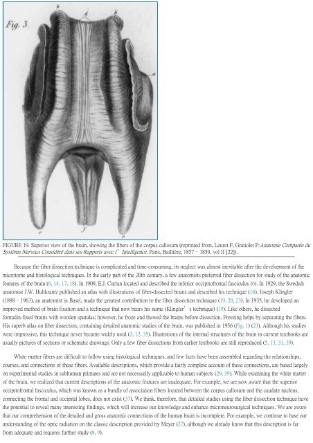
FIGURE 19. Superior view <strong>of</strong> the brain, showing the fibers <strong>of</strong> the corpus callosum (reprinted from, Leuret F, Gratiolet P:Anatomie Comparée du<br />
Système Nerveux Considéré dans ses Rapports avec l'Intelligence. Paris, Baillière, 1857–1859, vol II [22]).<br />
Because the fiber dissection technique is complicated and time-consuming, its neglect was almost inevitable after the development <strong>of</strong> the<br />
microtome and histological techniques. In the early part <strong>of</strong> the 20th century, a few anatomists preferred fiber dissection for study <strong>of</strong> the anatomic<br />
features <strong>of</strong> the brain (6, 14, 17, 18). In 1909, E.J. Curran located and described the inferior occipit<strong>of</strong>rontal fasciculus (6). In 1929, the Swedish<br />
anatomist J.W. Hultkrantz published an atlas with illustrations <strong>of</strong> fiber-dissected brains and described his technique (16). Joseph Klingler<br />
(1888–1963), an anatomist in Basel, made the greatest contribution to the fiber dissection technique (19, 20, 23). In 1935, he developed an<br />
improved method <strong>of</strong> brain fixation and a technique that now bears his name (Klingler's technique) (19). Like others, he dissected<br />
formalin-fixed brains with wooden spatulas; however, he froze and thawed the brains before dissection. Freezing helps by separating the fibers.<br />
His superb atlas on fiber dissection, containing detailed anatomic studies <strong>of</strong> the brain, was published in 1956 (Fig. 1) (23). Although his studies<br />
were impressive, this technique never became widely used (2, 12, 35). Illustrations <strong>of</strong> the internal structures <strong>of</strong> the brain in current textbooks are<br />
usually pictures <strong>of</strong> sections or schematic drawings. Only a few fiber dissections from earlier textbooks are still reproduced (5, 13, 31, 39).<br />
White matter fibers are difficult to follow using histological techniques, and few facts have been assembled regarding the relationships,<br />
courses, and connections <strong>of</strong> these fibers. Available descriptions, which provide a fairly complete account <strong>of</strong> these connections, are based largely<br />
on experimental studies in subhuman primates and are not necessarily applicable to human subjects (29, 39). While examining the white matter<br />
<strong>of</strong> the brain, we realized that current descriptions <strong>of</strong> the anatomic features are inadequate. For example, we are now aware that the superior<br />
occipit<strong>of</strong>rontal fasciculus, which was known as a bundle <strong>of</strong> association fibers located between the corpus callosum and the caudate nucleus,<br />
connecting the frontal and occipital lobes, does not exist (37). We think, therefore, that detailed studies using the fiber dissection technique have<br />
the potential to reveal many interesting findings, which will increase our knowledge and enhance microneurosurgical techniques. We are aware<br />
that our comprehension <strong>of</strong> the detailed and gross anatomic connections <strong>of</strong> the human brain is incomplete. For example, we continue to base our<br />
understanding <strong>of</strong> the optic radiation on the classic description provided by Meyer (27), although we already know that this description is far<br />
from adequate and requires further study (8, 9).


