

Surgical Anatomy of Supratentorial Midline Lesions
Surgical Anatomy of Supratentorial Midline Lesions
Surgical Anatomy of Supratentorial Midline Lesions

Create successful ePaper yourself
Turn your PDF publications into a flip-book with our unique Google optimized e-Paper software.
Arteries <strong>of</strong> the insula<br />
Parasylvian (M 4) and Terminal (M 5) Segments <strong>of</strong> the MCA<br />
The M 3 segments course laterally to exit the sylvian fissure<br />
and become M 4 segments on the lateral surface <strong>of</strong> the<br />
hemisphere. The M 4 and M 5 segments consist <strong>of</strong> 12 main<br />
arteries, which have been documented and named in earlier<br />
publications according to their territories <strong>of</strong> supply. 7,9,<br />
34,35 These include lateral orbit<strong>of</strong>rontal, prefrontal, precentral,<br />
central, anterior parietal, posterior parietal, angular,<br />
temporooccipital, posterior temporal, middle temporal,<br />
anterior temporal, and temporal polar arteries (Fig. 1). No<br />
branches from the M 4 and M 5 segments were observed to<br />
supply the insula.<br />
Arteries Supplying the Insula<br />
The insula receives its blood supply predominantly<br />
from the M2 segment. An examination <strong>of</strong> 40 hemispheres<br />
revealed 75 to 104 insular arteries originating from this<br />
segment. However, in 22 hemispheres (55%), between<br />
one and six insular arteries arose from the distal M1 segment<br />
and supplied the region <strong>of</strong> the limen insulae. In 10<br />
hemispheres (25%), one or two insular arteries arose from<br />
the M3 segment and supplied the region <strong>of</strong> either the superior<br />
or inferior periinsular sulcus. We observed no branches<br />
to the insula from the M4 and M5 segments. In each<br />
hemisphere, an average <strong>of</strong> 96 insular arteries (range 77–<br />
112 insular arteries) were found supplying the insula<br />
(Figs. 4 upper, 6, and 7). The average diameter <strong>of</strong> these arteries<br />
was 0.23 mm (range 0.1–0.8 mm). An average <strong>of</strong><br />
9.9 insular arteries (range four–14 insular arteries) in each<br />
hemisphere resembled perforator arteries, and their distribution<br />
occasionally reached as far as the corona radiata<br />
(Figs. 7 and 8). We sometimes observed a larger caliber<br />
insular artery that coursed along the surface <strong>of</strong> the insula<br />
and then looped laterally, providing branches to both<br />
the insula and the medial surface <strong>of</strong> the operculum. We<br />
named this artery the “insuloopercular artery” (Fig. 4 upper).<br />
Approximately 85 to 90% <strong>of</strong> insular arteries were<br />
short and supplied the insular cortex and extreme capsule;<br />
10% were medium sized and also supplied the claustrum<br />
and external capsule; and the remaining 3 to 5% were long<br />
and extended as far as the corona radiata (Fig. 8). The long<br />
insular arteries were perforator-like and mostly located in<br />
the posterior region <strong>of</strong> the insula. The putamen, globus<br />
pallidus, and internal capsule were vascularized by the<br />
LLAs (Fig. 4 lower). The external capsule was found to be<br />
the border <strong>of</strong> territories supplied by the LLAs and the insular<br />
arteries. We observed no gross communications between<br />
the insular arteries and LLAs.<br />
Discussion<br />
The MCA is the most complex <strong>of</strong> the cerebral vessels.<br />
This artery supplies almost the entire lateral surface <strong>of</strong> the<br />
hemisphere, as well as the insula, the lentiform nucleus,<br />
and the internal capsule. Microsurgical anatomy <strong>of</strong> the<br />
MCA, especially that <strong>of</strong> the M1 segment and the LLAs,<br />
has been examined and analyzed in detail in many studies.<br />
2–4,6–14,16,18–22,24–36 It is well known that chronic hypertension<br />
induces pathological changes in cerebral vessels,<br />
resulting in either their occlusion or rupture, which leads<br />
to lacunar infarctions or intracerebral hemorrhages, re-<br />
J. Neurosurg. / Volume 92 / April, 2000<br />
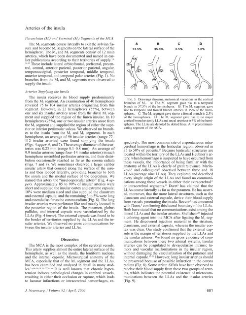
FIG. 3. Drawings showing anatomical variations in the cortical<br />
branches <strong>of</strong> M 1. A: The M 1 segment gave rise to a temporal<br />
branch in 57.5% <strong>of</strong> the hemispheres. B: The M 1 segment gave<br />
rise to temporal and frontal branch arteries in 35% <strong>of</strong> the hemispheres.<br />
C: The M 1 segment gave rise to a frontal branch in 2.5%<br />
<strong>of</strong> the hemispheres. D: The M 1 segment gave rise to no major<br />
cortical branches (only LLAs and uncal arteries) in 5% <strong>of</strong> the hemispheres.<br />
The LLAs are denoted by dotted lines. A 1 = precommunicating<br />
segment <strong>of</strong> the ACA.<br />
spectively. The most common site <strong>of</strong> a spontaneous intracerebral<br />
hemorrhage is the lenticular region, observed in<br />
35 to 50% <strong>of</strong> patients. 25 Because lenticular structures are<br />
located within the territory <strong>of</strong> the LLAs and Heubner’s artery,<br />
when hemorrhage is suspected to have occurred from<br />
these vessels, the importance <strong>of</strong> being familiar with the<br />
anatomy <strong>of</strong> the LLAs is clearly <strong>of</strong> great relevance. Marinković<br />
and colleagues 12 observed between three and 18<br />
LLAs (average nine LLAs). They explored and described<br />
every single origin <strong>of</strong> the LLAs and found no communications<br />
among these vessels in either their extracerebral 12<br />
or intracerebral segments. 13 Duret 3 has claimed that the<br />
LLAs course laterally as far as the putamen. He has asserted,<br />
moreover, that the more lateral structures, such as the<br />
claustrum and external capsule, derive their blood supply<br />
from vessels penetrating the insula. Beevor 2 has concurred<br />
with Duret, 3 confirming this lateral boundary <strong>of</strong> the LLAs.<br />
Both have stated that no communications exist among the<br />
lateral LLAs and the insular arteries. Shellshear 23 injected<br />
a coloring agent into the MCA after ligating the M 2 segment.<br />
He discovered injection material in the striatum,<br />
claustrum, and external capsule, whereas the insular cortex<br />
was clear. Our study confirmed that the external capsule<br />
is the margin <strong>of</strong> territories supplied by the LLAs and<br />
the insular arteries. We found no gross evidence <strong>of</strong> communications<br />
between these two arterial systems. Insular<br />
arteries can be coagulated to devascularize intrinsic tumors<br />
and vascular malformations in the insular region,<br />
without damaging the vascularization <strong>of</strong> the putamen and<br />
internal capsule. 37–39 However, long insular arteries should<br />
be preserved because <strong>of</strong> possible infarction in the corona<br />
radiata (Fig. 8). Some striate AVMs have been observed to<br />
receive their blood supply from these two groups <strong>of</strong> arteries,<br />
which indicates the potential existence <strong>of</strong> microcommunications<br />
between the LLAs and the insular arteries<br />
(Fig. 9).<br />
681


