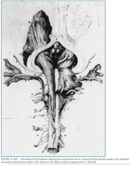
FIGURE 14. Bell's illustration <strong>of</strong> the brainstem, depicting the corticospinal tract as it passes from the internal capsule to the pyramidal decussation (reprinted from, Bell C:The <strong>Anatomy</strong> <strong>of</strong> the Brain. London, Longman and Co., 1802 [3]).
FIGURE 15. Drawing from one <strong>of</strong> Reil's dissections, demonstrating the white matter tracts in the insular region (reprinted from, McHenry LC Jr:Garrison's History <strong>of</strong> Neurology. Springfield, Charles C Thomas, 1969, p 141 [26]). In 1827, English anatomist Herbert Mayo, who was a student <strong>of</strong> Bell, published a book that included several <strong>of</strong> the best illustrations <strong>of</strong> dissected brains available at that time (Fig. 16) (25). He demonstrated the corona radiata, internal capsule, superior and inferior cerebellar peduncles, fasciculus uncinatus, fasciculus longitudinalis superior, outer surface <strong>of</strong> the lenticular nucleus, tapetum, mamillothalamic tractus, and anterior commissure. Two years later, the Italian anatomist Luigi Rolando (1773–1831) was the first to accurately portray the cerebral sulci and convolutions, including the central sulcus, which bears his name (34). His atlas contained several drawings <strong>of</strong> dissected brains. Rolando described and illustrated the continuity <strong>of</strong> fibers, starting with the medial olfactory stria and proceeding through the subcallosal area and cingulate and parahippocampal gyri, forming a nearly complete circle, and ending in the uncus (Fig. 17). In 1838, German anatomist Friedrich Arnold (1803–1890) first demonstrated the frontopontine tract (known as Arnold's tract), which extends from the frontal cortex through the anterior limb <strong>of</strong> the internal capsule, via the medial part <strong>of</strong> the cerebral peduncle, to the pons (1). In 1844, German anatomist and physiologist Karl Friedrich Burdach (1776–1847) demonstrated, using the fiber dissection technique, and named the cuneate fasciculus <strong>of</strong> Burdach (4). The same year, French neurologist Achille L. Foville (1799–1878) produced a major work on the nervous system, accompanied by an atlas that illustrated many admirable dissections (10). Although not well known, his atlas is probably the most accurate, the most artistic, and the highest quality publication in the neuroscience literature (Fig. 18). Italian anatomist Bartholomeo Panizza (1785–1867) demonstrated the visual pathway from the eye to the occipital cortex, using the fiber dissection technique, in 1855 (30). In 1857, French anatomist Louis Pierre Gratiolet (1815–1865), collaborating with his teacher and friend Francois Leuret (1797–1851), published an atlas that depicted fiber-dissected brains (Fig. 19) (22). Gratiolet also identified the optic radiation (initially called Gratiolet's radiation), from the lateral geniculate body to the occipital cortex, in detail. In 1872 in Vienna, Theodor H. Meynert (1833–1892), a pr<strong>of</strong>essor <strong>of</strong> neurology and psychiatry, refined the relatively crude division <strong>of</strong> fiber systems <strong>of</strong> the brain introduced by Gall and, for the first time, used the terms “association" and “projection" fibers in their modern sense (28). His studies <strong>of</strong> human brains convinced him that the corpus callosum consists primarily <strong>of</strong> decussating cortical fibers, which course downward to the basal ganglia. Meynert also described the habenulointerpeduncular tract or fasciculus retr<strong>of</strong>lexus (Meynert's bundle). In 1895, French neurologist Joseph J. Dejerine (1849–1917) described the occipit<strong>of</strong>rontal fasciculus (7). Our study, however, demonstrated that the location he described for this structure was inaccurate (37). In 1896, Swedish anatomist and anthropologist Magnus G. Retzius (1842–1919) was the first to use photographs to illustrate brain dissections (33).