

Surgical Anatomy of Supratentorial Midline Lesions
Surgical Anatomy of Supratentorial Midline Lesions
Surgical Anatomy of Supratentorial Midline Lesions

You also want an ePaper? Increase the reach of your titles
YUMPU automatically turns print PDFs into web optimized ePapers that Google loves.
frontal and occipital lobes, forming the superior occipit<strong>of</strong>rontal fasciculus (Fig. 1, A and B) (7, 8, 18, 23, 24, 30, 32). Thus, we focused on the<br />
clarification <strong>of</strong> the anatomic structures <strong>of</strong> this region.<br />
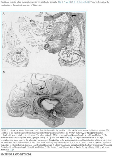
FIGURE 1. A, coronal section through the center <strong>of</strong> the third ventricle, the mamillary body, and the hippocampus. In this panel, number 22 is<br />
identified as the superior occipit<strong>of</strong>rontal fasciculus (arrow) (our dissection identified the structure marked 22 as the superior thalamic<br />
peduncle).23, stria terminalis; 32, optic tract;33, cerebral peduncle; 35, hippocampus (from, Nieuwenhuys R, Voogd J, van Huijzen C: The<br />
Human Central Nervous System. Berlin, Springer-Verlag, 1988, p 101, with permission [23]). B, long association bundles <strong>of</strong> the right<br />
hemisphere in a lateral view. In this schematic figure, number 1 is identified as the superior occipit<strong>of</strong>rontal fasciculus (arrow) (in our dissection,<br />
we observed no fasciculus composed <strong>of</strong> association fibers following the pattern as shown in 1).2, site <strong>of</strong> corona radiata; 3, superior longitudinal<br />
fasciculus; 6, outline <strong>of</strong> insula;7, inferior occipit<strong>of</strong>rontal fasciculus; 8, inferior longitudinal fasciculus; 9, site <strong>of</strong> anterior commissure;10, uncinate<br />
fasciculus (from, Nieuwenhuys R, Voogd J, van Huijzen C: The Human Central Nervous System. Berlin, Springer-Verlag, 1988, p 367, with<br />
permission [23]).<br />
MATERIALS AND METHODS


