


You also want an ePaper? Increase the reach of your titles
YUMPU automatically turns print PDFs into web optimized ePapers that Google loves.
5. Zenith - for the central and canine, the position of the zenith should be towards<br />
distal and for the lateral, it should be in the middle (Figure 11)<br />
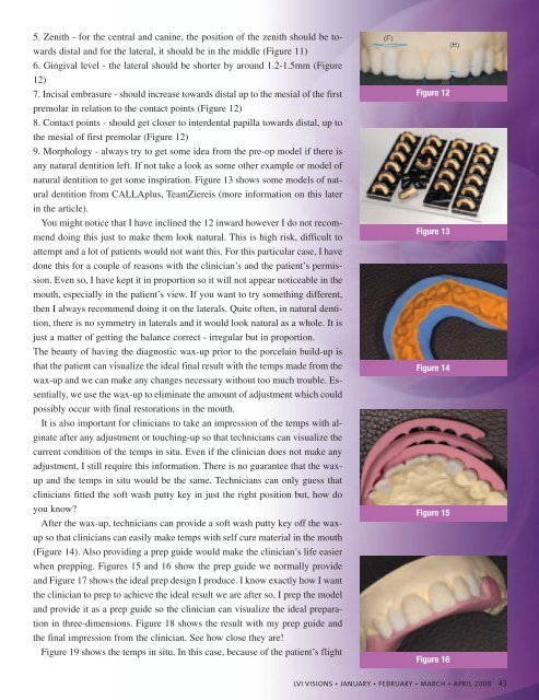
6. Gingival level - the lateral should be shorter by around 1.2-1.5mm (Figure<br />
12)<br />
7. Incisal embrasure - should increase towards distal up to the mesial of the first<br />
premolar in relation to the contact points (Figure 12)<br />
8. Contact points - should get closer to interdental papilla towards distal, up to<br />
the mesial of first premolar (Figure 12)<br />
9. Morphology - always try to get some idea from the pre-op model if there is<br />
any natural dentition left. If not take a look as some other example or model of<br />
natural dentition to get some inspiration. Figure 13 shows some models of natural<br />
dentition from CALLAplus, TeamZiereis (more information on this later<br />
in the article).<br />
You might notice that I have inclined the 12 inward however I do not recommend<br />
doing this just to make them look natural. This is high risk, difficult to<br />
attempt and a lot of patients would not want this. For this particular case, I have<br />
done this for a couple of reasons with the clinician’s and the patient’s permission.<br />
Even so, I have kept it in proportion so it will not appear noticeable in the<br />
mouth, especially in the patient’s view. If you want to try something different,<br />
then I always recommend doing it on the laterals. Quite often, in natural dentition,<br />
there is no symmetry in laterals and it would look natural as a whole. It is<br />
just a matter of getting the balance correct - irregular but in proportion.<br />
The beauty of having the diagnostic wax-up prior to the porcelain build-up is<br />
that the patient can visualize the ideal final result with the temps made from the<br />
wax-up and we can make any changes necessary without too much trouble. Essentially,<br />
we use the wax-up to eliminate the amount of adjustment which could<br />
possibly occur with final restorations in the mouth.<br />
It is also important for clinicians to take an impression of the temps with alginate<br />
after any adjustment or touching-up so that technicians can visualize the<br />
current condition of the temps in situ. Even if the clinician does not make any<br />
adjustment, I still require this information. There is no guarantee that the waxup<br />
and the temps in situ would be the same. Technicians can only guess that<br />
clinicians fitted the soft wash putty key in just the right position but, how do<br />
you know?<br />
After the wax-up, technicians can provide a soft wash putty key off the waxup<br />
so that clinicians can easily make temps with self cure material in the mouth<br />
(Figure 14). Also providing a prep guide would make the clinician’s life easier<br />
when prepping. Figures 15 and 16 show the prep guide we normally provide<br />
and Figure 17 shows the ideal prep design I produce. I know exactly how I want<br />
the clinician to prep to achieve the ideal result we are after so, I prep the model<br />
and provide it as a prep guide so the clinician can visualize the ideal preparation<br />
in three-dimensions. Figure 18 shows the result with my prep guide and<br />
the final impression from the clinician. See how close they are!<br />
Figure 19 shows the temps in situ. In this case, because of the patient’s flight<br />
Figure 12<br />
Figure 13<br />
Figure 14<br />
Figure 15<br />
Figure 16<br />
<strong>LVI</strong> VISIONS • JANUARY • FEBRUARY • MARCH • APRIL 2008 43