A Simple, Disposable End Tidal Carbon Dioxide Detector
A Simple, Disposable End Tidal Carbon Dioxide Detector
A Simple, Disposable End Tidal Carbon Dioxide Detector

You also want an ePaper? Increase the reach of your titles
YUMPU automatically turns print PDFs into web optimized ePapers that Google loves.
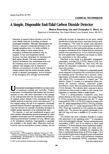
Anesth Prog 38:24-26 1991<br />
CLINICAL TECHNIQUES<br />
A <strong>Simple</strong>, <strong>Disposable</strong> <strong>End</strong> <strong>Tidal</strong> <strong>Carbon</strong> <strong>Dioxide</strong> <strong>Detector</strong><br />
Detection of expired carbon dioxide is one of the<br />
most reliable methods of avoiding accidental<br />
esophageal intubation. Although capnography has<br />
become a standard monitoring technique in the<br />
hospital operating room, it is rarely available in<br />
the office setting or other arenas where<br />
emergency endotracheal intubation may be<br />
required. A new and inexpensive device,<br />
however, has been developed for assessing endtidal<br />
carbon dioxide. This semi-quantitative<br />
detector fits between the endotracheal tube and<br />
the breathing circuit and uses a pH-sensitive<br />
indicator that changes color in response to<br />
different concentrations of carbon dioxide. Clinical<br />
studies indicate that this device provides similar<br />
results to standard capnography, and its inclusion<br />
in the emergency kit is strongly recommended.<br />
nrecognized esophageal intubation is a major cause<br />
of anesthesia mortality and morbidity.1 Positive<br />
confirmation of endotracheal tube position has become a<br />
standard of care, with direct observation of the passage<br />
of the endotracheal tube into the trachea and detection<br />
of expired carbon dioxide (CO2) considered the most<br />
reliable methods of assuring correct tube placement.2,3 In<br />
the hospital setting, the use of an expensive, electrically<br />
powered infrared detection capnograph or mass spectroscopy<br />
has become an essential monitoring technique to<br />
confirm proper placement of an endotracheal tube.<br />
The majority of oral and maxillofacial surgeons and<br />
other dentists administering general anesthesia for office<br />
outpatient procedures use non-endotracheal techniques,<br />
reserving endotracheal intubation for long, difficult cases<br />
or for emergency situations. Capnography is generally<br />
not available in the dental office. Alternative methods,<br />
however, such as auscultation or pulse oximetry, are not<br />
Received July 1, 1990; accepted for publication February 27, 1991.<br />
Address correspondence to Morton Rosenberg, DMD, Department<br />
of Anesthesiology, 750 Washington St., New England Medical Center<br />
Hospitals, Boston, MA 02111.<br />
© 1991 by the American Dental Society of Anesthesiology<br />
Morton Rosenberg, DMD, and Christopher S. Block, MD<br />
Department of Anesthesiology, New England Medical Center Hospitals, Boston, MA 02111<br />
24<br />
sufficiently accurate or responsive for the quick, reliable<br />
identification of tube placement, especially during a medical<br />
emergency.4 This need for proper tube placement<br />
confirmation may occur in the unanticipated intubation in<br />
the dental office or other prehospital settings, as well as in<br />
emergency intubations at the hospital that are performed<br />
outside of the operating room. It is our belief that auscultation<br />
and questionable visualization are simply not definitive,<br />
particularly in the office setting.<br />
Described in this article is a disposable, inexpensive,<br />
colorimetric, end-tidal CO2 (ETCO2) detector (FEF <strong>End</strong>-<br />
<strong>Tidal</strong> CO2 <strong>Detector</strong>, FENEM, Inc., New York, NY) that<br />
permits the immediate confirmation of proper endotracheal<br />
tube position.<br />
The FEF <strong>End</strong>-<strong>Tidal</strong> CO2 <strong>Detector</strong> is a disposable plastic<br />
device designed to fit between the endotracheal tube and<br />
the breathing circuit. The device has a nontoxic, chemically<br />
treated, pH-sensitive indicator strip that colorimetrically<br />
reflects CO2 concentrations in expired gas.5 The<br />
detector (Figure 1) consists of a transparent dome containing<br />
the chemical indicator, metacresol purple,<br />
mounted on a mesh. Inlet and outlet ports are the standard<br />
15 mm diameter. The indicator strip's response is almost<br />
instantaneous; fast enough to reflect a color change from<br />
inspiration to expiration within a single breath.<br />
There is a semiquantitative comparison color chart<br />
printed directly on the dome in three color ranges (Figure<br />
2). If CO2 is present, the initial purple color, the "A" range<br />
(representing 0.03% to 0.3% CO2) fades to a beige color<br />
(the "B" range representing 0.5% to 1.0% CO2) and then<br />
to pale yellow (the "C" range representing 2.0% to 5.0%<br />
CO2). Fresh gas containing no CO2 restores the original<br />
purple color. The devices are packed individually in airtight<br />
foil bags and have a shelflife of 15 months if unopened.<br />
The detector has been evaluated clinically to confirm<br />
tube placement in the operating room,6-8 in emergency<br />
room and prehospital settings,4 during cardiopulmonary<br />
resuscitation (CPR),9 and as an adjunct to blind nasal<br />
intubation. 10 In those patients where inadvertent esophageal<br />
intubation occurred, diagnosis was easily accomplished.<br />
After emergency intubation for respiratory distress<br />
or cardiopulmonary arrest, an ETCO2 range- 15.2<br />
torr recorded by this device indicated correct endotracheal<br />
tube placement.9 In a study of 62 intubations comparing<br />
the efficacy of colorimetric ETCO2 assessment versus<br />
ISSN 0003-3006/91/$3.50
~~~~~~~~~~~~~~~~~~~~~~~~~~~~~~~~~~~~~~~~~~~~~~~~~~~~~~~~~~~~~~~~~~~~~~~~~~~~~~~~~~~~~~~~~~~~~~~~~~~~~~~~<br />
:.<br />
~~~~~~~~~~~~~~~~~~~~~~~~~~~~~~~~~~~~~~~~..~. .. ..<br />
Anesth Prog 38:24-26 1991 Rosenberg and Block 25<br />
Figure 1. Side view of the FEF <strong>End</strong>-<strong>Tidal</strong> CO2 <strong>Detector</strong>.<br />
chest auscultation and capnography, colorimetric CO2 detection<br />
of esophageal intubation was more rapid than<br />
auscultation and equivalent to the use of capnography.<br />
There were no false-positive or false-negative results with<br />
,. ,.,~~~ ~~~~~~~~~~~~~~~~~~~~~~~~~~~~~~~~~~~~~~~~~~~~~~~~~~~~~~~~~~~~~......<br />
..:..i:i the ETCO2 detector.8 The manufacturer recommends that<br />
Figure 2. Top view of the FEF <strong>End</strong>-<strong>Tidal</strong> CO2 <strong>Detector</strong>.<br />
".<br />
six breaths be assessed before interpreting a color change.<br />
This will avoid errors due to the forcing of CO2 into the<br />
stomach during mask ventilation or the generation of CO2<br />
in the stomach by prophylactic antacids or carbonated<br />
beverage ingestion.11l<br />
Effective CPR has been shown to produce ETCO2 values<br />
of 0.5% to 2.5%, well within the range of this device<br />
to demonstrate detectable color changes. 12 If the detector<br />
does not change color in the cardiac arrest setting, there<br />
still is a possibility that the endotracheal tube may be in<br />
the trachea. This limitation, also experienced with capnography,<br />
occurs when the cardiac output is extremely low,<br />
resulting in decreased pulmonary blood flow and reduced<br />
delivery of CO2 to the alveoli. As both devices may occasionally<br />
fail to confirm a successful endotracheal intubation<br />
in extremely low flow states, clinicians should immediately<br />
verify endotracheal tube position by other means<br />
and attempt to increase perfusion.<br />
Changes in ETCO2 may serve as a prognostic indicator<br />
of the effectiveness of therapy in cardiac arrest.4,13,14 If the<br />
ETCO2 range detected by this device during CPR remains<br />
low (< 15.2 torr despite correct endotracheal intubation<br />
and optimization of perfusion), successful resuscitation is<br />
unlikely. 14 An increase in ETCO2 provides an immediate<br />
bedside validation of the efficacy of the resuscitative effort.<br />
The FEF <strong>Detector</strong> is extremely sensitive to the presence<br />
of CO2; the mean CO2 concentration required for detection<br />
of a color change has been shown to be 0.54% (4.1<br />
torr). 11<br />
In summary, a disposable, compact ETCO2 detector<br />
has been developed that can be extremely valuable in<br />
the office setting to verify endotracheal tube placement.<br />
Among its attributes are that it fits all standard airway<br />
connections and requires no warm-up time, calibration,<br />
or servicing. In locations where intubation is performed<br />
routinely, capnography is an essential monitor. It will not<br />
only confirm endotracheal tube placement, but give valuable<br />
information in the diagnosis of other conditions, such<br />
as malignant hyperthermia and rebreathing. For practices<br />
where intubation is the exception rather than the rule,<br />
we strongly recommend the inclusion of a colorimetric<br />
ETCO2 detector as a safe, reliable, and rapidly acting<br />
device in the emergency kit to verify tube placement.<br />
REFERENCES<br />
1. Brahams D: Anaesthesia and the law. Two cases of oesophageal<br />
intubation. Anaesthesia 1989;44:64-65.<br />
2. Link K, Summa TK, Fretund BL: Capnography for detection<br />
of accidental oesophageal intubation. Acta Anaesthesiol<br />
Scand 1983;27:199-202.<br />
3. Birmingham PK, Cheney FW, Ward RJ: Esophageal<br />
intubation: A review of detection techniques. Anesth Analg<br />
1986;64:886-891.
26 <strong>Disposable</strong> <strong>End</strong>-<strong>Tidal</strong> CO2 <strong>Detector</strong> Anesth Prog 38:24-26 1991<br />
4. Gerard J, MacLeod BA, Heller MB, Yealy DM: Verification<br />
of endotracheal intubation using a disposable end-tidal CO2<br />
detector. Prehosp Disast Med 1989;4:74.<br />
5. O'Callaghan JP, Williams RT: Confirmation of tracheal<br />
intubation using a chemical device. Can Anaesth Soc J<br />
1988;35:s59.<br />
6. Strunin L, Williams T: The FEF end-tidal carbon dioxide<br />
detector. Anesthesiology 1989;71:621-622.<br />
7. Feinstein R, White PF, Westerfield SZ: Intraoperative<br />
evaluation of a disposable end-tidal CO2 detector. Anesthesiology<br />
1989;71:A461.<br />
8. Goldberg JS, Rawle PR, Zehnder JL, Sladen RN: Colorimetric<br />
end-tidal carbon dioxide monitoring for tracheal intubation.<br />
Anesth Analg 1990;70:191-194.<br />
9. Varon AJ, Morrina J, Civetta JM: Clinical utility of a<br />
colorimetric end-tidal carbon dioxide detector in emergency intubation.<br />
Anesthesiology 1990;73:A413.<br />
10. King H, Wooten DJ: Blind nasal intubation by monitoring<br />
end-tidal CO2. Anesth Analg 1989;69:412-413.<br />
11. Jones BR, Dorsey MJ: <strong>Disposable</strong> end-tidal CO2 detector:<br />
minimal CO2 requirements. Anesthesiology 1989;71:A359.<br />
12. Gamett AR, Ornato JP, Gonzalez ER, Johnson EB: <strong>End</strong>tidal<br />
carbon dioxide monitoring during cardiopulmonary resuscitation.<br />
JAMA 1987;257:512-515.<br />
13. Trevino RP, Bisera J, Linna DK: <strong>End</strong>-tidal CO2 as a<br />
guide to successful cardiopulmonary resuscitation: A preliminary<br />
report. Crit Care Med 1985;13:910-911.<br />
14. Varon AJ, Morrina JM, Civetta JM: Use of colorimetric<br />
end-tidal carbon dioxide monitoring to pronosticate immediate<br />
resuscitation from cardiac arrest. Anesthesiology 1990; 73:A412.