Chapter 21 - Lymphatic and Immune Systems
Chapter 21 - Lymphatic and Immune Systems
Chapter 21 - Lymphatic and Immune Systems
Create successful ePaper yourself
Turn your PDF publications into a flip-book with our unique Google optimized e-Paper software.
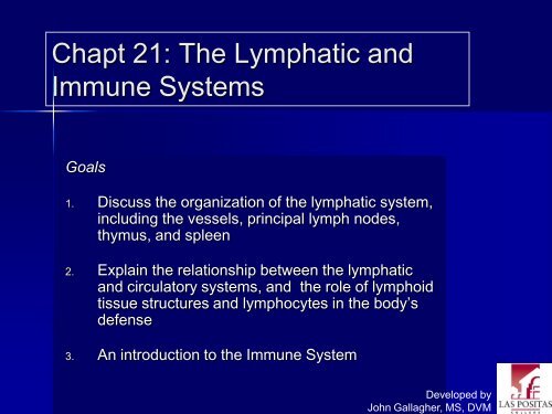
Chapt <strong>21</strong>: The <strong>Lymphatic</strong> <strong>and</strong><br />
<strong>Immune</strong> <strong>Systems</strong><br />
Goals<br />
1. Discuss the organization of the lymphatic system,<br />
including the vessels, principal lymph nodes,<br />
thymus, <strong>and</strong> spleen<br />
2. Explain the relationship between the lymphatic<br />
<strong>and</strong> circulatory systems, <strong>and</strong> the role of lymphoid<br />
tissue structures <strong>and</strong> lymphocytes in the body’s<br />
defense<br />
3. An introduction to the <strong>Immune</strong> System<br />
Developed by<br />
John Gallagher, MS, DVM
Overview of the <strong>Lymphatic</strong><br />
System<br />
Includes, vessels, fluid, <strong>and</strong> nodes or<br />
nonsecreting "gl<strong>and</strong>s".<br />
<strong>Lymphatic</strong> vessels convey fluid from<br />
the periphery to the veins.<br />
The fluid, lymph (=clear water), is<br />
what seeps out of the blood at the<br />
peripheral capillaries. Composition<br />
is similar to plasma without as<br />
much protein<br />
Fig 20.1
Overview of the <strong>Lymphatic</strong><br />
System<br />
<strong>Lymphatic</strong> organs or tissues ("gl<strong>and</strong>s”<br />
is a misnomer) are filtering areas<br />
<strong>and</strong> arenas of lymphocyte<br />
maturation <strong>and</strong> competency.<br />
Accessory to cardiovascular system,<br />
thus there are two drainage<br />
systems.<br />
Fig 20.1
Major Functions of the<br />
<strong>Lymphatic</strong> System<br />
1. Filtration of lymph<br />
2. Return of leaked fluid to<br />
cardiovascular system<br />
3. “Education” <strong>and</strong><br />
production of immune<br />
system lymphocytes<br />
4. Transport of digested<br />
lipids from small intestinal<br />
lacteals
Lymph Capillaries<br />
Thin walled endothelium (no<br />
BM) with periodic one way<br />
valves. In general ,they<br />
parallel veins.<br />
– Usually not visible on tissue<br />
sections<br />
Lymph capillaries converge<br />
into collecting vessels
Lymph Capillaries<br />
Closed ends allow fluid flow<br />
inward only<br />
– Also bacteria, viruses, cancer<br />
cells<br />
Pick up <strong>and</strong> recycle extra<br />
tissue fluid<br />
The fluid flows to lymph<br />
nodes
Lymph Capillaries<br />
Located everywhere, except<br />
for CNS, bone marrow, cornea<br />
<strong>and</strong> cartilage.<br />
– (XS fluid in CNS becomes part of<br />
CSF)<br />
Special set of lymph<br />
capillaries in villi of small<br />
intestine = Lacteals<br />
– Fat Absorption<br />
– To liver
<strong>Lymphatic</strong> vessels<br />
Comparable in structure to<br />
capillaries, <strong>and</strong> in turn, veins.<br />
Thin walls!<br />
Hard to find in a general<br />
dissection<br />
Damaged valves or blocked<br />
lymph vessels edema
Lymph capillaries<br />
converge to become<br />
collecting vessels<br />
<strong>and</strong> end up as either<br />
Thoracic duct or<br />
right lymphatic duct<br />
Right<br />
lymphatic<br />
duct<br />
Right<br />
subclavian<br />
vein<br />
Cysterna Chyli<br />
Left subclavian vein<br />
Thoracic (left lymphatic) duct
Lymphoid Organs<br />
1. Lymph Nodes<br />
1. Lymph Nodules<br />
1. Tonsils<br />
2. Thymus<br />
3. Spleen<br />
4. Bone Marrow<br />
1. Stem cells
1. Lymph Nodes<br />
~ 500 ( 1mm to 25 mm)<br />
Bean-shaped with hilus, cortex <strong>and</strong><br />
medulla<br />
Several afferent vessels, one efferent<br />
vessel<br />
Function: filter<br />
Popular term “lymph gl<strong>and</strong>” is<br />
misnomer. Why?<br />
Contain lots of Lymphocytes,<br />
Macrophages <strong>and</strong> Plasma Cells<br />
Clinical application: Swollen lymph<br />
nodes<br />
Fig 23.9
Distribution of LNs<br />
Cervical lymph nodes - drain head <strong>and</strong> neck<br />
Axillary lymph nodes - drain arms <strong>and</strong> breasts<br />
Popliteal lymph nodes - drain legs<br />
Inguinal lymph nodes - drain lower limb<br />
Thoracic lymph nodes - drain thoracic viscera<br />
Abdominal lymph nodes - drain pelvic region<br />
Intestinal <strong>and</strong> mesenteric lymph nodes - drain abdominal<br />
viscera
Important example:<br />
Axillary Drainage<br />
Drainage from<br />
breast <strong>and</strong> arm
Tonsils, p629<br />
Simple lymphoid organs<br />
– “Lymph nodules”<br />
In the mouth/pharynx<br />
– Lingual tonsil on posterior<br />
aspect of tongue<br />
– Palatine -- lateral pharynx,<br />
removed in tonsillectomy<br />
– Pharyngeal – AKA adenoids<br />
– Tubal, behind Eustachian<br />
Tubes<br />
Fig 22.3, page 639
Lymphoid Tissue<br />
Connective tissue is loaded with<br />
lymphocytes<br />
Lymphoid nodules are unencapsulated<br />
clusters of lymphocytes ( ~ 1mm).<br />
Found beneath epithelial lining of<br />
respiratory, digestive & urinary tracts,<br />
etc.<br />
Mucosa Associated <strong>Lymphatic</strong> Tissue<br />
(MALT) in GI tract<br />
– 5 tonsils<br />
– aggregate lymphoid nodules in<br />
small intestine (= Peyer’s patches)<br />
– appendix - walls contain lymphoid<br />
tissue<br />
= adenoids
2. Thymus<br />
Location above heart, posterior to sternum<br />
Divided into lobules<br />
Only lymphoid organ that does NOT fight antigens, it<br />
functions as “T-cell academy”<br />
Involution after puberty<br />
Epithelial cells produce thymic hormones, thymosin <strong>and</strong><br />
thymopoetin
3. Spleen<br />
Largest lymphoid organ, located in LUQ<br />
Soft <strong>and</strong> very blood rich<br />
Red pulp sinusoids containing RBCs<br />
white pulp lymphoid tissue<br />
Major Functions:<br />
1. Initiation of <strong>Immune</strong> response to<br />
antigens in blood<br />
2. Removal of aged <strong>and</strong> defective RBCs, Fe<br />
salvaging<br />
3. Reservoir for new RBCs
4. Bone Marrow<br />
Mostly in red marrow<br />
From pluripotent<br />
stem cells<br />
Fig. 17.8
Bone Marrow:<br />
Lymphopoiesis<br />
Lymphocytes are also<br />
produced in thymus,<br />
spleen <strong>and</strong> tonsils
The <strong>Immune</strong> System<br />
The primary defense against<br />
disease<br />
– Infectious, especially<br />
– Specific<br />
Centered around the activity of<br />
lymphocytes<br />
– Other cells, too.<br />
Neutrophil with Bacillus anthracis
Lymphocytes<br />
Agranulocytes - large nuclei <strong>and</strong> small amount of cytoplasm<br />
Function in identification <strong>and</strong> inactivation/destruction of<br />
pathogens<br />
Types of Lymphocytes<br />
1. T Cells - cellular immunity - specific for previously identified<br />
pathogens. AKA “cytotoxic cells.”<br />
2. B Cells - humoral immunity – become plasma cells, which<br />
produce antibodies specific to the antigen or pathogen;<br />
memory cells for future exposures<br />
3. NK (Natural Killer) cells - non-specific, provide immunological<br />
surveillance, recognition of “non-self”
Antibodies (Ab)<br />
AKA immunoglobulins (Ig)<br />
Proteins produced by plasma cells in<br />
response to a specific antigen (Ag)<br />
– Plasma cells are derived from Blymphocytes<br />
Antibodies [Ab] frequently measured<br />
as a diagnostic tool<br />
– Serology<br />
IgG
5 subclasses of Igs:<br />
1. IgG: main Ab (75%) in serum; + main Ab<br />
during 2 o response<br />
2. IgA: main Ab in external secretions<br />
3. IgE: main Ab in allergic reactions<br />
4. IgM: Ab on virgin B-cells; + main Ab during 1 o<br />
response<br />
5. IgD: Ab on virgin B-cells
Sunset on the Atlantic Ocean


