The Aerobiology Pathway
The Aerobiology Pathway
The Aerobiology Pathway

You also want an ePaper? Increase the reach of your titles
YUMPU automatically turns print PDFs into web optimized ePapers that Google loves.
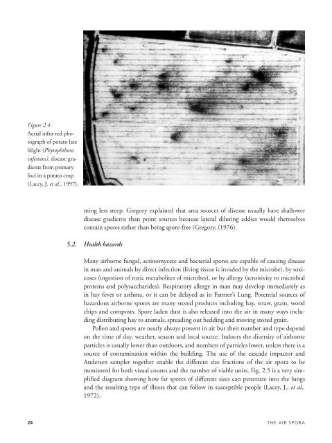
Figure 2.4<br />
Aerial infra-red photograph<br />
of potato late<br />
blight (Phytophthora<br />
infestans), disease gradients<br />
from primary<br />
foci in a potato crop<br />
(Lacey, J. et al., 1997).<br />
ming less steep. Gregory explained that area sources of disease usually have shallower<br />
disease gradients than point sources because lateral diluting eddies would themselves<br />
contain spores rather than being spore-free (Gregory, (1976).<br />
5.2. Health hazards<br />
Many airborne fungal, actinomycete and bacterial spores are capable of causing disease<br />
in man and animals by direct infection (living tissue is invaded by the microbe), by toxicoses<br />
(ingestion of toxic metabolites of microbes), or by allergy (sensitivity to microbial<br />
proteins and polysaccharides). Respiratory allergy in man may develop immediately as<br />
in hay fever or asthma, or it can be delayed as in Farmer’s Lung. Potential sources of<br />
hazardous airborne spores are many stored products including hay, straw, grain, wood<br />
chips and composts. Spore laden dust is also released into the air in many ways including<br />
distributing hay to animals, spreading out bedding and moving stored grain.<br />
Pollen and spores are nearly always present in air but their number and type depend<br />
on the time of day, weather, season and local source. Indoors the diversity of airborne<br />
particles is usually lower than outdoors, and numbers of particles lower, unless there is a<br />
source of contamination within the building. <strong>The</strong> use of the cascade impactor and<br />
Andersen sampler together enable the different size fractions of the air spora to be<br />
monitored for both visual counts and the number of viable units. Fig. 2.5 is a very simplified<br />
diagram showing how far spores of different sizes can penetrate into the lungs<br />
and the resulting type of illness that can follow in susceptible people (Lacey, J., et al.,<br />
1972).<br />
24 THE AIR SPORA

![Estructuras secretoras internas [4.64 MB]](https://img.yumpu.com/14294979/1/190x143/estructuras-secretoras-internas-464-mb.jpg?quality=85)
![anatomía y exomorfología [7.14 MB]](https://img.yumpu.com/12744163/1/190x143/anatomia-y-exomorfologia-714-mb.jpg?quality=85)