Description of primordial germ cells, oogonia, oocytes and ... - Digitum
Description of primordial germ cells, oogonia, oocytes and ... - Digitum
Description of primordial germ cells, oogonia, oocytes and ... - Digitum
Create successful ePaper yourself
Turn your PDF publications into a flip-book with our unique Google optimized e-Paper software.
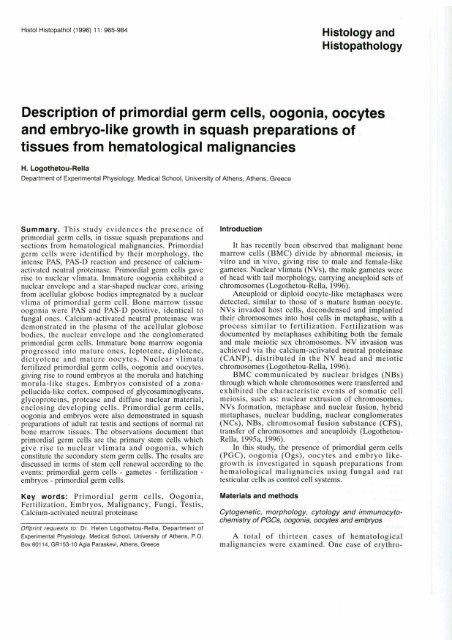
Histol Histopathol (1 996) 1 1 : 965-984 Histology <strong>and</strong><br />
Histo pathology<br />
<strong>Description</strong> <strong>of</strong> <strong>primordial</strong> <strong>germ</strong> <strong>cells</strong>, <strong>oogonia</strong>, <strong>oocytes</strong><br />
<strong>and</strong> embryo-like growth in squash preparations <strong>of</strong><br />
tissues from hem-atologica malignancies<br />
,C<br />
H. Logothetou-Rella<br />
Department <strong>of</strong> Experimental Physiology, Medical School, University <strong>of</strong> Athens, Athens, Greece<br />
Summary. This study evidences the presence <strong>of</strong><br />
<strong>primordial</strong> <strong>germ</strong> <strong>cells</strong>, in tissue squash preparations <strong>and</strong><br />
sections from hematological malignancies. Primordial<br />
<strong>germ</strong> <strong>cells</strong> were identified by their morphology, the<br />
intense PAS, PAS-D reaction <strong>and</strong> presence <strong>of</strong> calcium-<br />
activated neutral proteinase. Primordial <strong>germ</strong> <strong>cells</strong> gave<br />
rise to nuclear vlimata. Immature <strong>oogonia</strong> exhibited a<br />
nuclear envelope <strong>and</strong> a star-shaped nuclear core, arising<br />
from acellular globose bodies impregnated by a nuclear<br />
vlima <strong>of</strong> <strong>primordial</strong> <strong>germ</strong> cell. Bone marrow tissue<br />
<strong>oogonia</strong> were PAS <strong>and</strong> PAS-D positive, identical to<br />
fungal ones. Calcium-activated neutral proteinase was<br />
demonstrated in the plasma <strong>of</strong> the acellular globose<br />
bodies, the nuclear envelope <strong>and</strong> the conglomerated<br />
<strong>primordial</strong> <strong>germ</strong> <strong>cells</strong>. Immature bone marrow <strong>oogonia</strong><br />
progressed into mature ones, leptotene, diplotene,<br />
dictyotene <strong>and</strong> mature <strong>oocytes</strong>. Nuclear vlimata<br />
fertilized <strong>primordial</strong> <strong>germ</strong> <strong>cells</strong>, <strong>oogonia</strong> <strong>and</strong> <strong>oocytes</strong>,<br />
giving rise to round embryos at the morula <strong>and</strong> hatching<br />
morula-like stages. Embryos consisted <strong>of</strong> a zona-<br />
pellucida-like cortex, composed <strong>of</strong> glycosaminoglycans,<br />
glycoproteins, protease <strong>and</strong> diffuse nuclear material,<br />
enclosing developing <strong>cells</strong>. Primordial <strong>germ</strong> <strong>cells</strong>,<br />
<strong>oogonia</strong> <strong>and</strong> embryos were also demonstrated in squash<br />
preparations <strong>of</strong> adult rat testis <strong>and</strong> sections <strong>of</strong> normal rat<br />
bone marrow tissues. The observations document that<br />
<strong>primordial</strong> <strong>germ</strong> <strong>cells</strong> are the primary stem <strong>cells</strong> which<br />
give rise to nuclear vlimata <strong>and</strong> <strong>oogonia</strong>, which<br />
constitute the secondary stem <strong>germ</strong> <strong>cells</strong>. The results are<br />
discussed in terms <strong>of</strong> stem cell renewal according to the<br />
events: <strong>primordial</strong> <strong>germ</strong> <strong>cells</strong> - gametes - fertilization -<br />
embryos - <strong>primordial</strong> <strong>germ</strong> <strong>cells</strong>.<br />
Key words: Primordial <strong>germ</strong> <strong>cells</strong>, Oogonia,<br />
Fertilization, Embryos, Malignancy, Fungi, Testis,<br />
Calcium-activated neutral proteinase<br />
Offprint requests to: Dr. Helen Logothetou-Rella, Department <strong>of</strong><br />
Experimental Physiology, Medical School, University <strong>of</strong> Athens, P.O.<br />
Box 601 14, GR153-10 Agia Paraskevi, Athens, Greece<br />
Introduction<br />
It has recently been observed that malignant bone<br />
marrow <strong>cells</strong> (BMC) divide by abnormal meiosis, in<br />
vitro <strong>and</strong> in vivo, giving rise to male <strong>and</strong> female-like<br />
gametes. Nuclear vlimata (NVs), the male gametes were<br />
<strong>of</strong> head with tail morphology, carrying aneuploid sets <strong>of</strong><br />
chromosomes (Logothetou-Rella, 1996).<br />
Aneuploid or diploid oocyte-like metaphases were<br />
detected, similar to those <strong>of</strong> a mature human oocyte.<br />
NVs invaded host <strong>cells</strong>, decondensed <strong>and</strong> implanted<br />
their chromosomes into host <strong>cells</strong> in metaphase, with a<br />
process similar to fertilization. Fertilization was<br />
documented by metaphases exhibiting both the female<br />
<strong>and</strong> male meiotic sex chromosomes. NV invasion was<br />
achieved via the calcium-activated neutral proteinase<br />
(CANP), distributed in the NV head <strong>and</strong> meiotic<br />
chromosomes (Logothetou-Rella, 1996).<br />
BMC communicated by nuclear bridges (NBs)<br />
through which whole chromosomes were transferred <strong>and</strong><br />
exhibited the characteristic events <strong>of</strong> somatic cell<br />
meiosis, such as: nuclear extrusion <strong>of</strong> chromosomes,<br />
NVs formation, metaphase <strong>and</strong> nuclear fusion, hybrid<br />
metaphases, nuclear budding, nuclear conglomerates<br />
(NCs), NBs, chromosomal fusion substance (CFS),<br />
transfer <strong>of</strong> chromosomes <strong>and</strong> aneuploidy (Logothetou-<br />
Rella, 1995a, 1996).<br />
In this study, the presence <strong>of</strong> <strong>primordial</strong> <strong>germ</strong> <strong>cells</strong><br />
(PGC), <strong>oogonia</strong> (Ogs), <strong>oocytes</strong> <strong>and</strong> embryo like-<br />
growth is investigated in squash preparations from<br />
hematological malignancies using fungal <strong>and</strong> rat<br />
testicular <strong>cells</strong> as control cell systems.<br />
Materials <strong>and</strong> methods<br />
Cytogenetic, morphology, cytology <strong>and</strong> immunocyto-<br />
chemistry <strong>of</strong> PGCs, <strong>oogonia</strong>, <strong>oocytes</strong> <strong>and</strong> embryos<br />
A total <strong>of</strong> thirteen cases <strong>of</strong> hematological<br />
malignancies were examined. One case <strong>of</strong> erythro-
leukemia (EL), four cases <strong>of</strong> acute myeloid leukemia<br />
(AML-1, AML-2), two cases <strong>of</strong> acute myelomonocytic<br />
leukemia (AMMol-M4) two cases <strong>of</strong> undifferentiated<br />
leukemia (UL-1, UL-2), one case <strong>of</strong> chronic myelo-<br />
genous leukemia (CML) <strong>and</strong> three cases <strong>of</strong> acute<br />
lymphoblastic leukemia (ALL). Patients had received no<br />
treatment prior to aspirate collection. Small bone<br />
marrow tissue (BMT) pieces, washed in phosphate-<br />
buffered saline (PBS), were treated with KC1 (0.075M)<br />
for 10 min, fixed in 3:l ethano1:acetic acid for 24 hours,<br />
followed by 6:4 acetic acid:distilled water for 5 min,<br />
squashed on glass slides, dried <strong>and</strong> stained with Giemsa,<br />
PAS, PAS-D for cytogenetic morphology. Squashed<br />
BMT, after KC1 treatment, fixed in 4% formaldehyde in<br />
PBS were stained with Feulgen (without counterstain),<br />
or immunostained for al-antichymotrypsin.<br />
Isolated BMC from two cases <strong>of</strong> AML, growing<br />
attached on the culture vessel surface in the presence <strong>of</strong><br />
phytohaemaglutinin (PHA) (Logothetou-Rella, 1996),<br />
were treated with KC1 for 10 min (HT), fixed in 4%<br />
formaldehyde in PBS for PAS, PAS-D staining <strong>and</strong><br />
immunocytochemistry. For immunocytochemical studies<br />
the avidin-biotin peroxidase complex method was<br />
applied (Hsu et al., 1981) using the antiserum against<br />
al-chymotrypsin (1:100, A022, Dako Corp.). Positive<br />
<strong>and</strong> negative controls were used.<br />
Testis, <strong>of</strong> l-8-week-old Wistar rats were removed,<br />
sliced, treated with KC1 (0.075M) for 10 min squashed<br />
on slides <strong>and</strong> stained with Giemsa, PAS <strong>and</strong> PAS-D.<br />
Three types <strong>of</strong> fungus, Dermatophyte microsporum<br />
sp., Aspergillus fumigatus <strong>and</strong> C<strong>and</strong>ida albicans were<br />
used for cytogenetic morphology, PAS <strong>and</strong> PAS-D<br />
staining <strong>and</strong> immunocytochemistry as previously<br />
described (Logothetou-Rella, 1996). Ten serial BMT<br />
paraffin sections from 10 cases <strong>of</strong> CML, 10 cases <strong>of</strong><br />
AML <strong>and</strong> 5 cases <strong>of</strong> MDS were stained with Giemsa,<br />
PAS, PAS-D, <strong>and</strong> immunostained using the antiserum<br />
against al-chymotrypsin (1:1000, A022, Dako Corp.),<br />
using negative <strong>and</strong> positive controls (Hsu et al., 1981).<br />
Results<br />
Cytogenetic, morphology, cytology <strong>and</strong> immunocyto-<br />
chemistry <strong>of</strong> <strong>primordial</strong> <strong>germ</strong> <strong>cells</strong>, <strong>oogonia</strong>, <strong>oocytes</strong> <strong>and</strong><br />
embryos<br />
All tissue squash preparations exhibited immature<br />
Primordial <strong>germ</strong> <strong>cells</strong> in hematological malignancies<br />
<strong>and</strong> mature exfoliated Ogs (Fig. 1). Various-sized<br />
acellular globose bodies (AGBs) (Fig. la), embraced by<br />
nuclear segments arising from crescentic conglomerated<br />
nuclei (Fig. lb) or impregnated by a NV (Fig. lc,d), or<br />
containing a nuclear star-shaped core with or without<br />
spokes (Fig. le,f), were the immature forms <strong>of</strong> Og.<br />
Mature Ogs were characterized by a complete, intact<br />
nucleus. Some mature Ogs contained a round, hyperchromatic,<br />
condensed nucleus (Fig. lg), while others a<br />
larger one with dispersed chromatin (Fig. lh,i). The<br />
various-sized Og contained various numbers <strong>of</strong> double<br />
minutes (DMs), minute <strong>and</strong> ring chromosomes,<br />
surrounded by a thin or thick nuclear envelope (NE)<br />
(Fig. lj,k). Ogs <strong>of</strong> a full nucleus were surrounded <strong>and</strong><br />
invaded by NVs (Fig. 11,m). Og appeared single or in<br />
clusters encircled by nuclear segments arising from<br />
conglomerated crescentic nuclei (Fig. ln,o). Large<br />
immature Og divided by budding to smaller daughter<br />
<strong>cells</strong> containing DMs (Fig. 2a). Og also arose within<br />
large NCs (Fig. 2b) which extended nuclear segments<br />
embracing <strong>and</strong> forming the NE <strong>of</strong> Og (Fig. 2c).<br />
Conglomerated nuclei free <strong>of</strong> Ogs were present (Fig.<br />
2d).<br />
Progression <strong>of</strong> Og to <strong>oocytes</strong> was documented by the<br />
presence <strong>of</strong> leptotene (Fig. 2e,f), diplotene (Fig. 2g) <strong>and</strong><br />
dictyotene (Fig. 2h) stages <strong>of</strong> the first meiotic prophase.<br />
Dictyotene-stage <strong>oocytes</strong> were in various sizes within<br />
the same sample, been characterized by the lacy network<br />
<strong>of</strong> chromatin.<br />
Oocytic metaphases were identified by chromosomes<br />
identical to those <strong>of</strong> a human mature oocyte (Fig.<br />
2i) (Plachot et al., 1987). Most <strong>oocytes</strong> consisted <strong>of</strong><br />
meiotic condensed prochromosomes (Fig. 2j-n). Some<br />
<strong>oocytes</strong> kept the NE consisting <strong>of</strong> DMs <strong>and</strong> minute<br />
chromosomes (Fig. 2k). Abnormal metaphases <strong>of</strong><br />
fertilized oocyte (Plachot et al., 1987) were present,<br />
showing two female a0>> chromosomes, one c>, one<br />
bivalent CXYN <strong>and</strong> a marker chromosome in one<br />
metaphase from a male patient (Fig. 20).<br />
Presence <strong>and</strong> formation <strong>of</strong> Og was confirmed by<br />
PAS <strong>and</strong> PAS-D staining (Fig. 3). The plasma <strong>of</strong> the<br />
AGBs, immature <strong>and</strong> mature Ogs, were PAS <strong>and</strong> PAS-D<br />
positive, consisting <strong>of</strong> glycogen, glycoproteins <strong>and</strong><br />
glycosaminoglycans (GAG). Ogs were in clusters,<br />
embraced by thick nuclear segments, implanted in the<br />
tissue, or exfoliated. Upon exfoliation large round<br />
unstained vacuoles remained on tissues, surrounded by<br />
7<br />
Fig. l. AGB, immature <strong>and</strong> mature Og. lnset a: various-sized exfoliated AGB. lnset b: crescentic, conglomerated nucleus extends nuclear segments<br />
embracing the AGB, forming a NE. lnsets c <strong>and</strong> d: AGB impregnated by a NV. lnsets e <strong>and</strong> 1: immature Og with star-shaped nuclear core <strong>and</strong> NE.<br />
lnset g: mature Og with a small, round, condensed nucleus. lnsets h <strong>and</strong> i: mature Og with large, round nucleus with dispersed chromatin. lnsets j<br />
<strong>and</strong> k: immature Og with DMs, ring <strong>and</strong> minute chromosomes. lnset I: figure focused on NV embracing Og. lnset m: mature Og fertilized by two NVs.<br />
lnsets n <strong>and</strong> o: Ogs in clusters, held together by nuclear segments arisen from conglomerated, crescentic <strong>cells</strong>. Giemsa. X 1,000<br />
Flg. 2. Female gametes in BMT squash preparations, lnset a; dividing Og by budding. Daughter Og (arrow) contains a DM. lnset b: conglomerated<br />
nucleus bearing an Og. Giemsa. X 1,000. lnset c: nuclear conglomerate extends thick nuclear segments embracing Ogs. Giemsa. X 400.<br />
lnset d: conglomerated nucleus free <strong>of</strong> Og. Giemsa X 1,000. lnset e <strong>and</strong> 1: leptotene-stage <strong>oocytes</strong>. lnset g: diplotene-stage oocyte. Giemsa. X 1,000.<br />
lnset h: dictyotene-stage <strong>oocytes</strong> <strong>of</strong> various size <strong>and</strong> lacy network <strong>of</strong> chromatin. Giemsa. X 100. lnset I: metaphase with chromosomes <strong>of</strong> a mature<br />
human oocyte. lnsets j-n: <strong>oocytes</strong> with meiotic, condensed prochromosomes with or without a NE. Giemsa. X 1,000. lnset o: a metaphase <strong>of</strong> a<br />
fertilized oocyte showing two female chromosomes [SO*, one "Y., one bivalent (
thick nuclear segments (Fig. 3c). It was obvious that<br />
no fungal <strong>cells</strong> were associated with the Ogs.<br />
Conglomerated nuclei gave rise to nuclear segments<br />
embracing the AGB <strong>and</strong>for nuclear spokes or NVs which<br />
impregnated the AGB forming the nuclear core <strong>of</strong> the<br />
immature Og (Fig. 4). Some AGB showed stained <strong>and</strong><br />
unstained PAS-D areas (Fig. 4, 4a). Mature Og <strong>of</strong><br />
complete, intact, large or small round nucleus were<br />
observed (Fig. 4b). The various possible morphologies<br />
<strong>of</strong> AGB, immature <strong>and</strong> mature Og were shown in Fig. 5.<br />
AGB impregnated by a NV (Fig. 5a) or immature Og<br />
embraced (Fig. 5b) or impregnated by a NV (Fig. 5c)<br />
were obvious. The content <strong>of</strong> AGBs showed PAS-D<br />
negative or mixed negative with positive or positive<br />
areas (Fig. 5d-f).<br />
Og formation was also apparent in Feulgen stained<br />
preparations. Unstained AGB (Fig. 6, 6a) were<br />
impregnated by Feulgen-positive nuclear spokes or NVs<br />
arising from the NE, transfering DNA to the center <strong>of</strong><br />
the AGB (Fig. 6b). Feulgen stained the NE, the core <strong>of</strong><br />
Ogs (Fig. 6, 6b) <strong>and</strong> the nuclear rings <strong>and</strong> segments, red<br />
with dark blue or dark blue in spite <strong>of</strong> the omission <strong>of</strong><br />
counterstain (Fig. 6c). Regular chromosomes were<br />
stained red (Fig. 6d) <strong>and</strong> pachytene ones red with dark<br />
blue chromomeres (Fig. 6e).<br />
In squash PAS-stained preparations, PGCs were<br />
identified by: the two prominent nucleoli (Fig. 7a),<br />
round, large nucleus (Fig. 7b-d), the amoeboid<br />
movement (Fig. 7a, e-g), the oval (Fig. 7b) or elliptical<br />
morphology (Fig. 7h) <strong>and</strong> mainly by the dark PAS stain<br />
reaction. A PGC <strong>of</strong> large, round nucleus, was surrounded<br />
<strong>and</strong> invaded by a NV (Fig. 7i-k). The NVs arose from<br />
surrounding crescentic, conglomerated nucleus. PGC<br />
were intensively PAS-D positive, some showing<br />
irregular conglomerated nucleus, indistinct from the<br />
cytoplasm (Fig. 71), <strong>of</strong>ten with a NE, keeping a darker<br />
red colour than the Og. PGC embryos were large<br />
globose, PAS-D positive, <strong>of</strong> 2-3 nuclei (Fig. 7m-o)<br />
surrounded by a NE.<br />
PAS-D-stained BMT smears showed conglomerated<br />
PGCs surrounding an 0g (Fig. 8a), a NV embracing an<br />
immature Og (Fig. 8b), PAS-D negative AGB with<br />
positive NE (Fig. 8c), clusters <strong>of</strong> various-sized Ogs (Fig.<br />
8d), various intensity <strong>of</strong> PAS-D reaction, nuclear size<br />
<strong>and</strong> morphology (Fig. 8d-i).<br />
AGB, all forms <strong>of</strong> immature <strong>and</strong> mature Ogs <strong>and</strong><br />
PGCs were identified in PAS-, PAS-D-stained paraffin<br />
tissue sections, from ALL, CML, MDS <strong>and</strong> AML (Fig.<br />
9). Amoeboid shape (Fig. 9a) <strong>and</strong> conglomerated PGCs<br />
(Fig. 9b-e) with prominent nucleoli (Fig. 9d) associated<br />
closely with AGB <strong>and</strong> Og were observed, identical to<br />
Primordial <strong>germ</strong> <strong>cells</strong> in hematological malignancies<br />
those in squash preparations. AGB embraced by NV<br />
(Fig. 9f), NE <strong>of</strong> immature Og (Fig. 9g), Og with a star-<br />
shaped nuclear core (Fig. 9h) or NV (Fig. 9i) <strong>and</strong> mature<br />
Og (Fig. 9j) were apparent, all surrounded by crescentic<br />
or round BMC. The easy exfoliation <strong>of</strong> PGC <strong>and</strong> Og<br />
from tissues left behind large vacuoles with the<br />
surrounding <strong>cells</strong>.<br />
The function <strong>of</strong> BMT <strong>oocytes</strong> <strong>and</strong> their fertilization<br />
by NVs was evidenced by the presence <strong>of</strong> embryo-like<br />
growth in squash preparations. Embryos were round <strong>and</strong><br />
<strong>of</strong> various size <strong>and</strong> cell composition (Fig. 10), consisting<br />
<strong>of</strong> a zona-pellucida-like cortex enclosing NCs in<br />
Flg. 3. GAG content <strong>of</strong> EMT Ogs. lnset a: AGE, immature <strong>and</strong> mature<br />
Og, embraced by nuclear segments, implanted in the tissue. PAS.<br />
X 200. lnsets b <strong>and</strong> c: exfoliated Og leave empty round vacuoles<br />
(arrow) surrounded by nuclear segments. PAS-D. X 100<br />
W<br />
Fig. 4. AGES with PAS-D stained <strong>and</strong> unstained areas, impregnated (arrows) by nuclear segments or NVs arisen from the surrounding <strong>cells</strong> or the<br />
nuclear envelope. PAS-D. X 1,000. lnset a: PASD negative AGBs. PAS-D. X 100. lnset b: mature Og <strong>of</strong> small or large round nucleus <strong>and</strong> clear, bright<br />
red cytoplasm. PAS. X 1,000<br />
Fig. 5. Various morphologies <strong>of</strong> AGE, <strong>and</strong> Ogs. AGE with negative or negative <strong>and</strong> positive or positive PAS-D areas. lnset a: AGE impregnated by a<br />
NV. lnset b: an immature Og embraced by a NV. lnset c: an immature Og impregnated by a NV. Insets d <strong>and</strong> e: AGE with stained <strong>and</strong> unstained<br />
areas. Inset 1: two AGBs one stained the other unstained. PAS-D, X 1,000
dissoluted nuclear material (Fig. lOa), or NCs, NVs <strong>and</strong><br />
intact <strong>cells</strong> (Fig. lob-f). Some embryos resembled uteral<br />
embryos at the morula stage (Fig. 10 c,d) <strong>and</strong> others<br />
resembled hatching morula (Fig. lOe), releasing the <strong>cells</strong><br />
extraembryonically. Some embryos showed intact nuclei<br />
in the cortex (Fig. 10f). The cortex kept the <strong>cells</strong><br />
enclosed even after tissue hypotonic treatment <strong>and</strong><br />
squashing. Within the cortex <strong>of</strong> early embryos,<br />
dissoluted nuclear material, with or without circular<br />
nuclear segments (Fig. lla,b), was condensing <strong>and</strong><br />
organising itself into nuclei, which led to new nucleus<br />
Primordial <strong>germ</strong> <strong>cells</strong> in hematological malignancies<br />
<strong>and</strong> cell genesis (Fig. llc,d). Formed <strong>cells</strong> were released<br />
towards the center <strong>of</strong> the developing embryos (Fig. lle).<br />
It was clear that diffuse nuclear material gradually<br />
condensed giving rise to hyperchromatic intact nuclei, in<br />
towards the center <strong>and</strong> away from the embryo cortex<br />
(Fig. 12a). This observation was not considered<br />
artifactual since there was a continuance <strong>of</strong> the nuclear<br />
material from the cortex to the intact nuclei which co-<br />
existed with diffuse nuclear material (Fig. 12a).<br />
Developing <strong>cells</strong> within embryos exhibited the meiotic<br />
properties previously described (Logothetou-Rella,<br />
Flg. 6. Feulgen-stained nuclear rings, segments, NE <strong>and</strong> Og nuclear core red with dark Mue or blue colour. lnset a: AGE free <strong>of</strong> DNA, surrounded by<br />
nuclear segments. lnset b: NV <strong>and</strong>lor spokes transverse <strong>and</strong> carry DNA from the NE to the center <strong>of</strong> AGB. Inset c: Og wlh nuclear core <strong>of</strong> blue<br />
colour. Imet d: regular chromosomes <strong>of</strong> red colour. Ineel e: pachytene chromosomes with dark blue chromomeres. Feulgen (without counterstain).<br />
X 1,000<br />
Fig. 7. Various morphologies <strong>of</strong> PGCs. lnsat a: migrating PGC with hvo small prominent nucleoli. Insets W: oval-shaped PGC <strong>of</strong> large nucleus.<br />
Ingets e-g: migrating PGC <strong>of</strong> NV morphology. Inset h: PGC <strong>of</strong> elliptical shape. PAS. X 1,000. Inset I: A PGC embraced by a NV. Insets j <strong>and</strong> k: PGC<br />
with NE, fertilized by a NV. Inset I: thrw conglomerated PGCs <strong>of</strong> Irregular, d i e nucleus, indinct from the cytoplasm. lnset m+: PGC globose<br />
embryos <strong>of</strong> two-three nuclei <strong>and</strong> NE. PAS-D. X 1,000<br />
Fig. 8. PAS-D reaction <strong>of</strong> BMT smears. lnset a: conglomerated PGC surrounding an immature Og. Inset b: A NV embracing an Og. Inset c: negative<br />
AGB with positive NE. PAS-D. X 1,000. lnsat d: Cluster <strong>of</strong> Ogs. PAS-D. X 400. Inseta oh: clusters <strong>of</strong> various-sized Ogs, various intensity <strong>of</strong> PAS-D<br />
reaction, nuclear size <strong>and</strong> morphology. PAS-D. X 1,000
1996).<br />
In cultured AML <strong>cells</strong>, fertilized Og (Fig. 12b), <strong>and</strong><br />
small round embryos, with minute chromosomes inside<br />
the thick (Fig. 12c) or thin cortex (Fig. 12d) containing<br />
NCs, were evident. These embryos correlated well with<br />
those in histological sections from AML, consisting <strong>of</strong><br />
globose NCs with a distinct cortex, embraced by a NV<br />
(Fig. 12e,f).<br />
Within developing embryos, various-sized AGB<br />
Primordial <strong>germ</strong> <strong>cells</strong> in hematological malignancies<br />
(Fig. 13a), impregnated by NV <strong>and</strong> surrounded by<br />
scattered chromosomes were frequent (Fig. 13b,c).<br />
Intranuclear or extracellular nuclear rings, hyphae (Fig.<br />
13d-f) <strong>and</strong> NCs giving rise to Og (Fig. 13g) or scattered<br />
chromosomes (Fig. 13h) were obvious.<br />
The cortex <strong>of</strong> embryos was PAS <strong>and</strong> PAS-D<br />
positive, consisting <strong>of</strong> glycoproteins <strong>and</strong> GAG (Fig. 14).<br />
Also, round embryos in cultured AML <strong>cells</strong> showed PAS<br />
<strong>and</strong> PAS-D positive newly forming nuclei (Fig. 14a),<br />
Fig. 9. QAG content <strong>of</strong> PGC, AGB <strong>and</strong> Og in AML tissue sections. lnset a: irregular morphology <strong>of</strong> migrating PGC <strong>of</strong> darker colour closely associated<br />
with Og. Insets b <strong>and</strong> c: conglomerated PGCs <strong>of</strong> darker colour, closely associated with Ogs. PAS. X 1,000. lnset d: conglomerated PGC or with<br />
prominent nucleoli <strong>and</strong> cluster <strong>of</strong> various-sized Ogs. PAS. X 400. lnset e: single conglomerated PGC associated with one Og (focus on PGC). PAS-D.<br />
X 1,000. lnset f: AGB embraced by a NV. Inset g: AGBs <strong>and</strong> Ogs with NE, implanted in the tissue (focus on NE). lnset h: a huge Og embraced by<br />
crescentic <strong>cells</strong>. Inset l: One AGB <strong>and</strong> one Og side by side surrounded by round epithelia1 <strong>cells</strong>. The Og shows a nuclear core <strong>of</strong> NV morphology. PAS.<br />
X 1,000. Inset 1: a mature Og with large round nucleus similar to those in squash preparations. PAS-D. X 1,000<br />
7<br />
Flg. 10. Various-sized embryos showing a zona-pellucida-like cortex. Inset a: an embryo <strong>of</strong> a central NC surrounded by dissoluted nuclear material<br />
Giemsa. X 100. lnset b: embryo with NC, NVs <strong>and</strong> <strong>cells</strong>. Giemsa. X 400. lnset c: Giemsa. X 200. lnset d: embryo cortex contains faint nuclei (focus on<br />
cortex). Giemsa r 200. lnset e: hatching morula-like embryo, releasing cell extraembryonically. Giemsa. X 40. lnset f: embryo cortex contains <strong>cells</strong>.<br />
Giemsa. X 40<br />
Fig. 11. Early embryos showing nucleus <strong>and</strong> cell genesis. lnset a: embryo shows circular nuclear segments in dissoluted nudear material. There are<br />
no nuclei present. Glemsa. X 100. Inset b: dissoluted nuclear material in embryo reaching sligM round shape (arrow). lnset c: embryo shows<br />
completed round-shaped nuclei with boundaries, inside the cortex. Surrounding nuclei are intact. Giemsa. X 200.lnsel d: nucleus formed by dissoluted<br />
nuclear material. Giemsa. X 1,000
I:. 7<br />
qq, F<br />
. ..<br />
;p: .F! :
Primordial <strong>germ</strong> <strong>cells</strong> in hematological malignancies<br />
positive cortex with positive DMs <strong>and</strong> minute<br />
chromosomes (Fig. 15). Identical round embryos were<br />
demonstrated in AML histological section showing a<br />
discrete PAS <strong>and</strong> PAS-D cortex (Fig. 15a), enclosing<br />
<strong>cells</strong> <strong>of</strong> indistinct boundaries <strong>and</strong> prominent nuclei; these<br />
embryos morphologically correlate well with a 2-cell<br />
embryo in rat uteral tissue sections (Fig. 15b).<br />
Cultured AML <strong>cells</strong> showed conglomerated PGC,<br />
NE, NVs <strong>and</strong> embryos highly immunoreactive with a -<br />
antichymotrypsin (Fig. 16a-d). The content <strong>of</strong> A G ~<br />
exhibited negative, weak to very strong immuno-<br />
reactivity (Fig. 16b-d). Squash preparations showed<br />
positive nuclear segments from positive NCs or <strong>cells</strong>,<br />
embracing negative to weak positive AGBs (Fig. 17a-c).<br />
The nuclear core <strong>of</strong> immature Ogs was positive to<br />
negative, the nucleus <strong>of</strong> mature Ogs negative, <strong>and</strong> the<br />
NE positive (Fig. 17d). Tissue sections from AML, MDS<br />
<strong>and</strong> CML, also showed strong a -antichymotrypsin<br />
immunoreactivity in PGCs, NE, Nbs <strong>and</strong> AGBs (Fig.<br />
18). PGC with prominent nucleoli producing a NV (Fig.<br />
18a), conglomerated PGC extending NVs or nuclear<br />
segments forming the NE <strong>of</strong> AGB (Fig. 18b-e) were<br />
events involving high protease activity. The negative to<br />
strong immunoreactivity <strong>of</strong> AGB was evident in<br />
malignant tissues (Fig. 18b-e). Migrating, immuno-<br />
stained PGC <strong>of</strong> prominent nucleoli (Fig. 18f) or<br />
Flg. 12. lnset a: progressbe condensation <strong>of</strong> diffuse nuclear material leads to nucleus genesis starting from the embryo cortex, towards the center <strong>of</strong><br />
the embryo. DHbe nuclear material co-exists with intact nuclei. Giemsa. X 1,000. lnset b: an Og fertilized by a fourchromosome NV. The Og shows<br />
conglomeratrJd chromosomes <strong>and</strong> NE, in cultured AML <strong>cells</strong>. Insets c <strong>and</strong> d: early embryos <strong>of</strong> thick or thin cortex with chromosomes containing a<br />
central NC, in cultured AML <strong>cells</strong>. Inset e: globose conglomerated embryo in MDS histological picture, showing a disttnct cortex, embraced by a NV.<br />
Giemsa. X 1.000. lnset t: same as e. H-E. X 1,000<br />
Flg. 13. Content <strong>of</strong> embryos. Insets a-c: AGB surrounded by scattered chromosomes <strong>and</strong>tor impregnated by a NV. Insets d-t: intranuciear or<br />
extracellular nuclear rings, hyphae <strong>and</strong> nuclel in dissoluted nuclear matrk. Giemsa. X 1,000. Inset g: embryo with NC bearing Og. Giemsa. X 200. lnset<br />
h: NC releasing chromosomes. Giemsa. X 1,000<br />
Flg. 14. GAG content <strong>of</strong> embryos. PAS-D. X 40. Inget a: cultured AML embryo with newly forming nuclei. PAS-D. X 1,000
conglomerated nucleus (Fig. 18g) <strong>of</strong> NV morphology,<br />
oocyte-like <strong>cells</strong> with immunostained NE <strong>and</strong> NCs (Fig.<br />
18h), globose embryos with immunostained NE (Fig.<br />
18i) <strong>and</strong>lor <strong>cells</strong> (Fig. 18j) were obvious. Mature Og<br />
showed negative plasma but positive NE <strong>and</strong><br />
surrounding <strong>cells</strong> (Fig. 18k). Megakaryocytes were<br />
devoid <strong>of</strong> a NE <strong>and</strong> a]-antichymotrypsin immuno-<br />
reactivity (Fig. 181), distinguishing them from embryos.<br />
Primordial <strong>germ</strong> <strong>cells</strong> in hematological malignancies<br />
Assembly <strong>of</strong> immunoreactive chromosomes around an<br />
AGB could be seen in tissues (Fig. 18m).<br />
Normal rat BMT also showed PAS <strong>and</strong> PAS-D PGCs<br />
<strong>and</strong> Ogs which were implanted <strong>and</strong> surrounded by tissue<br />
matrix only (Fig. 19). Cytogenetic morphology showed a<br />
few hollow embryos, devoid <strong>of</strong> <strong>cells</strong>.<br />
The cytogenetic morphology <strong>and</strong> formation <strong>of</strong><br />
fungal Og was identical to those <strong>of</strong> BMT (Fig. 20a).<br />
Fig. 15. Embryos in cultured AML <strong>cells</strong><br />
show PAS-D positive DMs <strong>and</strong> minute<br />
chromosomes in the cortex. In situ PAS-<br />
D. X 1,000. Inset a: embryo in AML<br />
histological section showing a discrete<br />
cortex. PAS-D. X 1,000. lnset b: 2-cell<br />
rat embryo in uterus PAS-D. X 400<br />
Fig. 16. al-antichymotrypsin in cultured<br />
AML <strong>cells</strong>. lnset a: conglomerated<br />
positive PGCs. al-antichymotrypsin.<br />
X 200. lnset b: positive PGC <strong>and</strong> NE <strong>of</strong><br />
weak positive AGB. al-antichymo-<br />
trypsin. X 400. lnset c: strong immuno-<br />
reactivity in the content <strong>of</strong> AGB <strong>and</strong> NE.<br />
al-antichymotrypsin. X 200. lnset d:<br />
positive NV surrounding negative AGB<br />
in squash preparations. al-antichymo-<br />
trypsin. X 400. lnset e: embryo with<br />
positive <strong>cells</strong> <strong>and</strong> NE. a,-antichymo-<br />
I ttypsin X 1,000
Fungal Og was fertilized by a fungal NV (Fig. 20b). The<br />
fungal Og formation was clear in PAS, PAS-D <strong>and</strong><br />
immunostained squash preparations. Immunostained<br />
fungal <strong>cells</strong> or NCs extended nuclear segments around<br />
negative AGB, impregnated by positive NV <strong>and</strong> showed<br />
strongly positive conidiophores <strong>and</strong> conidiospores (ring<br />
chromosomes) (Fig. 20c). Fungal AGB, Og conidio-<br />
spores <strong>and</strong> hyphae were PAS <strong>and</strong> PAS-D positive (Fig.<br />
21). Fungal NCs (Fig. 21a) <strong>and</strong> globose 2-cell embryos<br />
(Fig. 21b) <strong>of</strong> positive PAS, PAS-D reaction resemble<br />
those <strong>of</strong> PGCs.<br />
Rat testicular tissue squash preparations, also<br />
showed Og formation. Conglomerated PGC extended<br />
nuclear segments, forming the NE <strong>of</strong> Og (Fig. 22a-c).<br />
AGB impregnated by a spermatocytic NV was evident<br />
(Fig. 22a). Ogs <strong>of</strong> DMs <strong>and</strong> minute chromosomes were<br />
present (Fig. 22d). Ogs were abundant in clusters<br />
connected by nuclear segments in five-week-old<br />
rat testis (Fig. 22e). The general morphology <strong>of</strong><br />
conglomerated PGC <strong>and</strong> Og in PAS-D-stained<br />
Primordial <strong>germ</strong> <strong>cells</strong> in hematological malignancies<br />
preparations resembled those in BMT (Fig. 229. PGCs<br />
were present in all examined rat ages, dividing<br />
<strong>and</strong> migrating in two-week- (Fig. 23a), in the form <strong>of</strong><br />
NV in five-week- (Fig. 23b) or with two large prominent<br />
nucleoli in eight-week-old rat testis (Fig. 23c). Invasion<br />
<strong>of</strong> testicular PGCs by a NV was observed (Fig. 23d).<br />
Testicular AGB also showed stained <strong>and</strong> unstained PAS-<br />
D areas (Fig. 23e). PAS positive AGB <strong>and</strong> Ogs were<br />
connected by nuclear segments in clusters (Fig. 239, or<br />
encircled by crescentic epithelia1 <strong>cells</strong> (Fig. 23g), in five-<br />
week-old rat testis. Spermatocytic NVs fusing (invading<br />
or fertilizing) with another spermatocyte was<br />
demonstrated in five-week-old rat testis (Fig. 24a).<br />
Developing round embryos with a cortex, dissoluted<br />
nuclear material, NCs <strong>and</strong> nuclei or spermatocytes in<br />
meiosis observed in 4-week-old testis <strong>and</strong> smaller<br />
globose embryos in l-week-old testis (Fig. 24c,d)<br />
document a common mechanism <strong>of</strong> cell renewal<br />
via embryo formation, in rapidly growing <strong>cells</strong> <strong>and</strong><br />
tissues.<br />
Flg. 17. Distribution <strong>of</strong> a,-antichymotrypsin in quash preparations in AML. Weak positive or negative nuclear core <strong>of</strong> immature Ogs. Inset a*: amngly<br />
irnmunoatained nuclear segments embrace <strong>and</strong> form the NE <strong>of</strong> negative to weak positive Ki8s. Inset d: mature Og with positive NE, negeMve plasma<br />
<strong>and</strong> nucleus. a,-antichymotrypsin. X 1,000
Discussion<br />
Before accepting the existence <strong>of</strong> PGCs, Ogs <strong>and</strong><br />
embryos, it is necessary to rule out the possibility <strong>of</strong><br />
artifactual observations. Firstly, talcum powder from<br />
rubber gloves <strong>and</strong> fungal contamination. Talcum powder<br />
slightly resembles immature Og <strong>and</strong> can contaminate<br />
samples during aspirate collection, but it cannot<br />
penetrate tissues in depth. Ogs identified in deep serial<br />
tissue section, being implanted <strong>and</strong> embraced by tissue<br />
<strong>cells</strong> or nuclear segments, excludes presence <strong>of</strong> talcum<br />
powder. Ogs due to fungal contamination are also<br />
excluded because all PAS-stained tissues were free <strong>of</strong><br />
fungal <strong>cells</strong> <strong>and</strong> BMT aspirates tested on sabouraud agar<br />
gave no fungal growth. Secondly, it is necessary to rule<br />
out artifacts due to hypotonic treatment <strong>of</strong> tissue pieces.<br />
Hypotonic treatment breaks the extracellular matrix <strong>and</strong><br />
cell membrane without affecting the integrity <strong>of</strong> nuclei<br />
Primordial <strong>germ</strong> <strong>cells</strong> in hematological malignancies<br />
<strong>and</strong> nuclear material. This is obvious in squash<br />
preparations where diffuse nuclear material CO-exists<br />
with intact nuclei. Moreover, the repetitive observations<br />
shown in the three systems, squash preparations,<br />
cultured <strong>cells</strong> <strong>and</strong> tissue sections exclude the possibility<br />
<strong>of</strong> artifacts.<br />
The main observation in this study is the existence<br />
<strong>of</strong> PGCs, Ogs, <strong>oocytes</strong>, fertilization <strong>and</strong> embryos in<br />
hematological malignancies in vivo, confirming previous<br />
in vitro study (Logothetou-Rella, 1996). It was<br />
evidenced that PGCs give rise to male (NVs) <strong>and</strong> female<br />
(<strong>oocytes</strong>) gametes. The female gamete originating from<br />
AGB developed through immature <strong>and</strong> mature Og, into a<br />
mature oocyte. NV, the male gamete, daughter cell <strong>of</strong><br />
meiosis has been extensively discussed previously<br />
(Logothetou-Rella, 1994a,c, 1995a, 1996). Oocytes were<br />
fertilized by NV giving rise to embryo-like growth in the<br />
overall process <strong>of</strong> PGC-gametes-fertilization-embryo-<br />
r'lg. 18. uistribution <strong>of</strong> a,-antichymotrypsin m AML t~ssue sections. lnset a: a positive PGC with prominent nucleoli giving rise to a positive W. Insets<br />
b-e: conglomerated PGCs extend NV or nuclear segments embracing negative to positive AGBs. Insets f <strong>and</strong> g: PGCs in the shape <strong>of</strong> a NV with<br />
prominent nucleoli or conglomerated. lnset h: an oocyte-like cell with positive NE <strong>and</strong> plasma. Nearby NC is strongly positive. lnsets I <strong>and</strong> 1: small<br />
embryos with posEtwe NE <strong>and</strong>lor <strong>cells</strong>. lnset k: a mature Og with positive surrounding <strong>cells</strong>, NE, negative nucleus <strong>and</strong> plasma. lnset I: megakaryocyte<br />
is negative <strong>and</strong> shows no NE. lnset m: scattered immunoreactive chromosomes assemble around an AGB. a,-antichymotrypsin. X 1,000
Primordid <strong>germ</strong> <strong>cells</strong> in hematoiogical mfignancies<br />
Flg. 19. GAG content in normal rat BMT.<br />
A PGC embraced by a NV (arrow) within<br />
a cluster <strong>of</strong> various-sized AGBs <strong>and</strong><br />
immature Ogs. Single PGCs <strong>and</strong> im-<br />
mature Ogs are implanted in tissue<br />
matrix without adjacent <strong>cells</strong>. PAS.<br />
X 1,000. insets a <strong>and</strong> b: PGCs. PAS-D.<br />
X 1,000<br />
Fig. 20. Fungal <strong>cells</strong>. Inset a: Og with a round or star-shaped nuclear core <strong>and</strong> NE. In- b: A NV fertilizing an Og. Giemsa. X 1,000. in- c: positive<br />
<strong>cells</strong> or NC extend positive nuclear segments around negative AGB. Positive conidiophores <strong>and</strong> conidiospores (arrow). a,-antichymotrypsin. X 1,000
PGC.<br />
Very little information is available in the literature<br />
on PGCs, mainly from embryology. PGCs are first<br />
recognised in the yolk-sac endoderm, in early stages <strong>of</strong><br />
embryonic development <strong>and</strong> eventually they migrate into<br />
the gonadal anlage where they differentiate <strong>and</strong><br />
transform into spermatogonia (in male embryo) or<br />
<strong>oogonia</strong> (in female embryo) (Rabinovici <strong>and</strong> Jaffe,<br />
1990). Because <strong>of</strong> this transformation, it was, until now,<br />
believed that PGCs disappeared upon fetal development<br />
<strong>of</strong> the gonads. Surprisingly enough, PGCs exist in<br />
normal BMT, malignant BMT <strong>and</strong> adult testis, as shown<br />
in this study.<br />
Primordial <strong>germ</strong> <strong>cells</strong> in hematological malignancies<br />
It has been reported that PGCs can be recognized by<br />
their morphology, the positive PAS reaction <strong>of</strong> the<br />
cytoplasm <strong>and</strong> the high alkaline phosphatase activity in<br />
the cell membrane (Chiquoine, 1954; Falin, 1969;<br />
Rabinovici <strong>and</strong> Jaffe, 1990).<br />
PGCs in BMT were evidenced by their large,<br />
migrating irregular, globose shape, the two prominent<br />
nucleoli, their encirclement by adjacent <strong>cells</strong>, single or<br />
syncytial <strong>and</strong> mainly by the intense PAS reaction (Falin,<br />
1969; Kwang <strong>and</strong> Kennedy, 1973; Zamboni <strong>and</strong><br />
Merchant, 1973; Fujimoto et al., 1977). These<br />
characteristics, when observed, clearly distinguish PGCs<br />
from all other <strong>cells</strong> in squash preparations, even.at low<br />
Fig. 21. GAG content <strong>of</strong> fungal <strong>cells</strong>. PAS-D positive Og, AGB, conidiophores <strong>and</strong> conidiospores (arrow). lnset a: fungal NC. lnset b: 2-cell fungal,<br />
globose embryo. PAS-D. X 1,000<br />
*<br />
Flg. 22. Testicular squash cell preparations from five- <strong>and</strong> six-week-old rats. Insets a-c: conglomerated (short arrow) PGC extends nuclear segments<br />
to Og. Sperrnatocytic NV has impregnated an AGB (long arrow). lnset d: immature Og with DMs <strong>and</strong> minute chromosomes. Glemsa. X 1,000. lnset e:<br />
Ogs in dusters connected with nuclear segments. Giemsa. X 200. lnset 1: conglomerated PGC (arrows) with various Ogs in clusters. PAS-D. X 1,000<br />
Flg. 23. Testicular squash cell preparations. Insets a: dividing <strong>and</strong> migrating PGCs in two-week-old rat. lnset b: PGC in the shape <strong>of</strong> NV in five-week-<br />
old rat. lnset c: PGC with WO large prominent nucleoli in eight-week-old rat. lnset d: An NV invadlng a PGC. lnset e: AGB with stained <strong>and</strong> unstained<br />
areas embraced by nuclear segment. lnset f: Cluster <strong>of</strong> Ogs connected by nuclear segments. Figure focused on nuclear segments. PAS-D. X 1,000.<br />
Inset g: cluster <strong>of</strong> Ogs surrounded by crescentic <strong>cells</strong>. PAS-D. X 400<br />
v-.
magnification.<br />
The use <strong>of</strong> alkaline phosphatase as a marker for PGC<br />
(Chiquoine, 1954) has not been clear, because specific<br />
localization on PGCs has not been detected (Kwang <strong>and</strong><br />
Kennedy, 1973) <strong>and</strong> it was therefore not used in this<br />
study. Instead, the antiserum against al-chymotrypsin<br />
was used to detect CANP activity due to unavailability<br />
<strong>of</strong> CANP antiserum <strong>and</strong> mainly because this antiserum<br />
cross-reacted with CANP in human meiotic malignant<br />
(Logothetou-Rella et al., 1992; Logothetou-Rella, 1996)<br />
<strong>and</strong> embryonic <strong>cells</strong> (Logothetou-Rella, 1995a). The fact<br />
that BMC have been sensitive to the inhibitor <strong>of</strong> CANP<br />
<strong>and</strong> insensitive to the inhibitor <strong>of</strong> trypsin-chymotrypsin<br />
(Logothetou-Rella, 1994b), supports the presence <strong>of</strong><br />
CANP in BMT PGCs, AGB <strong>and</strong> nuclear segments.<br />
Existence <strong>of</strong> protease in PGCs is in agreement with<br />
others (Chiquoine, 1954; Fujimoto et al., 1977)<br />
regardless <strong>of</strong> it being alkaline phosphatase or CANP or<br />
other proteases. The high presence <strong>of</strong> protease in AGBs<br />
<strong>and</strong> absence in the cytoplasm <strong>of</strong> mature Ogs suggests<br />
Primordial <strong>germ</strong> <strong>cells</strong> in hematological malignancies<br />
involvement <strong>of</strong> protease in cell differentiation.<br />
The positive PAS reaction <strong>of</strong> BMT <strong>and</strong> PGCs is in<br />
agreement with others reporting that embryonic PGCs<br />
contain high amounts <strong>of</strong> glycogen (Falin, 1969).<br />
However, glycogen has not been detected at the<br />
ultrastructural level (Zamboni <strong>and</strong> Merchant, 1973). For<br />
the first time, this study shows that PGCs contain GAG.<br />
Embryonic rat PGCs were also rich in GAG (un-<br />
published data) documenting the resemblance <strong>of</strong> the<br />
fetal gonadal progenitors to those in BMT.<br />
Fetal PGCs are the progenitors <strong>of</strong> spermatozoa <strong>and</strong><br />
<strong>oocytes</strong> each developed in different male or female<br />
organisms. This study shows that PGCs give rise to NVs<br />
<strong>and</strong> <strong>oocytes</strong> within the same tissue which shows<br />
hermaphroditus behaviour.<br />
It is not the purpose <strong>of</strong> the present investigation to<br />
survey in detail the field <strong>of</strong> spermatogenesis. However,<br />
testicular tissue was used in this study as a control<br />
system <strong>of</strong> high meiotic activity, possessing the self-<br />
renewal property. Testicular tissue is composed <strong>of</strong> stem<br />
Fig. M Testicular squash cell preparations. lnset a: a spermatocytic NV fertilizing another spermatocyte. Giemsa. X 1,000. lnset b: early embryo with<br />
cortex <strong>of</strong> diffuse nudeer material <strong>and</strong> intact nuclei in the center, in four-week-old rat. Giemsa. X 100. lnset c: embryo with thin cortex <strong>and</strong> <strong>cells</strong> in<br />
meiosis, in four-week-old rats. Giemsa X 200. lnset d: small globose embryos in one-week-old rat Giemsa. X 200
<strong>cells</strong> (spermatogonia) <strong>and</strong> differentiating <strong>cells</strong><br />
(spermatocytes, spermatozoa). The exact mode <strong>of</strong> stem<br />
cell renewal in testicular tissue, in many rapidly growing<br />
tissues, in embryos, in regeneration or in tumors has<br />
only recently been observed (unpublished data).<br />
This study evidenced the existence <strong>of</strong> PGCs, NVs<br />
<strong>and</strong> embryo-like growth in adult rat testis, with a well<br />
correlated general morphology to those in BMT. This<br />
observation documents that the mode <strong>of</strong> stem cell<br />
renewal in rapidly growing tissues is identical,<br />
accomplished by the events: PGCs-gametes-fertilization-<br />
embryos. These events explain the arrangement in<br />
groups <strong>of</strong> (> <strong>and</strong> the<br />
((nodal* divisions (embryos) resulting in the equivalent<br />
in nature to production <strong>of</strong> new stem <strong>and</strong> differentiating<br />
<strong>cells</strong> (Clermont, 1972). The observations <strong>of</strong> this study<br />
are <strong>of</strong> direct relevance <strong>and</strong> strengthened by previous<br />
reports (Zamboni <strong>and</strong> Upadhya, 1983). It has been<br />
documented that PGCs in late fetal mouse adrenal gl<strong>and</strong>s<br />
always differentiated into <strong>oocytes</strong>, irrespective <strong>of</strong> the sex<br />
<strong>of</strong> the animal (Zamboni <strong>and</strong> Upadhya, 1983). However,<br />
these <strong>germ</strong> <strong>cells</strong> were characterized as ectopic mainly<br />
because the NV, the somatic male gamete, was unknown<br />
at the time <strong>and</strong> described only recently (Logothetou-<br />
Rella, 1994a, 1995a, 1996). CO-existence <strong>of</strong> <strong>oocytes</strong>,<br />
NVs <strong>and</strong> embryos in the same tissue in this study,<br />
suggests that <strong>germ</strong> <strong>cells</strong> observed in mouse adrenal<br />
gl<strong>and</strong>s were not ectopic gonadal PGC (Zamboni <strong>and</strong><br />
Upadhya, 1983) but progenitors <strong>of</strong> adrenal gl<strong>and</strong> <strong>cells</strong>,<br />
leading to development <strong>and</strong> growth <strong>of</strong> the fetal adrenal<br />
gl<strong>and</strong>.<br />
The present observations cannot be compared with<br />
previous studies on <strong>germ</strong> <strong>cells</strong> because the use <strong>of</strong><br />
cytogenetic morphology is very recent (Logothetou-<br />
Rella, 1994a,c, 1995a, 1996) <strong>and</strong> the previous use <strong>of</strong><br />
PAS reaction without hematoxylin counterstaining<br />
(Falin, 1969) does not allow follow up <strong>of</strong> PGC nuclear<br />
events. In order to avoid subjective interpretation, only<br />
the repetitive morphological observations are used to<br />
draw conclusions in this study. NVs <strong>and</strong> Og, are closely<br />
associated with nuclear conglomerates, NE is common<br />
to PGCs, AGBs, Og, <strong>and</strong> <strong>oocytes</strong>, arising from<br />
surrounding conglomerated <strong>cells</strong> or PGCs. NE consists<br />
<strong>of</strong> linear nuclear segment or DMs, <strong>and</strong> minute<br />
chromosomes. Surrounding conglomerated <strong>cells</strong> also<br />
produce NVs which impregnate AGBs, fertilize Ogs <strong>and</strong><br />
<strong>oocytes</strong>, giving rise to various types <strong>of</strong> growing<br />
embryos. Embryos originated from fertilized PGCs<br />
(globose PGCs syncytia with NE), or Ogs <strong>and</strong>tor<br />
<strong>oocytes</strong>. Embryos show a cortex composed <strong>of</strong><br />
glycoproteins, GAG <strong>and</strong> diffuse nuclear material. The<br />
cortex develops <strong>cells</strong> towards the embryo center.<br />
Hatching embryos result in extraembryonic release <strong>and</strong><br />
dispersion <strong>of</strong> <strong>cells</strong>, which exhibit the growth<br />
characteristics described previously (Logothetou-Rella,<br />
1996). Transformation <strong>of</strong> PGCs into <strong>oocytes</strong> <strong>and</strong> NVs<br />
involves gain or loss <strong>of</strong> GAG, glycoproteins, protease<br />
<strong>and</strong> nuclear material.<br />
Due to the unavailability <strong>of</strong> normal human BMT<br />
Primordial <strong>germ</strong> <strong>cells</strong> in hematological malignancies<br />
(independent project in progress) direct comparison <strong>of</strong><br />
this mechanism between normal <strong>and</strong> malignant tissues<br />
cannot be presented. However, the detection <strong>of</strong> PGCs,<br />
Ogs <strong>and</strong> Ws in normal rat BMT confirms the presence<br />
<strong>of</strong> identical mechanism in normal BMT. One obvious<br />
difference between malignant human <strong>and</strong> normal rat<br />
BMT was that in the latter, PG&, AGBs <strong>and</strong> Ogs were<br />
not encircled by adjacent <strong>cells</strong> <strong>and</strong> the few embryos<br />
present were devoid (hollow) <strong>of</strong> <strong>cells</strong>.<br />
The present observations support the clonal<br />
evolution <strong>of</strong> malignant cell populations (McCulloch,<br />
1983) <strong>and</strong> provide further information on the<br />
morphology <strong>and</strong> behaviour <strong>of</strong> stem <strong>cells</strong>. PGCs are the<br />
primary stem <strong>cells</strong> giving rise to secondary <strong>germ</strong> <strong>cells</strong>,<br />
the <strong>oocytes</strong> <strong>and</strong> NVs. Cells in embryos are indeed the<br />
descendants <strong>of</strong> a single progenitor, the PGC. The origin<br />
<strong>of</strong> neoplasia may lie in the primary stem <strong>cells</strong> or in the<br />
secondary <strong>germ</strong> <strong>cells</strong> (McCulloch, 1983). PGCs <strong>and</strong><br />
<strong>germ</strong> <strong>cells</strong> are the cell renewal source <strong>of</strong> a neoplasm<br />
capable <strong>of</strong> serving as the seeds <strong>of</strong> metastatic spread <strong>of</strong><br />
cancer due to their migrating property <strong>and</strong> CANP<br />
content.<br />
The identification <strong>of</strong> PGCs in two different organs<br />
(testis <strong>and</strong> BMT) from different mammals (human-rat)<br />
documents that PGCs are tissue specific.<br />
NV formation by meiosis <strong>and</strong> fertilization has<br />
already been reported on rapidly growing embryonic<br />
<strong>cells</strong> (Logothetou-Rella, 1995a), virally-infected <strong>cells</strong><br />
(Logothetou-Rella, 1995b), PHA-activated lymphocytes<br />
(Logothetou-Rella, 1994c), malignant <strong>cells</strong> from solid<br />
(Logothetou-Rella, 1994a) <strong>and</strong> hematological tumors<br />
(Logothetou-Rella, 1996). The present observations<br />
reveal additional information on PGCs, Ogs <strong>and</strong><br />
embryos.<br />
The morphology <strong>of</strong> fungal <strong>germ</strong> <strong>cells</strong>, NCs <strong>and</strong><br />
embryos, closely resembling that <strong>of</strong> BMT <strong>and</strong> testicular<br />
tissues, can lead to negligence <strong>of</strong> <strong>and</strong> mislead the present<br />
observations. Tissue sectioning, at less than 5 pm<br />
thickness, displaces <strong>germ</strong> <strong>cells</strong> <strong>and</strong> embryos due to their<br />
globose shape; displaced <strong>germ</strong> <strong>cells</strong> can then be<br />
evaluated as fungal contamination or artifacts <strong>and</strong> be<br />
neglected. Moreover, the high morphological <strong>and</strong><br />
enzymatic resemblance <strong>of</strong> fungal with mammalian tissue<br />
<strong>germ</strong> <strong>cells</strong> <strong>and</strong> embryos, strongly documents identical<br />
life cycle involving meiosis.<br />
The cytogenetic morphology <strong>of</strong> fungal Ogs, arisen<br />
from AGBs within NCs, is identical in BMT, testicular<br />
<strong>and</strong> other tissues (unpublished data), invalving GAG,<br />
glycoproteins, protease <strong>and</strong> nuclear material. It is<br />
therefore evident that Ogs are fabricated de novo from<br />
the interaction <strong>of</strong> GAG <strong>and</strong> NV <strong>and</strong> do not pre-exist.<br />
The present observations raise many questions <strong>and</strong><br />
support the general hypothesis that residual, discarded<br />
bodies <strong>of</strong> differentiating <strong>cells</strong>, fused into NCs, provide<br />
the material for the novo fabrication <strong>of</strong> PGCs. NCs,<br />
previously considered to be inert products <strong>of</strong> cell<br />
degeneration, are nuclear pools formed by discarded<br />
meiotic <strong>cells</strong> (Logothetou-Rella, 1995a, 1996) <strong>and</strong><br />
mainly composed <strong>of</strong> blended nuclear material with
GAG, glycoproteins <strong>and</strong> CANP. The fact that NCs <strong>of</strong><br />
PGCs give rise to Ogs <strong>and</strong> NVs, documents that NCs are<br />
functional.<br />
More work is in progress for further information on<br />
the stem cell renewal mechanism.<br />
Acknowledgements. The author is indebted to Mrs. M. Skoura <strong>and</strong> Mrs.<br />
E. Katsarou for their expert technical assistance <strong>and</strong> Mr. C.E. Harissis<br />
for his kind provision <strong>of</strong> a Nikon phase contrast microscope. The author<br />
wishes to thank Pr<strong>of</strong>essor C. Kittas <strong>and</strong> S. Patsouris for providing bone<br />
marrow paraffin-embedded tissues, <strong>and</strong> the General Secretary <strong>of</strong><br />
Research <strong>and</strong> Technology for supporting this project with the grant No<br />
288.<br />
References<br />
Chiquoine D.A. (1954). The identification, origin <strong>and</strong> migration <strong>of</strong> the<br />
<strong>primordial</strong> <strong>germ</strong> <strong>cells</strong> in the mouse embryo. Anat. Rec. 118, 135-<br />
145.<br />
Clermont Y. (1972). Kinetics <strong>of</strong> spermatogenesis in mammals:<br />
seminiferous epithelium cycle <strong>and</strong> sperrnatogonial renewal. Physiol.<br />
Rev. 52, 198-236.<br />
Falin LI. (1969). The development <strong>of</strong> genital gl<strong>and</strong>s <strong>and</strong> the origin <strong>of</strong><br />
<strong>germ</strong> <strong>cells</strong> in human embryogenesis. Acta Anat. 72, 195-232.<br />
Fujimoto T., Miyayama Y. <strong>and</strong> Fuyuta M. (1977). The origin, migration<br />
<strong>and</strong> fine morphology <strong>of</strong> human <strong>primordial</strong> <strong>germ</strong> <strong>cells</strong>. Anat. Rec.<br />
188,315-330.<br />
Hsu S.M., Raine L. <strong>and</strong> Fanger H. (1981). A comparative study <strong>of</strong> the<br />
peroxidase-antiperoxidase method <strong>and</strong> an avidin-biotin complex<br />
method for studying polypeptide hormones with radioiummunoassay<br />
antibodies. Am. J. Clin. Pathol. 75, 734-738.<br />
Kwang W.J. <strong>and</strong> Kennedy J.R. (1973). The <strong>primordial</strong> <strong>germ</strong> <strong>cells</strong> in early<br />
mouse embryos: light <strong>and</strong> electron microscopic studies. Dev. Biol.<br />
31,275-284.<br />
Logothetou-Rella H., Karayannis A., Vamvassakis E., Livaditou A.,<br />
Pikramenos D., Nesl<strong>and</strong> J.M. <strong>and</strong> Dimopoulos C. (1992). A<br />
cytologic, ultrastructural <strong>and</strong> immunocytochemical comparison <strong>of</strong><br />
Primordial <strong>germ</strong> <strong>cells</strong> in hematological malignancies<br />
tumor <strong>cells</strong> <strong>and</strong> cell cultures originating in invasive bladder<br />
carcinoma. Eur. Urol. 21, 146-150.<br />
Logothetou-Rella H. (1994a). Cytogenetic analysis <strong>and</strong> morphology <strong>of</strong><br />
malignant nuclear vlimata. The life cycle <strong>of</strong> malignant <strong>cells</strong>. Histol.<br />
Histopathol. 9, 747-763.<br />
Logothetou-Rella H. (1994b). The selective anticancer activity <strong>of</strong> the<br />
endogenous inhibitor <strong>of</strong> calcium-activated neutral proteinase. A<br />
histological, cytological <strong>and</strong> chemosensitivity study. Histol.<br />
Histopathol. 9,485-493.<br />
Logothetou-Rella H. (1994~). Differentiation <strong>of</strong> human lymphocytes<br />
into nuclear vlimata by meiosis. The cytotoxic effect <strong>of</strong> calcium-<br />
activated neutral proteinase inhibitor. Histol. Histopathol. 9, 469-<br />
484.<br />
Logothetou-Rella H. (1995a). Nuclear vlimata <strong>and</strong> aneuploidy in<br />
embryonic <strong>cells</strong> is caused by meiosis. Behavior <strong>and</strong> properties <strong>of</strong><br />
meiotic <strong>cells</strong>. Histol. Histopathol. 10. 541-566.<br />
Logothetou-Rella H. (1995b). The inhibitor <strong>of</strong> calcium-activated neutral<br />
proteinase is an anti-meiotic agent. The spermicidal <strong>and</strong> anti-viral<br />
action. Histol. Histopathol. 10, 271 -282.<br />
Logothetou-Rella H. (1996). Meiosis in hematologic malignancies. In<br />
situ cytogenetic morphology. Histol. Histopathd. 11,943-963.<br />
McCulloch E.A. (1983). Stem <strong>cells</strong> in normal <strong>and</strong> leukemic hemopoiesis.<br />
Blood 62, 1-13.<br />
Plachot M., de Grouchy J., Junca A.M., M<strong>and</strong>elbaum J., Turleau C.,<br />
Coullin P., Cohen J. <strong>and</strong> Salat-Baroux J. (1987). From oocyte to<br />
embryo: A model, deduced from in vitro fertilization, for natural<br />
seclection against chromosome abnormalities. Ann. Genet. 30, 22-<br />
32.<br />
Rabinovici J. <strong>and</strong> Jaffe R.B. (1990). Development <strong>and</strong> regulation <strong>of</strong><br />
growth <strong>and</strong> differentiated function in human <strong>and</strong> subhuman primate<br />
fetal gonads. Endocr. Rev. 1 1, 532-557.<br />
Zamboni L. <strong>and</strong> Merchant H. (1973). The fine morphology <strong>of</strong> mouse<br />
<strong>primordial</strong> <strong>germ</strong> <strong>cells</strong> in extragonadal locations. Am. J. Anat. 137,<br />
236-299.<br />
Zamboni L. <strong>and</strong> Upadhya Y. (1983). Germ cell differentation in mouse<br />
adrenal gl<strong>and</strong>s. J. Exp. Zool. 228, 173-183.<br />
Accepted June 10,1996


