DNA Extraction - Sucrose Lysis Method
DNA Extraction - Sucrose Lysis Method
DNA Extraction - Sucrose Lysis Method

You also want an ePaper? Increase the reach of your titles
YUMPU automatically turns print PDFs into web optimized ePapers that Google loves.
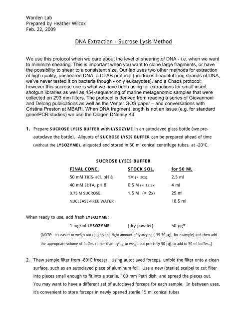
Worden Lab<br />
Prepared by Heather Wilcox<br />
Feb. 22, 2009<br />
<strong>DNA</strong> <strong>Extraction</strong> - <strong>Sucrose</strong> <strong>Lysis</strong> <strong>Method</strong><br />
We use this protocol when we care about the level of shearing of <strong>DNA</strong> - i.e. when we want<br />
to minimize shearing. This is important when you want to clone large fragments, or have<br />
the possibility to shear to a consistent size. Our lab uses two other methods for extraction<br />
of high quality, unsheared <strong>DNA</strong>, a CTAB protocol (produces beautiful long strands of <strong>DNA</strong>,<br />
we’ve never tested it on bacteria though - only eukaryotes), and a Chaos protocol;<br />
however this sucrose one is what we have been using for extractions for small insert<br />
shotgun libraries as well as 454-sequencing of marine metagenomic samples that were<br />
collected on 293 mm filters. The protocol is derived from reading a series of Giovannoni<br />
and Delong publications as well as the Venter GOS paper – and conversations with<br />
Cristina Preston at MBARI. When <strong>DNA</strong> fragment length is not an issue (e.g. for standard<br />
gene/PCR studies) we use the Qiagen DNeasy Kit.<br />
1. Prepare SUCROSE LYSIS BUFFER with LYSOZYME in an autoclaved glass bottle (we pre-<br />
autoclave the bottle). Aliquots of SUCROSE LYSIS BUFFER can be prepared ahead of time<br />
(without the LYSOZYME), aliquoted and stored in 50 ml conical centrifuge tubes, at -20°C.<br />
When ready to use, add fresh LYSOZYME:<br />
SUCROSE LYSIS BUFFER<br />
FINAL CONC. STOCK SOL. for 50 ML<br />
50 mM TRIS-HCl, pH 8 1M (= 20x) 2.5 ml<br />
40 mM EDTA, pH 8 0.5 M (= 12.5x) 4 ml<br />
0.75 M SUCROSE 1.5 M (= 2x) 25 ml<br />
NUCLEASE-FREE WATER 18.5 ml<br />
1 mg/ml LYSOZYME (dry powder) 50 μg*<br />
[NOTE: it’s easier to weigh out roughly the right amount of lysozyme ( 35-50 μg, for example) and then add<br />
the appropriate volume of buffer, rather than trying to weigh out precisely 50 μg to add to 50 ml buffer…]<br />
2. Thaw sample filter from -80°C freezer. Using autoclaved forceps, unfold the filter onto a clean<br />
surface, such as an autoclaved piece of aluminum foil. Use a new (sterile) scalpel to cut filter<br />
into pieces small enough to fit into a sterile, 100 mm Petri dish, and spread the pieces out.<br />
You may want to have a different set of autoclaved forceps for each sample. In between uses,<br />
it’s convenient to store forceps in newly opened sterile 15 ml conical tubes
3. Add 6 ml SUCROSE LYSIS BUFFER with LYSOZYME, making sure that all filter pieces are<br />
covered. If desired, set the 100 mm Petri dish inside a 150 mm Petri, to avoid setting dish on<br />
dirty surfaces. Keep left over buffer on ice for later use.<br />
4. Incubate Petri dish at 37°C for 60 min, gently shaking to mix every 10 min. Flip entire pile of<br />
filter with clean forceps at halfway point.<br />
5. When the hour has nearly elapsed, prepare 5X PROTEINASE K/SDS SOLUTION. For each 6<br />
ml sample, prepare 1.5 ml of:<br />
5X PROTEINASE K/SDS SOLUTION<br />
1x [final]* 5x [final]* STOCK for 1.5 ml of 5x<br />
PROTEINASE K 0.5 mg/ml 2.5 mg/ml 20 mg/ml 187.5 μl<br />
SDS 1% 5% 20% 375 μl<br />
BUFFER**<br />
+ 562.5 μl LYSIS<br />
* [final] refers to the concentration of reagents after mixing with the existing lysate<br />
** if available, use SUCROSE LYSIS BUFFER with LYSOZYME set aside earlier<br />
6. Mix the 5X PROTEINASE K/SDS SOLUTION well. Then, tilting each Petri dish, mix 1.5 ml 5X<br />
PROTEINASE K/SDS SOLUTION with the 6 ml buffer already present. When the solution is<br />
homogeneous, set the dishes flat again, and gently shake/rock each dish to ensure all filter<br />
pieces are covered.<br />
7. Incubate dishes at 55°C for (1 to) 2 hours, with mixing and filter flipping as before.<br />
8. Tilt Petri dish; use a serological pipette to gently collect as much lysate as possible. (For<br />
maximal lysate recovery, use forceps to push filter pieces into the barrel of a 60 ml syringe<br />
(without a needle), and use the plunger to squeeze out all liquid).<br />
This is Fraction A.<br />
NOTE: If <strong>DNA</strong> yield is not a concern, you may decide not to collect more fractions. In our<br />
experience, Fraction B typically yields about 25% the <strong>DNA</strong> of Fraction A. Collecting a<br />
Fraction C yields approximately 5% the <strong>DNA</strong> of Fraction A. Conceivably, Fractions B and C
could be enriched for organisms that are difficult to lyse. To date, we haven’t analyzed the<br />
differences.<br />
9. Set Fraction A aside on ice. Add lysate for Fraction B:<br />
5.25 ml SUCROSE BUFFER with LYSOZYME<br />
300 μl 20% SDS<br />
150 μl 20 mg/ml PROTEINASE K<br />
10. Incubate Fraction B at 55°C for one or more hour(s) with periodic mixing, while Fraction A is<br />
processed.<br />
11. Split Fraction A into 1.5 or 2.0 ml nuclease-free microfuge tubes, with a volume of lysate less<br />
than half the tube’s capacity. [NOTE: USE CAUTION WHEN WORKING WITH PHENOL AND<br />
CHLOROFORM; PERFORM ALL STEPS IN THE FUME HOOD, AND SEPARATE BOTH WET AND DRY<br />
WASTE AS HAZARDOUS MATERIALS.]<br />
12. Prepare EQUILIBRATED PHENOL:CHLOROFORM:ISOAMYL ALCOHOL in a ratio of 25:24:1.<br />
13. Add a volume of EQUILIBRATED PHENOL:CHLOROFORM:ISOAMYL ALCOHOL equal to the<br />
volume of lysate. Mix by gently inverting, or by twirling slowly on a rotisserie, end over end for<br />
5 min. Spin in microcentrifuge @ 12,000 x g for 10 min at 4°C. While spinning, label new<br />
tubes A1, B1, etc.<br />
14. Carefully transfer aqueous (top) phase to new tubes, using a P200 pipettor. Avoid the<br />
interface, but don’t worry too much about contamination here – the next steps will remove it.<br />
15. Repeat step 13, labeling new tubes A2, B2, C2, etc.<br />
16. Collect about 65-70% of aqueous phase (more conservative this time), and transfer into the<br />
new tubes (A2, B2, etc.). If doing back extractions, set these new tubes aside on ice -- you’ll<br />
add the back-extracted aqueous layers to these tubes.<br />
17. (optional: back extraction) To maximize both purity and yield, perform a back extraction.<br />
This is a way to ‘reclaim’ some of the <strong>DNA</strong> left behind. To the organic phase and interphase<br />
material in tubes A1, B1, etc, add a volume of TE (pH 8.0) or H20 equal to the volume in
each tube. Mix gently and spin, as in step 13. Conservatively collect aqueous phases, and add<br />
to the aqueous material in tubes A2, B2, etc<br />
18. To each of tubes A2, B2, etc, add a volume of CHLOROFORM:IAA (*without* PHENOL),<br />
and mix and centrifuge as before. Collect this fraction fairly conservatively, into tubes labeled<br />
A3, B3, etc. These samples can be left on ice while subsequent fractions are collected and<br />
processed.<br />
19. Prepare one or more AMICON-4 100,000 MWCO CENTRIFUGAL FILTER DEVICE<br />
(Millipore) for each sample. This molecular weight cut-off (MWCO) is appropriate for genomic<br />
<strong>DNA</strong>; other MWCOs may be better for smaller size fractions of material.<br />
20. To prepare filter devices, pre-rinse them by adding 4 ml TE (pH 8.0) or H20 to each sample<br />
reservoir, and spin at 3200 x g (4°C, 10 min) in the swinging bucket rotor of e.g. an<br />
Eppendorf 1510R or similar centrifuge. 3200 x g is the max speed for this rotor, but DON’T go<br />
over 2000 x g when spinning the genomic <strong>DNA</strong>. The pre-rinse solution should all go through<br />
into the lower chamber. If any wash solution remains in the upper chamber, extend the spin<br />
time for subsequent spins.<br />
21. Load up to 4 ml of your sample into the upper reservoir of your pre-rinsed column, and spin at<br />
2000 x g (4°C, 10 min). NOTE: After spinning your sample, there will still be liquid in the<br />
upper chamber -- this is the <strong>DNA</strong>-containing ‘retentate’, and the volume of retentate is<br />
proportional to the amount of <strong>DNA</strong> present. Remove the bottom reservoir of each filter, and<br />
either discard the filtrate (flow-through), or transfer it into clean 50 ml conical tubes, and set<br />
aside on ice.<br />
22. Repeat step 21 until all sample has been loaded. Add TE (pH 8.0) or H20 to sample if needed<br />
for a total volume of 4 ml.<br />
23. Wash the <strong>DNA</strong> in the column by adding TE (pH 8.0) or H20 to a total volume of 4 ml, spin;<br />
and discard filtrates as before.<br />
24. Repeat 4 ml washes until the volume of the retentate remains constant after consecutive runs.<br />
At this point, the retentate can be collected.
25. Collect retentate from filter column with a P200, using long, thin gel-loading tips, if available.<br />
To maximize recovery, you can add TE (pH 8.0) or H20 to the upper chamber, and carefully<br />
pipet it back out. You can also remove the basket-like filter from the column, invert it inside a<br />
50 ml conical tube, spin at 500 x g for 2 min, and collect any liquid that comes out.<br />
The <strong>DNA</strong> is now ready to use or store.<br />
26. Take a few microliters from each sample for quantitative and qualitative analysis. Aliquot the<br />
remaining material, and store in a -20°C or -80°C freezer until needed.<br />
NOTE: Nanodrop analysis of genomic <strong>DNA</strong>: because of the high MW of relatively<br />
unsheared genomic <strong>DNA</strong>, Nanodrop readings can be unreliable and variable. For<br />
accurate, reproducible measurements, take a 3-5μl aliquot, heat it to 65°C for 15 min<br />
(0.2 ml tubes in a PCR machine work well), then vortex on high speed for 30 sec.<br />
Make sure you’ve set aside separate aliquots for bioanalyzer or gel analysis!<br />
NOTE: Agarose gel analysis of genomic <strong>DNA</strong>: when run on a 1% agarose gel,<br />
with a HindIII-cut Lambda <strong>DNA</strong> ladder, minimally-sheared <strong>DNA</strong> will appear as a single<br />
band, right around the biggest ladder fragment, 21,226 kb.<br />
MATERIALS FOR <strong>DNA</strong> EXTRACTION<br />
BY SUCROSE LYSIS METHOD Worden lab<br />
REAGENT/PRODUCT Vendor CATALOG NO.<br />
TRIS-HCl, 1M, pH 8.0 AMBION AM9855G<br />
EDTA, pH 8.0, 0.5M AMBION AM9260G<br />
SUCROSE SIGMA S2395<br />
WATER, nuclease-free AMBION AM9937<br />
LYSOZYME CHLORIDE, grade VI SIGMA L2879<br />
PROTEINASE K QIAGEN 19131<br />
SDS, 20% solution AMBION AM9820<br />
TE, pH 8.0 (10mM Tris-HCl; 1mM EDTA) AMBION AM9858<br />
PHENOL solution, pH 8.0<br />
(with addition of included buffer)<br />
SIGMA P4557<br />
CHLOROFORM * SIGMA C2432<br />
ISOAMYL ALCOHOL (IAA) * (a.k.a. 3-methylbutanol) SIGMA I9392<br />
* you can also buy these last two components pre-mixed:<br />
CHLOROFORM:IAA (24:1) FLUKA (SIGMA) 25666<br />
FILTERED PIPETTOR TIPS;<br />
nuclease-free,<br />
aerosol resistant tips (ART)<br />
MOLECULAR<br />
BIOPRODUCTS<br />
p10: 2140-05<br />
p20: 2149P-05<br />
p200: 2069-05<br />
p1000: 2079E<br />
MICROCENTRIFUGE TUBES; 1.7 ml, nuclease-free AXYGEN MCT-175-C-S<br />
AMICON ULTRA-4 100,000 MWCO centrifugal filter devices<br />
(MWCO = molecular weight cut-off)<br />
MILLIPORE UFC810096