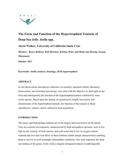
The Form and Function of the Hypertrophied Tentacle of Deep-Sea ...
The Form and Function of the Hypertrophied Tentacle of Deep-Sea ...
The Form and Function of the Hypertrophied Tentacle of Deep-Sea ...

Create successful ePaper yourself
Turn your PDF publications into a flip-book with our unique Google optimized e-Paper software.
<strong>The</strong> <strong>Form</strong> <strong>and</strong> <strong>Function</strong> <strong>of</strong> <strong>the</strong> <strong>Hypertrophied</strong> <strong>Tentacle</strong> <strong>of</strong><br />
<strong>Deep</strong>-<strong>Sea</strong> Jelly Atolla spp.<br />
Alexis Walker, University <strong>of</strong> California Santa Cruz<br />
Mentors: Bruce Robison, Rob Sherlock, Kristine Walz, <strong>and</strong> Henk-Jan Hoving, George<br />
Matsumoto<br />
Summer 2011<br />
Keywords: Atolla, tentacle, histology, SEM, hypertrophied<br />
ABSTRACT<br />
In situ observations <strong>and</strong> species collection via remotely operated vehicle, laboratory<br />
observations, <strong>and</strong> structural microscopy were used with <strong>the</strong> objective to shed light on <strong>the</strong><br />
form <strong>and</strong> subsequently <strong>the</strong> function <strong>of</strong> <strong>the</strong> hypertrophied tentacle exhibited by some<br />
Atolla species. Based upon <strong>the</strong> density <strong>of</strong> nematocysts, length, movement, <strong>and</strong><br />
ultrastructure <strong>of</strong> <strong>the</strong> hypertrophied tentacle, <strong>the</strong> function <strong>of</strong> <strong>the</strong> tentacle is likely<br />
reproductive, sensory, <strong>and</strong>/or utilized in food acquisition.<br />
INTRODUCTION<br />
<strong>The</strong> meso- <strong>and</strong> bathypelagic habitats are <strong>of</strong> <strong>the</strong> largest <strong>and</strong> least known on <strong>the</strong> planet.<br />
<strong>The</strong>y are extreme environments, characterized by high atmospheric pressure, zero to low<br />
light levels, scarcity <strong>of</strong> food sources, <strong>and</strong> cold water that is low in oxygen content.<br />
Animals that live <strong>and</strong> even thrive in <strong>the</strong>se habitats exhibit unique characteristics enabling<br />
<strong>the</strong>m to survive in such seemingly inhospitable conditions. One such organism, <strong>the</strong> deepsea<br />
medusa <strong>of</strong> <strong>the</strong> genus Atolla, trails a singular elongated tentacle, morphologically<br />
1
distinct from <strong>the</strong> marginal tentacles. This structure, <strong>of</strong>ten referred to as a trailing or<br />
hypertrophied tentacle, is unique within <strong>the</strong> cnidarian phylum.<br />
Ernst Haeckel described <strong>the</strong> first species <strong>of</strong> this deep pelagic jelly, Atolla wyvillei,<br />
during <strong>the</strong> 1872-1876 HMS Challenger Expedition. In <strong>the</strong> subsequent 135 years, <strong>the</strong><br />
genus Atolla has exp<strong>and</strong>ed to several species not yet genetically established, which have<br />
been observed in all <strong>of</strong> <strong>the</strong> worlds oceans (Russell 1970). Although <strong>the</strong> general<br />
morphology <strong>and</strong> external anatomy <strong>of</strong> Atolla, has been thoroughly described (Haeckel<br />
1881, Kramp, Russell 1959,1970), very little is known about <strong>the</strong> form <strong>and</strong> function <strong>of</strong> <strong>the</strong><br />
hypertrophied tentacle. This is due in part to <strong>the</strong> historical use <strong>of</strong> midwater trawling<br />
techniques employed to collect deep-sea organisms such as Atolla spp.<br />
Gelatinous organisms tend to be highly misrepresented when sampling with nets<br />
because s<strong>of</strong>t-bodied animals are easily damaged. But with <strong>the</strong> advent <strong>of</strong> remotely<br />
operated vehicles (ROVs), it is possible to observe <strong>and</strong> collect Atolla spp. in situ, without<br />
damaging <strong>the</strong> individual. One such observation was made by Hunt & Lindsay (1998),<br />
who witnessed an Atolla with its hypertrophied tentacle entwined with a siphonophore,<br />
Nanomia bijuga. <strong>The</strong> subsequent paper suggested that <strong>the</strong> hypertrophied tentacle is used<br />
in prey capture. However, it is unclear from this single observation, which animal was<br />
prey <strong>and</strong> <strong>the</strong> possibility exists that <strong>the</strong> two were simply entangled. <strong>The</strong> Hunt & Lindsay<br />
paper remains <strong>the</strong> only published observation <strong>of</strong> behavior concerning <strong>the</strong> hypertrophied<br />
tentacle <strong>of</strong> Atolla spp. Thus <strong>the</strong> purpose <strong>of</strong> this study was to fur<strong>the</strong>r investigate <strong>the</strong> form<br />
<strong>and</strong> potential function <strong>of</strong> <strong>the</strong> hypertrophied tentacle <strong>of</strong> <strong>the</strong> deep-sea jelly Atolla.<br />
Cnidarians are generally thought to be passive predators, feeding on prey that<br />
adhere to <strong>the</strong> tentacles where numerous nematocysts are deployed. Aside from aiding in<br />
prey capture, <strong>the</strong>se nematocyst-laden tentacles have a secondary function in defense.<br />
Provided that <strong>the</strong>se are typical functions <strong>of</strong> cnidarian tentacles, <strong>the</strong> hypertrophied tentacle<br />
<strong>of</strong> Atolla spp. may serve a similar function possibly as a lure, for prey specialization,<br />
<strong>and</strong>/or as <strong>the</strong> mechanism <strong>of</strong> food transport. Given <strong>the</strong> extreme habitat <strong>of</strong> Atolla spp. <strong>and</strong><br />
<strong>the</strong> uniqueness <strong>of</strong> <strong>the</strong> hypertrophied tentacle, it is reasonable to assume it is used in<br />
ano<strong>the</strong>r functional capacity. In an environment where it is difficult to find a mate, <strong>the</strong><br />
elongated tentacle could serve a reproductive function as an attachment point for<br />
potential mates or used in sperm transfer (Chad Widmer 2011 pers.comm.). Atolla spp.<br />
2
are dioecious <strong>and</strong> can be differentiated by <strong>the</strong> shape <strong>of</strong> <strong>the</strong> gonads <strong>and</strong> <strong>the</strong> presence or<br />
absence <strong>of</strong> eggs.<br />
<strong>The</strong>re is yet ano<strong>the</strong>r hypo<strong>the</strong>sis that is based upon <strong>the</strong> suggested function <strong>of</strong><br />
trailing filaments belonging to <strong>the</strong> squid Vampyroteuthis infernalis. Although this deepsea<br />
cephalopod has two trailing structures, <strong>the</strong>y are remarkably similar in appearance <strong>and</strong><br />
behavior to <strong>the</strong> hypertrophied tentacle <strong>of</strong> Atolla spp. Young (1967) <strong>and</strong> Dilly et. al.<br />
(2009) have suggested <strong>the</strong> trailing filaments <strong>of</strong> Vampyroteuthis, function both as feeding<br />
<strong>and</strong> sensory structures. <strong>The</strong> hypertrophied tentacle analogous to <strong>the</strong> trailing filaments,<br />
could serve a sensory function, detecting chemical cues from a mate or potential prey<br />
items. <strong>The</strong>refore, we investigated four possible functions <strong>of</strong> <strong>the</strong> hypertrophied tentacle <strong>of</strong><br />
Atolla spp.: reproduction, sensory, feeding, <strong>and</strong> defense.<br />
MATERIALS AND METHODS<br />
ROV COLLECTION<br />
30 Atolla specimens, 24 A. vanhoeffeni, 5 A. wyvillei, <strong>and</strong> 1 unidentified species, were<br />
collected at two deep-water sites <strong>of</strong>f Moss L<strong>and</strong>ing, CA in Monterey Bay during <strong>the</strong><br />
Midwater Ecology Expedition between Jun 13-19 th , 2011. <strong>The</strong> Midwater 1 (MW-1) <strong>and</strong><br />
Canyon Axis (3500m depth over axis <strong>of</strong> Monterey Submarine Canyon) sites are located<br />
approximately 12km <strong>and</strong> 100km <strong>of</strong>fshore respectively. Specimens were collected at<br />
depths between 406-2794m via <strong>the</strong> ROV Doc Ricketts, aboard <strong>the</strong> R/V Western Flyer,<br />
with low-impact suction <strong>and</strong> detritus samplers.<br />
VARS<br />
Behavioral, morphological, <strong>and</strong> distributional information on Atolla species were<br />
obtained through MBARI’s Video Annotation <strong>and</strong> Reference System (VARS) database.<br />
Behavioral observations, with respect to <strong>the</strong> marginal <strong>and</strong> hypertrophied tentacles,<br />
consisted <strong>of</strong> recording movement, orientation, reactions, <strong>and</strong> interactions within <strong>the</strong> water<br />
column. Morphological observations were based upon <strong>the</strong> presence <strong>and</strong> subsequent<br />
length <strong>of</strong> <strong>the</strong> hypertrophied tentacle. <strong>The</strong> approximate length <strong>of</strong> <strong>the</strong> elongated tentacle<br />
was determined using known bell diameter, from collected specimens, <strong>and</strong> <strong>the</strong><br />
3
corresponding video footage. Depth distribution was derived from VARS using<br />
observations made between 1989 –June 2011.<br />
LAB OBSERVATIONS<br />
Several types <strong>of</strong> lab observations were conducted during this study. <strong>The</strong>se observations<br />
included both live <strong>and</strong> preserved organisms. Specimens collected were measured,<br />
preserved in 5% formalin-seawater solution, or kept alive for fur<strong>the</strong>r observation <strong>and</strong><br />
experimentation. Lab observations were made aboard <strong>the</strong> R/V Western Flyer <strong>and</strong>, later,<br />
in <strong>the</strong> MBARI wetlab where surviving individuals were placed in three plankton kreisels.<br />
Those specimens that survived long enough <strong>and</strong> in good condition were placed in three<br />
separate kreisels. Kreisel I consisted <strong>of</strong> 3 Atolla wyvillei collected between 873-1582m,<br />
all female. Kreisel II contained 4 Atolla vanhoeffeni sampled from 499-533m, consisting<br />
<strong>of</strong> 3 gravid females <strong>and</strong> 1 male. Finally, in Kreisel III were 5 Atolla vanhoeffeni collected<br />
between 445-562m, consisting <strong>of</strong> 3 juveniles <strong>of</strong> unknown sex, 1 male, <strong>and</strong> 1 gravid<br />
female. Specimens were ultimately preserved in 5% formalin/ seawater solution.<br />
Daily qualitative observations were made <strong>of</strong> interactions between male <strong>and</strong><br />
female Atolla both on <strong>the</strong> research vessel as well as in kreisels II <strong>and</strong> III in <strong>the</strong> wetlab.<br />
Gonad state was monitored <strong>and</strong> any reproductive events noted.<br />
A total <strong>of</strong> 27 live Atolla spp. were involved in both passive <strong>and</strong> invasive feeding<br />
experiments. <strong>The</strong> passive experiments consisted <strong>of</strong> <strong>the</strong> addition <strong>of</strong> potential prey items to<br />
<strong>the</strong> Atolla kreisels without fur<strong>the</strong>r human involvement <strong>and</strong> included <strong>the</strong> feeding <strong>of</strong> frozen<br />
krill, live mysid shrimp, Artemia nauplii, Aurelia ephryrae, Aegina citrea, various<br />
copepods, unidentified polychaete, doliolids, ctenophores, chaetognaths, Cyclothone<br />
parts, <strong>and</strong> a marine snow simulation. Marine snow was obtained from <strong>the</strong> water collected<br />
with <strong>the</strong> Atolla from <strong>the</strong> ROV, <strong>and</strong> <strong>the</strong>n dispersed in kreisels I <strong>and</strong> II. <strong>The</strong> invasive<br />
feeding experiments involved <strong>the</strong> forced application <strong>of</strong> each prey organism previously<br />
mentioned, via forceps, to both <strong>the</strong> hypertrophied <strong>and</strong> marginal tentacles <strong>of</strong> <strong>the</strong> Atolla.<br />
Gut contents were also investigated to fur<strong>the</strong>r determine <strong>the</strong> diet <strong>of</strong> Atolla spp.<br />
<strong>and</strong> were analyzed only from individual specimens that were preserved immediately after<br />
collection aboard <strong>the</strong> Western Flyer. Experimentation testing <strong>the</strong> sensory hypo<strong>the</strong>sis<br />
consisted <strong>of</strong> mixing fluoresceine dye with three solutions: 1:1 Artemia-seawater , 1:1<br />
4
lended krill-seawater, <strong>and</strong> a seawater control. <strong>The</strong> solutions were added to <strong>the</strong> kreisels<br />
via pipette <strong>and</strong> responses were recorded.<br />
HISTOLOGY<br />
<strong>Tentacle</strong>s, both marginal <strong>and</strong> hypertrophied, were prepared for histological study. A<br />
distal, mid, <strong>and</strong> proximal segment <strong>of</strong> each tentacle was placed in 5% formalin/distilled<br />
seawater solution <strong>and</strong> sent to <strong>the</strong> histology lab at <strong>the</strong> Community Hospital <strong>of</strong> Monterey<br />
Peninsula (CHOMP). At CHOMP <strong>the</strong>y processed <strong>the</strong> tentacle samples from 5% <strong>Form</strong>alin<br />
solution through graded ethanol series to xylene, <strong>and</strong> finally paraffin wax. <strong>The</strong> sample<br />
was <strong>the</strong>n embedded in paraffin blocks <strong>and</strong> oriented for three longitudinal <strong>and</strong> three cross<br />
section cuts <strong>of</strong> each tentacle segment. Each section was <strong>the</strong>n stained with a st<strong>and</strong>ard<br />
Hematoxylin <strong>and</strong> Eosin stain (H&E), <strong>and</strong> mounted on labeled glass slides. <strong>The</strong>se slides<br />
were used to determine <strong>the</strong> presence or absences <strong>of</strong> nematocysts, cilia, along with o<strong>the</strong>r<br />
specialized ultrastructure <strong>and</strong> cells. Nematocysts densities for <strong>the</strong> marginal <strong>and</strong><br />
hypertrophied tentacles were also calculated using <strong>the</strong> histological slides. <strong>The</strong> area, mm 2 ,<br />
<strong>of</strong> each tentacle sample was determined under 10x objective on compound microscope<br />
using calibrated Infinity Analyze s<strong>of</strong>tware. <strong>The</strong>n unfired nematocysts, that were easily<br />
identifiable at 10x magnification, were counted for each tentacle segment <strong>and</strong> divided by<br />
<strong>the</strong> area <strong>of</strong> that segment. Nematocyst density was calculated for 3 individuals, 2 marginal<br />
<strong>and</strong> 1 hypertrophied tentacle segment for each individual, which were averaged for both<br />
tentacle types.<br />
SEM<br />
Marginal <strong>and</strong> hypertrophied tentacles were fixed in 2% Glutaraldeyhyde buffered<br />
solution <strong>and</strong> kept at room temperature for two hours. <strong>The</strong> samples were <strong>the</strong>n post-fixed<br />
for one hour in 1% osmium tetroxide. Each sample was <strong>the</strong>n rinsed three times each, for<br />
5 minutues, with DI water. Each sample was <strong>the</strong>n taken up to 100% ethanol through a<br />
series <strong>of</strong> graded steps, in increments <strong>of</strong> 10% ethanol, from a 10% ethanol/DI solution,<br />
remaining in each ethanol step for 15 minutes. After three washes at 100% ethanol for<br />
ano<strong>the</strong>r 15-minute interval, <strong>the</strong> chemical drying reagent HMDS was added to <strong>the</strong> ethanol<br />
mixture in 3:1 ethanolà HMDS ratio <strong>and</strong> taken up to 100% HMDS over one hour. <strong>The</strong><br />
5
samples were left for 16hrs under a chemical fume hood, mounted on stubs, grounded<br />
with gold spattering, <strong>and</strong> run through <strong>the</strong> scanning electron microscope (get model type).<br />
RESULTS<br />
LAB OBSERVATIONS / VARS<br />
<strong>The</strong> depth distribution for Atolla spp in Monterey Bay is 300 – 3000 m. Video footage<br />
over 71 dives <strong>and</strong> individuals, revealed that Atolla spp. displayed at least three general<br />
postures pertaining to <strong>the</strong> marginal tentacles: tucked aboral; where <strong>the</strong> base <strong>of</strong> <strong>the</strong><br />
tentacles are tucked against <strong>the</strong> exumbrellar bell, tucked oral; <strong>the</strong> tentacles are tucked<br />
tightly under <strong>the</strong> subumbrellar bell, <strong>and</strong> trailing oral; <strong>the</strong> tentacles are trailing unfurled<br />
behind <strong>the</strong> body.<br />
<strong>The</strong> presence <strong>of</strong> <strong>the</strong> hypertrophied tentacle was apparent over all video<br />
observations made <strong>of</strong> both A. vanhoeffeni <strong>and</strong> A. wyvillei <strong>and</strong> calculated to be 1.5- 36<br />
times <strong>the</strong> length <strong>of</strong> <strong>the</strong> bell diameter. Video also revealed that <strong>the</strong> hypertrophied tentacle<br />
is capable <strong>of</strong> retraction, coiling, <strong>and</strong> autotomy along <strong>the</strong> length <strong>of</strong> <strong>the</strong> tentacle (Fig.2).<br />
<strong>The</strong>se movements were later confirmed during lab observations, although it is important<br />
to note that it is not conclusive whe<strong>the</strong>r <strong>the</strong> tentacle is autotomizing or just detaching<br />
under strain.<br />
Two notable observations were made <strong>of</strong> female <strong>and</strong> male interactions. <strong>The</strong> first<br />
was aboard <strong>the</strong> Western Flyer involving one male <strong>and</strong> one female A.vanhoeffeni, with <strong>the</strong><br />
male attached to <strong>the</strong> female via hypertrophied tentacle. A whitish substance on <strong>the</strong><br />
tentacle appeared to move from <strong>the</strong> male to <strong>the</strong> female. Unfortunately we were unable to<br />
collect <strong>the</strong> substance. <strong>The</strong> second observation is really one that included several different<br />
days <strong>of</strong> observation <strong>of</strong> kreisel II (1 male <strong>and</strong> 3 females). On more than one occasion <strong>the</strong><br />
male A. vanhoeffeni was connected to a female via hypertrophied tentacle, however <strong>the</strong>re<br />
were no observations <strong>of</strong> female-female connections.<br />
<strong>The</strong> passive <strong>and</strong> invasive feeding experiments proved inconclusive for all types <strong>of</strong><br />
organisms used as prey. As indicated in Fig.3, <strong>the</strong>re were only 4 out <strong>of</strong> 12 prey items that<br />
were successfully adhesive amongst both <strong>the</strong> passive <strong>and</strong> invasive feeding methods.<br />
Aegina citrea <strong>and</strong> <strong>the</strong> Aurelia ephyrae adhered to <strong>the</strong> hypertrophied tentacle in <strong>the</strong><br />
6
invasive experiment only, but were not consumed <strong>and</strong> sloughed <strong>of</strong>f at a later point in<br />
time. <strong>The</strong> small, unidentified polychaete adhered to <strong>the</strong> marginal tentacles only during <strong>the</strong><br />
invasive feeding, <strong>and</strong> was again not consumed by <strong>the</strong> Atolla. <strong>The</strong> marine snow had <strong>the</strong><br />
highest success in adhesion to both <strong>the</strong> marginal <strong>and</strong> hypertrophied tentacles staying<br />
attached until removed or until death <strong>of</strong> <strong>the</strong> individual. None <strong>of</strong> <strong>the</strong> organisms fed to <strong>the</strong><br />
Atolla were observably consumed. As for gut contents, two <strong>of</strong> <strong>the</strong> specimens dissected<br />
for analysis yielded unidentified organisms, which were too deteriorated to determine.<br />
HISTOLOGY<br />
Nematocysts were found on both <strong>the</strong> hypertrophied <strong>and</strong> marginal tentacles <strong>of</strong> Atolla.<br />
Although, not yet identified, it is likely that <strong>the</strong>re are at least two or more different types<br />
<strong>of</strong> nematocysts present within Atolla tentacles. Marginal tentacles had significantly<br />
greater numbers <strong>of</strong> nematocysts than did hypertrophied tentacles, on average 226<br />
nematocysts/mm 2 <strong>and</strong> 42 nematocysts/ mm 2 respectively (Fig.5). Unidentified cells were<br />
also found within both types <strong>of</strong> tentacle. A prominent feature found at <strong>the</strong> base <strong>of</strong> <strong>the</strong><br />
hypertrophied tentacle, is a pronounced groove (Fig. 7)<br />
SEM<br />
<strong>The</strong> difference in ultrastructure between <strong>the</strong> hypertrophied tentacle <strong>and</strong> marginal tentacles<br />
can be observed in Fig. 6, which shows <strong>the</strong> distinct presence <strong>and</strong> absence <strong>of</strong> particular<br />
structures between tentacle types. Spherical structures, likely unfired nematocysts, that<br />
are prolific on <strong>the</strong> marginal tentacle, are sporadic on <strong>the</strong> hypertrophied tentacle.<br />
Conversely <strong>the</strong> hair-like structures, possible cilia, found covering <strong>the</strong> hypertrophied<br />
tentacle are sparse at best on <strong>the</strong> marginal tentacle. <strong>The</strong>se structures are yet to be<br />
identified. <strong>The</strong> SEM images <strong>of</strong> <strong>the</strong> base <strong>of</strong> <strong>the</strong> hypertrophied tentacle confirm <strong>the</strong> groovelike<br />
structure (Fig.7) observed on <strong>the</strong> histology slides<br />
DISCUSSION<br />
Thorough analyses <strong>of</strong> both <strong>the</strong> hypertrophied <strong>and</strong> marginal tentacles suggest that <strong>the</strong>y are<br />
functionally distinct, although it is important to note that <strong>the</strong>y may work in concert<br />
toward <strong>the</strong> same goal. Based upon <strong>the</strong> calculated nematocyst densities mentioned in<br />
7
Figure 5, it is likely that <strong>the</strong> marginal tentacles serve a defensive purpose, as well as aid<br />
in prey capture, as <strong>the</strong>y are equipped to immobilize potential predators <strong>and</strong> prey. <strong>The</strong><br />
hypertrophied tentacle, on <strong>the</strong> o<strong>the</strong>r h<strong>and</strong>, is almost certainly not used in a defensive<br />
capacity, as <strong>the</strong> nematocyst density was very low <strong>and</strong> sporadic. This does not rule out a<br />
potential function in feeding, however, as preliminary findings indicate a groove at <strong>the</strong><br />
base <strong>of</strong> <strong>the</strong> tentacle (Fig.7) <strong>and</strong> hair-like structures (Fig.6), which could aid in <strong>the</strong><br />
adhesion <strong>and</strong> transport <strong>of</strong> food materials to <strong>the</strong> manubrium. This, in conjunction with <strong>the</strong><br />
low nematocyst density, optimal placement <strong>of</strong> <strong>the</strong> hypertrophied tentacle, near <strong>the</strong><br />
opening <strong>of</strong> <strong>the</strong> manubrium, as well as <strong>the</strong> ability to coil/retract all suggest a potential for<br />
<strong>the</strong> tentacle to be utilized in food acquisition. Here, we use <strong>the</strong> term food acquisition<br />
instead <strong>of</strong> prey capture because <strong>the</strong> evidence suggests that, if used as a feeding structure,<br />
<strong>the</strong> tentacle specializes in <strong>the</strong> accumulation <strong>of</strong> prey that are relatively small, non-mobile,<br />
or slow-moving. Several video observations revealed that enough current <strong>and</strong> drag on <strong>the</strong><br />
hypertrophied tentacle resulted in detachment <strong>of</strong> <strong>the</strong> tentacle <strong>and</strong> thus loss <strong>of</strong> potential<br />
prey items, which as depicted in Figure 2e <strong>and</strong> 2f., could occur even with very small<br />
prey. <strong>The</strong>refore, it seems unlikely that Atolla would capture larger, highly mobile prey<br />
with <strong>the</strong> more delicate hypertrophied tentacle when it is armed with 17-36 thicker,<br />
nematocyst-laden marginal tentacles. If <strong>the</strong> hypertrophied tentacle is a feeding structure,<br />
we suspect that it is a transport mechanism for prey caught <strong>and</strong> paralyzed by <strong>the</strong> marginal<br />
tentacles as well as reserve food source akin to flypaper. As Atolla moves through <strong>the</strong><br />
water column with it’s hypertrophied tentacle extended, it accumulates small organisms<br />
<strong>and</strong> marine snow that adhere to it, providing <strong>the</strong> jelly with increased surface area <strong>and</strong> a<br />
supplementary diet, useful particularly if larger prey is scarce as is <strong>of</strong>ten <strong>the</strong> case in <strong>the</strong><br />
deep-sea (S<strong>and</strong>rini 1989, Sötje 2007, Robison 2010).<br />
In an immense, dark world where mates are difficult to come by, it may be that<br />
<strong>the</strong> hypertrophied tentacle “fishes” for <strong>and</strong> adheres to potential mates. Proximity during a<br />
spawning event would increase <strong>the</strong> odds <strong>of</strong> fertilization; particularly in <strong>the</strong> mesopelagic<br />
realm where Atolla live. Microscopy revealed a groove <strong>and</strong> hair-like structures on <strong>the</strong><br />
hypertrophied tentacle, which aside from food acquisition, could be utilized in<br />
reproduction. <strong>The</strong> placement <strong>of</strong> <strong>the</strong> hypertrophied tentacle is such that it lies not only<br />
near <strong>the</strong> manubrium, but close to <strong>the</strong> gonads <strong>and</strong> <strong>the</strong>refore may serve as a mechanism <strong>of</strong><br />
8
sperm transport as purposed by Chad Widmer (2011). <strong>The</strong> hypertrophied tentacle <strong>of</strong> a<br />
male Atolla could potentially attach near <strong>the</strong> gonads <strong>of</strong> a female to transfer sperm. Very<br />
little is known about <strong>the</strong> life history <strong>of</strong> Atolla, however scyphomedusae are generally<br />
accepted as broadcast spawners, thus making it remarkable if <strong>the</strong> hypertrophied tentacle<br />
is utilized in sperm transport, but that would require fur<strong>the</strong>r investigation.<br />
<strong>The</strong>re is <strong>the</strong> least amount <strong>of</strong> evidence available to support or refute <strong>the</strong> sensory<br />
hypo<strong>the</strong>sis, although <strong>the</strong> hair-like structures <strong>and</strong> unidentified cells on <strong>the</strong> hypertrophied<br />
tentacle may serve as a start for fur<strong>the</strong>r investigation. <strong>The</strong> hair-like structures could aid in<br />
chemical or mechanoreception as suggested for <strong>the</strong> trailing filaments <strong>of</strong> Vampyroteuthis<br />
aforementioned (Young 1969, Dilly 2009). A study conducted on <strong>the</strong> mesopelagic<br />
scyphomedusa, Mitrocoma cellularia revealed that <strong>the</strong>se jellies could sense <strong>the</strong> presence<br />
<strong>of</strong> prey via waterborne chemical signals <strong>and</strong> even pursue scent trails (Tamburri 2000). It<br />
is important to note, however, that <strong>the</strong> mechanism <strong>of</strong> chemoreception has not yet been<br />
identified. Being that <strong>the</strong> hypertrophied tentacle can extended over 30 times <strong>the</strong> length <strong>of</strong><br />
<strong>the</strong> bell diameter, it would make an ideal sensory structure with <strong>the</strong> ability to “taste”<br />
waters far from <strong>the</strong> organism itself in order to assess nearby locales for potential food,<br />
mates, or predators.<br />
CONCLUSIONS/RECOMMENDATIONS<br />
It is clear that <strong>the</strong> hypertrophied tentacle is a unique structure that is morphologically,<br />
behaviorally, <strong>and</strong> anatomically distinct from <strong>the</strong> marginal tentacles <strong>of</strong> Atolla spp. Where<br />
marginal tentacles are primarily for feeding <strong>and</strong> defense, <strong>the</strong> hypertrophied tentacle, may<br />
have multiple uses. Whe<strong>the</strong>r a primary or secondary function, it is likely that <strong>the</strong><br />
hypertrophied tentacle has some sensory capabilities to aid in ei<strong>the</strong>r food acquisition or<br />
reproduction. Fur<strong>the</strong>r in situ, morphological, behavioral, microscopic, experimental, <strong>and</strong><br />
gut content analysis is needed. During this investigation more questions were produced<br />
than answered. Aside from <strong>the</strong> mystery surrounding <strong>the</strong> form <strong>and</strong> function <strong>of</strong> <strong>the</strong><br />
hypertrophied tentacle, <strong>the</strong>re are many interesting questions we have about Atolla, such<br />
as genetic <strong>and</strong> morphological differences between species, patterns <strong>of</strong> bioluminescence,<br />
photosensitivity, <strong>and</strong> details <strong>of</strong> its life history that remain obscure. Thus, Atolla are an<br />
9
optimal study organism for future research. <strong>The</strong> deep- sea jelly Atolla however, remains,<br />
as many <strong>of</strong> <strong>the</strong>ir midwater counterparts, highly enigmatic.<br />
ACKNOWLEDGEMENTS<br />
A special thanks to <strong>the</strong> midwater ecology lab at MBARI: Bruce Robison, Rob Sherlock,<br />
Kris Walz, Henk-Jan Hoving, <strong>and</strong> Kim Reisenbichler. Thank you for giving me this<br />
incredible opportunity <strong>and</strong> helping me immensely, throughout this amazing process. Rob,<br />
you are an extraordinary mentor. I also want to give a huge thanks to Kurt Buck, Josi<br />
Taylor, <strong>and</strong> Sara Tanner for <strong>the</strong>ir help with processing my samples for SEM work. All <strong>of</strong><br />
who were incredibly generous with <strong>the</strong>ir time <strong>and</strong> effort. <strong>The</strong> VARS lab, Susan Von<br />
Thun <strong>and</strong> Kyra Schlining, were cornerstones <strong>of</strong> this process, as <strong>the</strong>y are <strong>the</strong> guardians <strong>of</strong><br />
all things digital <strong>and</strong> video related, thank you. And last, but definitely not least, my<br />
sincerest gratitude to Linda Kuhnz <strong>and</strong> George Matsumoto, who made being an intern<br />
worth while. I want to especially thank George for his constant efforts in assisting me<br />
with both <strong>the</strong> internship <strong>and</strong> with my project. Thank you.<br />
References<br />
Anderson P. A. V. & Schwab W. E. (1981) <strong>The</strong> organization <strong>and</strong> structure <strong>of</strong> nerve <strong>and</strong><br />
muscle in <strong>the</strong> jellyfish Cyanea capillata.J. Morphol. 170, 383–399.<br />
Blanquet, Richard S., Wetzel, Bruce. (1975) Surface ultrastructure <strong>of</strong> <strong>the</strong> schyphopolyp,<br />
Chrysaora quinquecirrha. <strong>The</strong> Biological Bulletin 148: 181-192.<br />
Chapman, D.M., Pantin, C.F.A. <strong>and</strong> Robson, E.A. (1962). Muscle in Coelenterates. Rev.<br />
Canad. Biol. 21: 267-278.<br />
Chapman, D.M. (1966). Evolution <strong>of</strong> <strong>the</strong> scyphistoma. Symp. Zool. Soc. Lond. 16:51-75.<br />
Chapman, D.M. (1968). A new type <strong>of</strong> muscle cell from <strong>the</strong> subumbrella <strong>of</strong> Obelia. J.<br />
Mar. Biol. Assoc. UK. 48: 667-688.<br />
Chapman, D.M. (1978). Microanatomy <strong>of</strong> <strong>the</strong> cubopolyp, Tripedalia cystophora (class<br />
Cubozoa). Helg. Wiss. Meeresunters. 31: 128-168.<br />
Chapman, D.M. (1999). Microanatomy <strong>of</strong> <strong>the</strong> bell rim <strong>of</strong> Aurelia aurita (Cnidaria:<br />
Scyphozoa). Can. J. Zool. 77: 34-46.<br />
10
Deopke, Hjike, Herrman, Karl, <strong>and</strong> Schuett, Christian. (2011). Endobacteria in <strong>the</strong><br />
tentacles <strong>of</strong> selected cnidarian species <strong>and</strong> in <strong>the</strong> cerata <strong>of</strong> <strong>the</strong>ir nudibranch predators.<br />
Helgol<strong>and</strong> Marine Reasearch. DOI: 10.1007/s10152-011-0245-4<br />
Dilly, D.N., Nixon, Marion, <strong>and</strong> Young J.Z. (2009). Mastigoteuthis–<strong>the</strong> whip-lash<br />
squid. Journal <strong>of</strong> Zoology. Vol 181: 4 1469-7998<br />
Glider, William V., Phipps, Donald W. Jr., Rosevelt, Pardy L. (1980). Localization <strong>of</strong><br />
Symbiotic Din<strong>of</strong>lagellate Cells within <strong>Tentacle</strong> Tissue <strong>of</strong> Aiptasia pallida (Coelenterata,<br />
Anthozoa). Transactions <strong>of</strong> <strong>the</strong> American Microscopical Society , Vol. 99, No. 4 (Oct.,<br />
1980), pp. 426-438<br />
Haeckel, E. (1881). Report on <strong>the</strong> deep-sea medusae dredged by H.M.S. Challenger,<br />
during <strong>the</strong> years 1873-1876.<br />
Hale, Garron (1999). <strong>The</strong> Classification <strong>and</strong> Distribution <strong>of</strong> <strong>the</strong> Class Scyphozoa.<br />
BI 375 - Biological Diversity. University <strong>of</strong> Oregon: 1-26.<br />
Kass-Simon, G., <strong>and</strong> Scappaticci, A.A. Jr. (2002). <strong>The</strong> behavioral <strong>and</strong> developmental<br />
physiology <strong>of</strong> nematocysts. Canadian Journal <strong>of</strong> Zoology, 80:(10) 1772-1794.<br />
Kramp.P.L. <strong>and</strong> Blanner.R.: 1972, 'Atollidae in <strong>the</strong> Zoological Museum <strong>of</strong> Copenhagen<br />
(Coelente-rata, Scyphomedusae)', Steenstrupia, 2, 157-165.<br />
Larson, R.J., Mills, C.E., <strong>and</strong> Harbison G.R. (1991). Western Atlantic midwater<br />
hydrozoan <strong>and</strong> scyphozoan medusae: in situ studies using manned submersibles.<br />
Hydrobiologia216/217: 311-317, 1991.<br />
Mauchline, J. <strong>and</strong> Harvey, P. F. (1983. <strong>The</strong> scyphomedusae <strong>of</strong> <strong>the</strong> Rockall Trough,<br />
Nor<strong>the</strong>astern Atlantic Ocean. J. Plankton Res., 5,881 –890.<br />
Mills, C.E., Boero, F., Migotto, A., <strong>and</strong> Gili, J.M. (2000). A guideline to nematocyst<br />
nomenclature <strong>and</strong> classification, <strong>and</strong> some notes on <strong>the</strong> systematic value <strong>of</strong> nematocysts.*<br />
SCIENTIA MARINA 64: (Supl. 1): 31-46.<br />
Osborn, D.A., Silver, M.W., Castro, C.G., Bros, S.M. Chavez, F.P. (2007). <strong>The</strong> habitat <strong>of</strong><br />
mesopelagic scyphomedusae in Monterey Bay, California. <strong>Deep</strong> <strong>Sea</strong> Research Part I:<br />
Oceanographic Research Papers Volume 54, Issue 8, 1241-1255.<br />
Robison, B.H., Sherlock, R.E, Reisenbichler, Kim (2010). <strong>The</strong> bathypelagic community<br />
<strong>of</strong> Monterey Canyon. <strong>Deep</strong> See Research Part II: Topical Studies in Oceanography. Vol.<br />
57, Issue 16: 1551-1556.<br />
Russell.F.S.: 1957, 'On a new species <strong>of</strong> scyphomedusa, Atolla vanhoeffeni n. sp.', J.<br />
Mar. Biol.Assoc. U.K., 36, 275-279. Russell.F.S.: 1958, 'A new species <strong>of</strong> Atolla',<br />
Nature, 181, 1811-1812.<br />
11
Russell.F.S.: 1959, 'Some observations on <strong>the</strong> scyphomedusa Atolla', J. Mar. Biol. Assoc.<br />
U.K., 38, 33-40.<br />
Russell, F.S. – 1970. <strong>The</strong> medusae <strong>of</strong> <strong>the</strong> British Isles, Vol. II, Pelagic Scyphozoa with a<br />
supplement to <strong>the</strong> first volume on hydromedusae. Cambridge University Press. London.<br />
S<strong>and</strong>rini, Rottini L. <strong>and</strong> Avian, M. (1989). Feeding mechanism <strong>of</strong> Pelagia noctiluca<br />
(Scyphozoa: Semaeostomeae); laboratory <strong>and</strong> open sea observations. Marine Biology<br />
102, 49-55.<br />
Seipel, Katja <strong>and</strong> Schmid Volker. (2006). Mesodermal anatomies in cnidarian polyps <strong>and</strong><br />
medusae. Int. J. Dev. Biol. 50: 589-599.<br />
Sötje I., Tiemann H., Båmstedt U.Trophic ecology <strong>and</strong> <strong>the</strong> related functional morphology<br />
<strong>of</strong> <strong>the</strong> deepwater medusa Periphylla periphylla (Scyphozoa, Coronata).Mar.<br />
Biol. 2007;150:329-343.<br />
Tamburri, M.N., M.N. Halt, <strong>and</strong> B.H. Robison (2000). Chemically regulated feeding by a<br />
midwater medusa. Limnology <strong>and</strong> Oceanography, 45: 1,661-1,666.<br />
Thomson, C.W. <strong>and</strong> J. Murray (eds.), Report on <strong>the</strong> scientific results <strong>of</strong> <strong>the</strong> voyage <strong>of</strong><br />
H.M.S. Challenger during <strong>the</strong> years 1873-1876. Zoology - Vol. IV, 2, pp. 1-154.<br />
Thuesen, Erik V. (2003) Crossota millsae (Cnidaria: Trachymedusae:<br />
Rhopalonematidae), a new species <strong>of</strong> viviparous hydromedusa from <strong>the</strong> deep sea <strong>of</strong>f<br />
California <strong>and</strong> Hawaii. Zootaxa 309: 1–12<br />
Widmer, Chad L (2008). How to keep jellyfish in aquariums : an introductory guide for<br />
maintaining healthy jellies. Tucson, AZ : Wheatmark, 2008. 192 p. : ill. ; 21 cm<br />
Yanagihara A.A, Kuroiwa J.M.Y, Oliver L.M, Kunkel D.D (2002) <strong>The</strong> ultrastructure <strong>of</strong><br />
nematocysts from <strong>the</strong> fishing tentacle <strong>of</strong> <strong>the</strong> Hawaiian bluebottle, Physalia<br />
utriculus (Cnidaria, Hydrozoa, Siphonophora). Hydrobiologia.489, 139–150.<br />
Young, R.E. 1967. Homology <strong>of</strong> retractile filaments <strong>of</strong> vampire squid. Science,<br />
156(3782):1633-1634.<br />
12
Fig.1. Sample Sites. This figure shows <strong>the</strong><br />
Midwater 1 <strong>and</strong> Canyon Axis sites, which<br />
were plotted based upon <strong>the</strong> GPS coordinates<br />
from <strong>the</strong> Midwater Research Expedition <strong>of</strong><br />
June 2011.<br />
Thursday, April 19, 2012
2a<br />
2b<br />
2c<br />
2d<br />
2e<br />
2f<br />
Fig.2. Movements <strong>of</strong> Marginal <strong>and</strong> <strong>Hypertrophied</strong> <strong>Tentacle</strong>s.<br />
Analysis <strong>of</strong> <strong>the</strong> video footage revealed three general postures with<br />
respect to <strong>the</strong> marginal tentacles: 2a. tucked aboral, 2b. tucked<br />
oral, <strong>and</strong> 2c trailing oral. <strong>The</strong> ability <strong>of</strong> <strong>the</strong> hypertrophied tentacle<br />
to coil <strong>and</strong> retract is depicted by pictures 2b <strong>and</strong> 2c. Two small<br />
organisms, a Solamaris medusa <strong>and</strong> a radiolarian (indicated by <strong>the</strong><br />
yellow circles), were observed stuck to <strong>the</strong> hypertrophied tentacle<br />
<strong>of</strong> <strong>the</strong> A.vanhoeffeni in picture 2e. While we were observing <strong>the</strong><br />
Atolla in 2e a current came through <strong>and</strong> <strong>the</strong> hypertrophied tentacle<br />
detached just above <strong>the</strong> Solamaris, <strong>the</strong> first organism circled<br />
above. <strong>The</strong> detached segment <strong>of</strong> <strong>the</strong> tentacle is pictured in 2f.<br />
Thursday, April 19, 2012
Prey Type<br />
(alive/dead)<br />
Passive Feeding<br />
Marginal <strong>Hypertrophied</strong><br />
Invasive Feeding<br />
Marginal <strong>Hypertrophied</strong><br />
Frozen Krill<br />
0<br />
0<br />
0<br />
0<br />
Mysid Shrimp<br />
0<br />
0<br />
0<br />
0<br />
Copepods<br />
0<br />
0<br />
0<br />
0<br />
Artemia nauplii<br />
/ /<br />
/ /<br />
Aurelia ephyrae<br />
0<br />
0<br />
0<br />
+<br />
Aegina citrea<br />
0 0<br />
0<br />
+<br />
Polychaetes<br />
0<br />
0<br />
+<br />
0<br />
Ctenophores<br />
0<br />
0<br />
0<br />
0<br />
Doliolids<br />
0<br />
0<br />
0<br />
0<br />
Chaetognath<br />
0<br />
0<br />
0<br />
0<br />
Cyclothone 0 0 0 0<br />
Marine Snow<br />
* * * *<br />
Fig.3. Passive <strong>and</strong> Invasive Feeding Experiments<br />
<strong>The</strong> table above shows which prey items successfully adhered to <strong>the</strong> marginal, hypertrophied, or<br />
both tentacles; none <strong>of</strong> <strong>the</strong> items were observably consumed. <strong>The</strong> 0 represents organisms that did<br />
not adhere to ei<strong>the</strong>r tentacle type. <strong>The</strong> / indicates that <strong>the</strong> adhesiveness or consumption <strong>of</strong><br />
organism is unknown. In this case <strong>the</strong> Artemia nauplii were too small to determine feeding success<br />
or even perform <strong>the</strong> invasive technique. <strong>The</strong> + represents organisms that attached to <strong>the</strong> tentacle<br />
but eventually detached without assistance, whereas <strong>the</strong> * indicated items that adhered <strong>and</strong><br />
remained on <strong>the</strong> tentacle until removal or death <strong>of</strong> <strong>the</strong> individual.<br />
Thursday, April 19, 2012
4a mt epm<br />
cn 4b 4c<br />
en<br />
300um<br />
4d mt<br />
epm<br />
cn<br />
me<br />
en<br />
4e<br />
300um<br />
Blank: Base MT<br />
300um<br />
Fig.4. Basic Mesodermal Anatomies <strong>of</strong> Atolla tentacles. <strong>The</strong> figure above is comparing <strong>the</strong><br />
general anatomies <strong>of</strong> <strong>the</strong> hypertrophied, a-c, to that <strong>of</strong> <strong>the</strong> marginal tentacle, d-f. Each column<br />
represents <strong>the</strong> corresponding segment for each tentacle, i.e. a <strong>and</strong> d are <strong>the</strong> distal, b <strong>and</strong> e <strong>the</strong><br />
mid, <strong>and</strong> c <strong>and</strong> e <strong>the</strong> proximal segments <strong>of</strong> both <strong>the</strong> hypertrophied <strong>and</strong> marginal tentacles from<br />
<strong>the</strong> same individual. <strong>The</strong> abbreviations are as follows: cn, cnidocytes; epm, epi<strong>the</strong>lial muscular<br />
cells; mt, muscle tissue; me, mesoglea; en, endo/gastrodermis.<br />
Thursday, April 19, 2012
en<br />
me<br />
cn epm mt<br />
5a<br />
300<br />
me<br />
epm mt<br />
cn<br />
en<br />
5b<br />
300<br />
<strong>Tentacle</strong> Type<br />
<strong>Hypertrophied</strong><br />
Marginal<br />
Nematocyst Density<br />
42 nem/mm 2<br />
226 nem/mm 2<br />
5c<br />
Fig. 5. Comparison <strong>of</strong> Atolla tentacles. <strong>The</strong><br />
photographs in this figure shows a longitudinal section<br />
<strong>of</strong> a distal segment <strong>of</strong> <strong>the</strong> (a) hypertrophied <strong>and</strong> (b)<br />
marginal tentacles at 20x magnification. Nematocyst<br />
densities (c) were calculated from similar photographs<br />
to be 42 nematocysts/mm 2 for <strong>the</strong> hypertrophied<br />
tentacles <strong>and</strong> 226 nematocysts/mm 2 for <strong>the</strong> marginal<br />
tentacles. See Fig.4. for abbreviations.<br />
Thursday, April 19, 2012
6a<br />
6c<br />
6b<br />
6e<br />
6c<br />
6f<br />
Fig. 6. SEM Ultrastructure <strong>of</strong> <strong>Hypertrophied</strong> <strong>and</strong> Marginal <strong>Tentacle</strong>. This figure shows<br />
both <strong>the</strong> ultrastructure <strong>of</strong> <strong>the</strong> marginal <strong>and</strong> hypertrophied tentacle, as well as a comparison<br />
between similar segments <strong>of</strong> tentacle types at <strong>the</strong> same magnification scale. Images 6a-6c are<br />
<strong>of</strong> <strong>the</strong> hypertrophied tentacle, while images 6d-6f are <strong>of</strong> <strong>the</strong> marginal tentacle. <strong>The</strong> marked<br />
differences between <strong>the</strong> two tentacles are: size, hypertrophied is smaller in width than <strong>the</strong><br />
marginal, hair-like structures blanketing <strong>the</strong> hypertrophied, <strong>and</strong> not <strong>the</strong> marginal, <strong>and</strong> <strong>the</strong><br />
appearance <strong>of</strong> obtuse spherical structures, likely nematocysts, in abundance on <strong>the</strong> marginal<br />
<strong>and</strong> not <strong>the</strong> hypertrophied tentacle.<br />
Thursday, April 19, 2012
7a<br />
7b<br />
Fig.7. A Groove in <strong>the</strong> <strong>Hypertrophied</strong> <strong>Tentacle</strong>. <strong>The</strong> cross section basal segment <strong>of</strong><br />
<strong>the</strong> hypertrophied tentacle, as shown in image 7a, has a very distinct invagination <strong>of</strong><br />
<strong>the</strong> epi- <strong>and</strong> endodermal tissues, indicating <strong>the</strong> presence <strong>of</strong> a groove. This is fur<strong>the</strong>r<br />
supported by <strong>the</strong> SEM image (7b) <strong>of</strong> <strong>the</strong> base <strong>of</strong> <strong>the</strong> hypertrophied tentacle on <strong>the</strong><br />
right.<br />
Thursday, April 19, 2012