Parasitic Copepods of Mackerel - and Tuna-like Fishes (Scombridae ...
Parasitic Copepods of Mackerel - and Tuna-like Fishes (Scombridae ...
Parasitic Copepods of Mackerel - and Tuna-like Fishes (Scombridae ...
You also want an ePaper? Increase the reach of your titles
YUMPU automatically turns print PDFs into web optimized ePapers that Google loves.
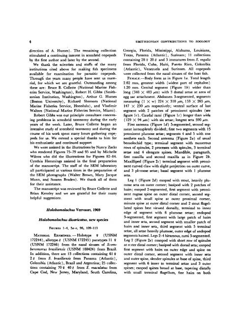
direction <strong>of</strong> A. Humes). The remaining collection<br />
stimulated a continuing interest in scombrid copepods<br />
by the first author <strong>and</strong> later by the second.<br />
We thank the scientists <strong>and</strong> staffs <strong>of</strong> the many<br />
institutions cited above for making fish specimens<br />
available for examination for parasitic copepods.<br />
Through the years many people have sent us material,<br />
for which we are grateful. Outst<strong>and</strong>ing among<br />
these are: Bruce B. Collette (National Marine Fisheries<br />
Service, Washington), Robert H. Gibbs (Smithsonian<br />
Institution, Washington), Arthur G. Humes<br />
(Boston University), Richard Shomora (National<br />
Marine Fisheries Service, Honolulu), <strong>and</strong> Vladimir<br />
Walters (National Marine Fisheries Service, Miami).<br />
Robert Gibbs was our principle consultant concerning<br />
problems in scombrid taxonomy during the early<br />
years <strong>of</strong> the work. Later, Bruce Collette began an<br />
intensive study <strong>of</strong> scombrid taxonomy <strong>and</strong> during the<br />
course <strong>of</strong> his work spent many hours gathering copepods<br />
for us. We extend a special thanks to him for<br />
his enthusiastic <strong>and</strong> continued support<br />
We were assisted in the illustrations by Nancy Zacks<br />
who rendered Figures 75-79 <strong>and</strong> 95 <strong>and</strong> by Michelle<br />
Wilcox who did the illustrations for Figures 82-84.<br />
Cynthia Hemmings assisted in the final preparation<br />
<strong>of</strong> the manuscript. The staff <strong>of</strong> the SEM laboratory<br />
all participated at various times in the preparation <strong>of</strong><br />
the SEM photographs (Walter Brown, Mary Jacque<br />
Mann, <strong>and</strong> Susann Braden). We thank all <strong>of</strong> them<br />
for their assistance.<br />
The manuscript was reviewed by Bruce Collette <strong>and</strong><br />
Brian Kensley <strong>and</strong> we are grateful for their many<br />
helpful suggestions.<br />
Holobomolochus Vervoort, 1969<br />
Holobomolochus divaricatus, new species<br />
FIGURES 1-4, ba-e, 96, 109-113<br />
MATERIAL EXAMINED. — Holotype 9 (USNM<br />
172244), allotype $ (USNM 172245) paratypes 11 9<br />
(USNM 172246) from the nasal sinuses <strong>of</strong> Scornberomorus<br />
brasiliensis (USNM 188424) from Brazil.<br />
In addition, there are 13 collections containing 61 $<br />
2$ from S. brasiliensis from Panama (Atlantic),<br />
Colombia (Atlantic), Brazil <strong>and</strong> Argentina; 25 collections<br />
containing 70 $ 40 $ from S. maculatus from<br />
Cape Cod, New Jersey, Maryl<strong>and</strong>, South Carolina,<br />
SMITHSONIAN CONTRIBUTIONS TO ZOOLOGY<br />
Georgia, Florida, Mississippi, Alabama, Louisiana,<br />
Texas, Panama (Atlantic), Surinam; 11 collections<br />
containing 28 9 20 $ <strong>and</strong> 3 immatures from S. regalis<br />
from Florida, Cuba, Haiti, Puerto Rico, Colombia<br />
(Atlantic), Venezuela <strong>and</strong> Surinam. All copepods<br />
were collected from the nasal sinuses <strong>of</strong> the host fish.<br />
FEMALE.—Body form as in Figure la. Total length<br />
2.62 mm, greatest width (widest part <strong>of</strong> cephalon)<br />
1.20 mm. Genital segment (Figure \b) wider than<br />
long (348 X 483 jxm) with 3 dorsal setae at area <strong>of</strong><br />
egg sac attachment. Abdomen 3-segmented, segments<br />
measuring (1 X w) 224 X 318 /*m, 153 X 265 ^m,<br />
147 X 230 /*m respectively; ventral surface <strong>of</strong> last<br />
segment with 2 patches <strong>of</strong> prominent spinules (see<br />
figure \c). Caudal rami (Figure \c) longer than wide<br />
(129 X 94 /*m) with six setae; longest seta 590 /*m.<br />
First antenna (Figure Id) 5-segmented, second segment<br />
incompletely divided; first two segments with 15<br />
prominent plumose setae; segments 4 <strong>and</strong> 5 with one<br />
aesthete each. Second antenna (Figure 2a) <strong>of</strong> usual<br />
bomolochid type; terminal segment with numerous<br />
rows <strong>of</strong> spinules, 2 processes with spinules, 3 terminal<br />
setae <strong>and</strong> 4 elongate spines. M<strong>and</strong>ible, paragnath,<br />
first maxilla <strong>and</strong> second maxilla as in Figure 2b.<br />
Maxilliped (Figure 2c) terminal segment with prominent<br />
curved claw with slight projection on outer curve,<br />
<strong>and</strong> 3 plumose setae; basal segment with 1 plumose<br />
seta.<br />
Leg 1 (Figure 2d) exopod with stout, heavily plumose<br />
seta on outer corner; basipod with 2 patches <strong>of</strong><br />
hairs; exopod 2-segmented, first segment with prominent<br />
rugose spine on outer distal corner, second segment<br />
with small spine at outer proximal comer,<br />
minute spine at outer distal corner <strong>and</strong> 2 stout flagellated<br />
spines best viewed dorsally, terminal to inner<br />
edge <strong>of</strong> segment with 6 plumose setae; endopod<br />
3-segmented, first segment with large patch <strong>of</strong> hairs<br />
<strong>and</strong> inner seta, second segment with smaller patch <strong>of</strong><br />
hairs <strong>and</strong> inner seta, third segment with 5 terminal<br />
setae, all setae heavily plumose, outer edge <strong>of</strong> endopod<br />
segments haired. Legs 2-4 biramous, rami 3-segmented.<br />
Leg 2 (Figure 2e) coxopod with short row <strong>of</strong> spinules<br />
at outer distal corner; basipod with dorsal seta; exopod<br />
first segment with hairs on outer edge <strong>and</strong> spine on<br />
outer distal corner, second segment with inner seta<br />
<strong>and</strong> outer spine, slender spinules at base <strong>of</strong> spine, third<br />
segment with 6 inner to terminal setae <strong>and</strong> 3 outer<br />
spines; exopod spines broad at base, tapering distally<br />
with small terminal flagellum, fine hairs on both


