Parasitic Copepods of Mackerel - and Tuna-like Fishes (Scombridae ...
Parasitic Copepods of Mackerel - and Tuna-like Fishes (Scombridae ...
Parasitic Copepods of Mackerel - and Tuna-like Fishes (Scombridae ...

Create successful ePaper yourself
Turn your PDF publications into a flip-book with our unique Google optimized e-Paper software.
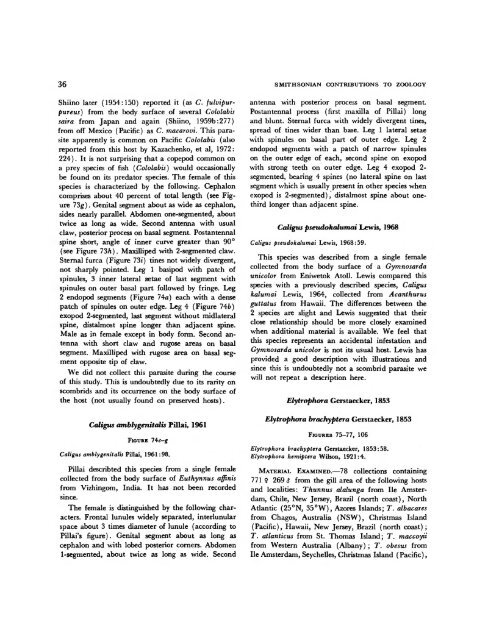
36 SMITHSONIAN CONTRIBUTIONS TO ZOOLOGY<br />
Shiino later (1954:150) reported it (as C. fulvipurpureus)<br />
from the body surface <strong>of</strong> several Cololabis<br />
saira from Japan <strong>and</strong> again (Shiino, 1959b:277)<br />
from <strong>of</strong>f Mexico (Pacific) as C. macarovi. This parasite<br />
apparently is common on Pacific Cololabis (also<br />
reported from this host by Kazachenko, et al, 1972:<br />
224). It is not surprising that a copepod common on<br />
a prey species <strong>of</strong> fish (Cololabis) would occasionally<br />
be found on its predator species. The female <strong>of</strong> this<br />
species is characterized by the following. Cephalon<br />
comprises about 40 percent <strong>of</strong> total length (see Figure<br />
73g). Genital segment about as wide as cephalon,<br />
sides nearly parallel. Abdomen one-segmented, about<br />
twice as long as wide. Second antenna with usual<br />
claw, posterior process on basal segment. Postantennal<br />
spine short, angle <strong>of</strong> inner curve greater than 90°<br />
(see Figure 73h). Maxilliped with 2-segmented claw.<br />
Sternal furca (Figure 73*) tines not widely divergent,<br />
not sharply pointed. Leg 1 basipod with patch <strong>of</strong><br />
spinules, 3 inner lateral setae <strong>of</strong> last segment with<br />
spinules on outer basal part followed by fringe. Leg<br />
2 endopod segments (Figure 74a) each with a dense<br />
patch <strong>of</strong> spinules on outer edge. Leg 4 (Figure 74fc)<br />
exopod 2-segmented, last segment without midlateral<br />
spine, distalmost spine longer than adjacent spine.<br />
Male as in female except in body form. Second antenna<br />
with short claw <strong>and</strong> rugose areas on basal<br />
segment. Maxilliped with rugose area on basal segment<br />
opposite tip <strong>of</strong> claw.<br />
We did not collect this parasite during the course<br />
<strong>of</strong> this study. This is undoubtedly due to its rarity on<br />
scombrids <strong>and</strong> its occurrence on the body surface <strong>of</strong><br />
the host (not usually found on preserved hosts).<br />
Caligns amblygenitalis Pill a i, 1961<br />
FIGURE l\c-g<br />
Caligns amblygenitalis Pillai, 1961:98.<br />
Pillai describted this species from a single female<br />
collected from the body surface <strong>of</strong> Euthynnus affinis<br />
from Vizhingom, India. It has not been recorded<br />
since.<br />
The female is distinguished by the following characters.<br />
Frontal lunules widely separated, interlunular<br />
space about 3 times diameter <strong>of</strong> lunule (according to<br />
Pillai's figure). Genital segment about as long as<br />
cephalon <strong>and</strong> with lobed posterior corners. Abdomen<br />
1-segmented, about twice as long as wide. Second<br />
antenna with posterior process on basal segment.<br />
Postantennal process (first maxilla <strong>of</strong> Pillai) long<br />
<strong>and</strong> blunt. Sternal furca with widely divergent tines,<br />
spread <strong>of</strong> tines wider than base. Leg 1 lateral setae<br />
with spinules on basal part <strong>of</strong> outer edge. Leg 2<br />
endopod segments with a patch <strong>of</strong> narrow spinules<br />
on the outer edge <strong>of</strong> each, second spine on exopod<br />
with strong teeth on outer edge. Leg 4 exopod 2segmented,<br />
bearing 4 spines (no lateral spine on last<br />
segment which is usually present in other species when<br />
exopod is 2-segmented), distalmost spine about onethird<br />
longer than adjacent spine.<br />
Caligus pseudokalumai Lewis, 1968<br />
Caligns pseudokalumai Lewis, 1968:59.<br />
This species was described from a single female<br />
collected from the body surface <strong>of</strong> a Gymnosarda<br />
unicolor from Eniwetok Atoll. Lewis compared this<br />
species with a previously described species, Caligus<br />
kalumai Lewis, 1964, collected from Acanthurus<br />
guttatus from Hawaii. The differences between the<br />
2 species are slight <strong>and</strong> Lewis suggested that their<br />
close relationship should be more closely examined<br />
when additional material is available. We feel that<br />
this species represents an accidental infestation <strong>and</strong><br />
Gymnosarda unicolor is not its usual host. Lewis has<br />
provided a good description with illustrations <strong>and</strong><br />
since this is undoubtedly not a scombrid parasite we<br />
will not repeat a description here.<br />
Elytrophora Gerstaecker, 1853<br />
Elytrophora brachyptera Gerstaecker, 1853<br />
FIGURES 75-77, 106<br />
Elytrophora brachyptera Gerstaecker, 1853:58.<br />
Elytrophora hemiptera Wilson, 1921:4.<br />
MATERIAL EXAMINED.—78 collections containing<br />
771 9 269 $ from the gill area <strong>of</strong> the following hosts<br />
<strong>and</strong> localities: Thunnus alalunga from He Amsterdam,<br />
Chile, New Jersey, Brazil (north coast), North<br />
Atlantic (25°N, 35°W), Azores Isl<strong>and</strong>s; T. albacares<br />
from Chagos, Australia (NSW), Christmas Isl<strong>and</strong><br />
(Pacific), Hawaii, New Jersey, Brazil (north coast) ;<br />
T. atlanticus from St. Thomas Isl<strong>and</strong>; T. maccoyii<br />
from Western Australia (Albany) ; T. obesus from<br />
He Amsterdam, Seychelles, Christmas Isl<strong>and</strong> (Pacific),