

1983 - Mycological Society of America
1983 - Mycological Society of America
1983 - Mycological Society of America

You also want an ePaper? Increase the reach of your titles
YUMPU automatically turns print PDFs into web optimized ePapers that Google loves.
<strong>Mycological</strong> <strong>Society</strong><br />
<strong>of</strong> <strong>America</strong><br />
NEWSLETTER<br />
Vol. 34 No. 1 June <strong>1983</strong>
ABBOTT LABORATORIES<br />
ANALYTAB PRODUCTS<br />
AYERST RESEARCH LABORATORIES<br />
BURROUGHS WELLCOME COMPANY<br />
CAROLINA BIOLOGICAL SWPPLY COMPANY<br />
DEKALB AGRESEARCH, INC.<br />
DI FCO LABORATORY PRODUCTS<br />
HOECHST-ROUSSEL PHARMACEUTICALS, INC.<br />
LANE SCIENCE EQUIPMENT COMPANY<br />
ELI LILLY AND COMPANY<br />
SUSTAINING MEMBERS<br />
MERCK SHARP AND DOHME RESEARCH LABORATORIES<br />
MILES LABORATORIES, INC.<br />
NEW BRUNSWICK SCIENTIFIC COMPANY<br />
PELCO<br />
CHARLES PFIZER AND COMPANY<br />
PIONEER HI-BRED INTERNATIONAL, INC.<br />
THE QUAKER OATS COMPANY<br />
ROHM AND HAAS COMPANY<br />
SCHIERING CORPORATION<br />
SPRINGER-VERLAG NEW YORK, INC.<br />
TRIARCH INCORPORATED<br />
WYETH LABORATORIES<br />
The <strong>Society</strong> is extremely grateful for the support <strong>of</strong> its Sustaining Mernbers.<br />
These organizations are 1 isted above in alphabetical order. Patronize them and let<br />
their representatives know <strong>of</strong> our appreciation whenever possible.<br />
OFFICERS OF THE MYCOLOGICAL SOCIETY OF AMERICA<br />
Harry D. Thiers, President Ian K. Ross, Councilor (1980-83)<br />
Henry C. Aldrich, Vice-president Walter J. Sundberg, Councilor (1980-83)<br />
Richard T. Hanl i n ,President-elect Joseph F. Ammi rati , Counci 1 or (1981-83)<br />
Roger Goos , Sec. -Treasurer 0' Nei 1 R. Col 1 ins, Counci 1 or ( 1980-83)<br />
Margaret Barr Bigel ow, Past Pres. (1982) Dona1 d T. W i ckl ow, Counci 1 o r (1980-83)<br />
Marie L. Farr, Past Pres. (1981) Donald J. S. Barr, Councilor (1981-84)<br />
F. A. Uecker, Councilor (<strong>1983</strong>-86)
MYCOLOGICAL SOCIETY OF AMERICA NEWSLETTER<br />
Sus t ai ni ng Members ....... i<br />
Officers <strong>of</strong> the MSA ....... i<br />
Table<strong>of</strong> Contents ........ 1<br />
Editor's Note .......... 1<br />
General Announcements ...... 2<br />
Calendar <strong>of</strong> Meetings and Forays . 3<br />
Forthcoming Courses ....... 4<br />
New <strong>Mycological</strong> Research .... 5<br />
Fungi for Distribution ..... 6<br />
Fungi Wanted .......... 7<br />
Identifications. ........ 9<br />
New Books by MSA Members .... 10<br />
Publications Available ..... 10<br />
Annual Meeting Program ..... 12<br />
Volume 34, No. 1 , June <strong>1983</strong><br />
Walter J. Sun4berg, Editor<br />
Department <strong>of</strong> Botany<br />
Southern I1 1 inois University<br />
(618) 536-2331<br />
TABLE OF CONTENTS<br />
Annual Meeting Abstracts ......... 12<br />
Pub1 icati ons Wanted ............ 41<br />
Positions Wanted ............. 42<br />
Vacancies for Mycologists ......... 42<br />
Postdoctoral Positions Available ..... 43<br />
Assistantships and Fellowships Available . 43<br />
Changes in Affiliation or Status ..... 44<br />
Travels and Visits ............ 45<br />
Papers, Seminars, Symposia, and Workshops . 46<br />
Honors, Awards, and Promotions ...... 48<br />
Personal News ............... 49<br />
News and Comments ............. 50<br />
Associations and Clubs .......... 52<br />
Affiliated Societies ........... 53<br />
One <strong>of</strong> the departures from MSA "routine", necessitated by this year's joint meeting<br />
<strong>of</strong> the MSA with the <strong>America</strong>n Phytopathological <strong>Society</strong> and the <strong>Society</strong> <strong>of</strong> Nematologists,<br />
was the printing and the distribution to all those attending <strong>of</strong> the abstracts <strong>of</strong> papers<br />
presented. Because <strong>of</strong> their potenti a1 value to the entire membership, the abstracts<br />
(from the MSA program) are included in this issue along with the usual news. Their lat-<br />
eral layout herein represents a one-time departure from the normal format and was neces-<br />
sitated by the special abstract size required for printing in the Official Meeting Publi-<br />
cation.<br />
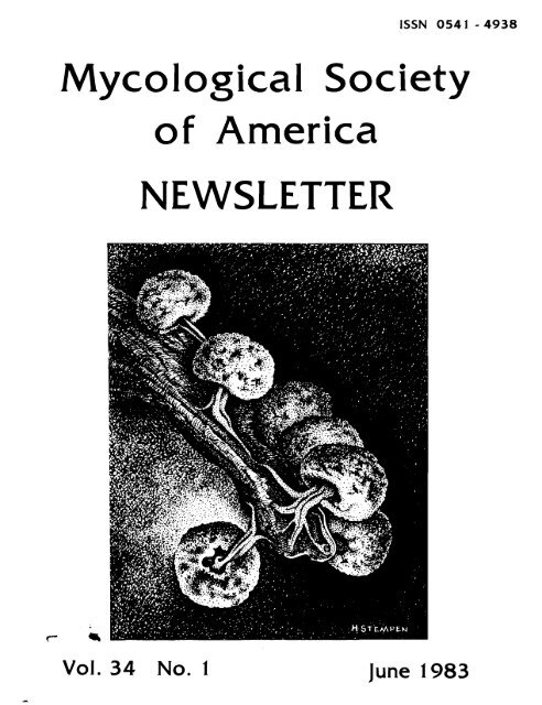
All <strong>of</strong> the illustrations included on these pages are <strong>of</strong> heret<strong>of</strong>ore unpublished, original<br />
art. The cover figure--Did mium nigripes--is the work <strong>of</strong> Henry Stempen (Biology<br />
Department, Rutgers Universi t y h y a1 so suppl ied the drawings <strong>of</strong> the "Fol iose Lichen<br />
Thallus On Pine Branch" (page 6) and Crucibulum laeve (page 49). The Astraeus hygrometricus,<br />
pictured on page 8, was done by Richard M. Hannan (Regional Plant Introduction<br />
Station, USDA, Washington State University) . Frank DiCosmo (Department <strong>of</strong> Botany, Uni -<br />
versi ty <strong>of</strong> British Columbia) submitted the il lustrations <strong>of</strong> Phacidium coniferarum on<br />
page 53.<br />
As evident on page 47, this issue marks the first to include advertising (see General<br />
Announcements for information).<br />
Finally, this Newsletter issue represents the first produced from the SIU mycologi-<br />
cal laboratory--wi th the he1 p (and patience!) <strong>of</strong> Shelly Briles and Jan Sundberg. Any<br />
errors or omissions, <strong>of</strong> course, are mine. My "initiation" into the editorship was made<br />
much easier by the efficient and detailed organization <strong>of</strong> the previous editors and, most<br />
<strong>of</strong> a1 1, by their will ingness to share their organizational strategy, information, and<br />
insights. The job done by Don Pfister, Geraldine Kaye, and their associates at the Far-<br />
low Herbarium over the past several years will indeed be a hard act to followl Herewith<br />
is my attempt.
GENERAL ANNOUNCEMENTS<br />
"Miss Edith Cash, now 92 years, sends her greetings to MSA members, and feels privi-<br />
ledged to have worked with several <strong>of</strong> us. She announces that she can no longer undertake<br />
translation <strong>of</strong> fungus diagnoses into Latin."<br />
STLINTZ MEMORIAL SCHOLARSHIP<br />
A Daniel Elliot Stuntz Memorial Scholarship Fund is being established to continue<br />
developing the area <strong>of</strong> mycology to which Pr<strong>of</strong>essor Stuntz contributed most <strong>of</strong> his life.<br />
It will support graduate students studying the systematics <strong>of</strong> fungi. For further informa-<br />
ti on, contact Joseph Ammi rati .<br />
AMERICAN MEN AND WOMEN OF SCIENCE<br />
The database for <strong>America</strong>n Men and Women <strong>of</strong> Science is currently being enlarged by the<br />
addition <strong>of</strong> new entrants who have the education and training equivalent to the doctorate<br />
and who have attained a position <strong>of</strong> responsibility in the sciences. Prospective entrants<br />
may request information or a questionnaire on which to submit information from The<br />
Editors, Jaques Cattell Press, P.O. Box 25001 , Tempe, AZ 85282.<br />
A NEW JOURNAL<br />
The new journal, Acta Mycologica Sinica, now in Vol. 2 and with abstracts in English,<br />
is now available from Science Press, 137 Chao-yang-men-nei Avenue, Bei jing, China.<br />
FORAY REPORTS WANTED<br />
Lists <strong>of</strong> fungi collected on MSA Forays in Arizona (1980), Indiana (1981), and Penn-<br />
sylvania (1982) will be appreciated by Wm. Bridge Cooke.<br />
GET YOUR ART IN PRINT<br />
W. J. Sundberg hereby requests submission <strong>of</strong> unpublished original mycological art<br />
work, especially for the Newletter cover and unpublished <strong>Mycological</strong> cartoons and humor<br />
to be used in the MSA Newsletter when feasible and as space permits. For art work, inked,<br />
"copy-ready" black and white illustrations are required. A1 1 original materials can be<br />
returned to the artist or author.<br />
NEWSLETTER ADVERTISING<br />
With the approval <strong>of</strong> the MSA Council (August 8, 1982) and starting with Volume 34,<br />
June <strong>1983</strong>, the MSA Newsletter will accept advertising (at the discretion <strong>of</strong> the editor)<br />
from commercial firms and individuals in private business. Advertisements may be full -<br />
page, half, or quarter-page size and must be prepared in photo-ready copy. He1 p support<br />
and defray the cost <strong>of</strong> the Newsletter by encouraging its use as an advertising vehicle<br />
for appropriate mycological, botanical , o r other biological materials. Contact W. J.<br />
Sundberg for detai 1 s.<br />
DON PFISTER HAS COPIES OF D. P. ROGERS' "A BRIEF HISTORY OF MYCOLOGY IN NORTH AMERICA"
July <strong>1983</strong><br />
-<br />
CALENDAR OF MEETINGS AND FORAYS<br />
16-17 The OHIO MYCOLOGICAL SOCIETY (OMS) SLIMMER FORAY, wi 11 be held at Penitentiary Glenn<br />
(NE Ohio). For details write to OMS/Walt Sturgeon, 288 E. North Ave., East<br />
Palestine, OH 44413.<br />
22-24 The THIRD WILD MUSHROOM FAIR at Museo Regional de Guadalajara Liceo e Hildalgo in<br />
Guadalajara City , Mexico. Contact F. Trujil lo-Flores, Facul tad de Ciencias,<br />
University <strong>of</strong> Guadalajara, Guadalajara, Jal isco, Mexico.<br />
August <strong>1983</strong><br />
7-12 The THIRD INTERNATIONAL SYMPOSIUM ON MYCROBIAL ECOLOGY will be held at Michigan<br />
State University. Program information and registration forms can be obtained<br />
from the Kel logg Center for Continuing Education, Michigan State University, East<br />
Lansing, MI 48824, USA or by telephoning (517) 355-4540.<br />
11-14 The University <strong>of</strong> Maine in Orono, Maine will be the site <strong>of</strong> the NORTHEAST MYCOLOGICAL<br />
FORAY. Get more data frorn Rokrt H. Peabody, RD $4, Box 281, Easton, PA 18342.<br />
25-28 The TELLURIDE MUSHROOM CONFERENCE, Tell uri de, Colorado, is for persons interested<br />
in mushroom identification and cultivation. For more information contact Emanuel<br />
Salzman, M. D., Box 5503, Denver, C3 80215-5503.<br />
28-S3 The THIRD INTERNATIONAL MYCOLOGICAL C3NGRESS will be held in Tokyo, Japan. For<br />
information write Secretariat: Pr<strong>of</strong>. K. Tubaki ; c/o International Congress Service,<br />
Inc. ; Chi kusen-Bldg. 2-7-4, Ni hombashi, Chuo-ku; Tokyo, Japan.<br />
Seotember <strong>1983</strong><br />
5-6 The FIRST I NTERNATIONAL SYMPOSIUM ON CHEMOBIODYNAMICS , entitled " Fi 1 amentous<br />
Mi crourani sms--Current Topics <strong>of</strong> Infection, Toxi cosi s and Control " is to be he1 d<br />
at Chi ba City, Japan. For details write (by Air Mail ) Pr<strong>of</strong>. Tetsuro Kuga; Gen.<br />
Secretary; 1 st Symposium on Chemobiodynamics; Research Institute for Chemobiody-<br />
namics; Chi ba University; 1-8-1, Inohana, Chi ba; 280 Japan.<br />
8-11 The ALEXANDER H. SMITH LAKE STATES FORAY for <strong>1983</strong> will be held at Southern Illinois<br />
University's outdoor laboratory and vicinity. Attendance is 1 imited and the cost<br />
is approximately $50.00. Contact W. J. Sundberg for further information.<br />
9-11 A FLESHY FUNGI WORKSHOP is being <strong>of</strong>fered by Walt Sturgeon at Terra Alta Mountain<br />
Camp in West Virginia. Fee: $40.00. Write to the Oglebay I~stitute, Brooks<br />
Nature Center, Oglebay Park, Wheeling, WV 26003 or call the Nature Center at<br />
(304) 242-6855.<br />
16-18 The 30th annual C. h. PECK MYCOLOGICAL FORAY will be held at the Edmund Niles Huyck<br />
Preserve in Rensselaervil le, New York. According to John Haines, "Rensselaervil le<br />
is the nost beautiful village in New York." Cost (sans meals) is $25.00 or free if<br />
youare a member <strong>of</strong> the Huyck Preserve Foundation ($10.00 membership fee by Sept.7).<br />
For information, membership blanks, etc., write John H. Haines, Rm. 3132 CEC, Plew<br />
York State Museum, A1 bany, NY 12230.<br />
October <strong>1983</strong><br />
2-23 A MUSHROOM STUDY TOUR, held in the People's Republic <strong>of</strong> China, is for persons<br />
interested in Oriental mushrooms and the use <strong>of</strong> fungi in traditional Chinese
4<br />
medicine. Contact Emanuel Sal zman, M. E. : Box 5503, Denver, CO 8021 5-5503.<br />
8-9 The OHIO MYCOLOGICAL SOCIETY FALL FORAY will be held in Hocking County, OH (SE<br />
OH). Write to Walt Sturgeon (see July 16-17 above) for details.<br />
May 1985<br />
19-24 The meeting <strong>of</strong> the I X COIVGRESS OF THE INTERNATIONAL SOCIETY FOR HUMAN AND ANIMAL<br />
MYCOLOGY will be held in Atlanta, Georgia. Direct inquiries to Dr. Libero Ajello,<br />
Division <strong>of</strong> Mycotic Diseases, Center for Disease Control , Atlanta, GA 30333.<br />
FORTHCOMING COURSES<br />
Through k'estern Washington University's suinmer session (July 29-August 19, <strong>1983</strong>),<br />
SEMINAR IN BIOLOGY--SURVEY OF ALPINE FUNGI AND LICHENS (Biology 417c, 3 credits) wi 11 be<br />
<strong>of</strong>fered. Contact Fred Rhoades , Biology Dept. , Western Washington University , Be1 1 ingham,<br />
WA 98225.<br />
A class in FIELD MYCOLOGY will be given in the Adirondacks via Cortland College, New<br />
York. Obtain further details from Timothy Baroni, Dept. <strong>of</strong> Biologjcal Sciences, P.O. Box<br />
2000, Cortl and Coll ege-SUNY, Cortl and, NY 13045.<br />
BASIC MUShKuOY IDENTIFICATION I--MACRO and MUSHROOM IDENTIFICATION I I--MICRO wi 11 be<br />
two courses giver1 at Cispus Environmental Center near Mt. St. Helens on June 24-26, <strong>1983</strong>.<br />
For more information contact Dr. David R. Hosford, Dept. <strong>of</strong> Biological Sciences, Central<br />
Washington University, El lensburg, WA 95926.<br />
A class entitled ECOLOGY OF THE FUNGI will be given at INIREB at Xalapa, Veracruz,<br />
Mexic/o from September-December, <strong>1983</strong> in the Master Degree curri cul um. Contact Dr. Gaston<br />
Guzman, INIREB, Apartado Postal 63, Xal apa, Veracruz, Mexico 91000.<br />
FLESHY FUNGI OF THE SIERRA wi 11 be <strong>of</strong>fered at the San Francisco State University<br />
Sierra, Nevada Field Camp June 13-17, <strong>1983</strong>. Contact Harry D. Thiers, Dept. <strong>of</strong> Bioiogical<br />
Sciences, San Francisco State University, San Francisco, CA 94132.<br />
In the Fa1 1, <strong>1983</strong>, IIVTRODUCTORY MYCOLOGY will be given at Auburn University. Contact<br />
Gareth Morgan-Jones, Dept. <strong>of</strong> Botany & Microbiology, Auburn University, Auburn, AL 36830.<br />
Instruction on the LABORATORY AND CLINICAL DIAGNOSES OF HUMAN AND ANIMAL MYCOSES will<br />
be presented on July 6-29, <strong>1983</strong>. For information, write Dr. Norman L. Goodman, Dept. <strong>of</strong><br />
Pathology, University <strong>of</strong> Kentucky, College <strong>of</strong> Medicine, Lexington, KY 40536.<br />
Fred Spiegel will teach a course in HIGHER FUNGI as Special Topics in Botany at the<br />
University <strong>of</strong> Arkansas during the spring semester, 1984. You may contact Dr. Spiegel at<br />
the Dept. <strong>of</strong> Botany and Microbiology, University <strong>of</strong> Arkansas, Fayettevil le, AR 72701.<br />
A continuing education course on METHODS FOR IDENTIFICATION OF PROFICIENCY-TESTING<br />
MOULDS will be held at the University <strong>of</strong> California Medical School in San Francisco on<br />
August 22-23, <strong>1983</strong> at a cost <strong>of</strong> $150.00. Write Dr. Carlyn Halde, Department <strong>of</strong> Micro-<br />
biology, UCSF, San Francisco, CA 94143 for further details.<br />
Systematics and ecology <strong>of</strong> resupinate fungi in sodtl:~!-n Illinois will be tile topic <strong>of</strong><br />
SENINARS IN BOTANY-MYCOLOGY (includes lab) in fa1 1 semester, <strong>1983</strong>. For information, write<br />
id. J. Sundberg, Dept. <strong>of</strong> Botany, Southern I1 1 inois University , Carbondale, IL 62901.<br />
Dexter H. Howard reports the <strong>of</strong>fering <strong>of</strong> DIAGNOSTIC MEDICAL MYCOLOGY (X497.2) at UCLA<br />
July 23-24, <strong>1983</strong>. Additional information is available from the Division <strong>of</strong> Allied Health,<br />
University Extension, UCLA, Los Angeles, CA 90024.
NEW MYCOLOGICAL RESEARCH<br />
I<br />
E. B. and M. F. ALLEN: The role <strong>of</strong> liiycorrhizae in competition between weeds and rangeland<br />
grasses.<br />
D. E. BIANCHI: Fungal infections <strong>of</strong> spiders.<br />
F. DICOSMO: Regulation <strong>of</strong> general secondary metabolism in cultured plant cells by fungal<br />
glycoproteins.<br />
6. DMITRIEFF: Study <strong>of</strong> nutrient media for Boletus cultivation<br />
A. FOUDIN: Developing new rapid identification techniques for fungal pathogens <strong>of</strong> corn<br />
and other crops. Initial focus on Drechslera maydis and - D. carbonum field<br />
identification technology.<br />
G. GULIYLN (with h. MARTINEZ):<br />
bagase.<br />
Subindustrial cultivation <strong>of</strong> Pleurotus ostreatus on c<strong>of</strong>fee<br />
R. T. HANLIN (with 0. TORTOLERO; B. JIIYEIVEZ, and E. S. LUTTRELL also participating):<br />
Taxonomi c studies on plant pathogenic Ascomycetes in Venezuela. Sponsored jointly<br />
by US,NSF, and CONICIT <strong>of</strong> Venezuela; primary objective is identification <strong>of</strong> crop<br />
pathogens <strong>of</strong> uncertain identity and currently prevalent in this country.<br />
G. KAYE: Received a Bryant Fellowship for a new project titled "Biographical notes on<br />
cryptogamic botanists: a computerized fi leu.<br />
R. P. KORF: Preparation <strong>of</strong> an annotated bibliography on taxonomy <strong>of</strong> non-lichenized<br />
discomycetes (5-year minimum projected completion).<br />
W. LITTEN: Effect <strong>of</strong> the herbicide hexazinone on growth <strong>of</strong> ericoid mycorrhizal species.<br />
Cortinarius in coastal Maine.<br />
G. MORGAN-JONES: Ecology <strong>of</strong> fungi associated with cysts and eggs <strong>of</strong> phytonematodes.<br />
Taxonomic studies in the genus Phoma.<br />
S. IVEWELL: The dynamics <strong>of</strong> biomass and productivity in the attached micr<strong>of</strong>lora <strong>of</strong> smooth<br />
cordgrass, Spartina a1 teriflora. Project involves radioimmunosorbent assay <strong>of</strong> fungal<br />
biomass dynamics; any advice or comments (including warnings) are welcome.<br />
D. PORTER: Macr<strong>of</strong>ungi <strong>of</strong> barrier island sand dunes (centered at Sapelo Island, GA).<br />
J. W. RIPPON: The relative virulence and thermotolerance <strong>of</strong> isolates <strong>of</strong> Trichosporon<br />
beige1 ii and Aspergi 1 lus flavus from a cluster <strong>of</strong> fatal human infections.<br />
-<br />
F. RHOADES: Niche space occupation by Mycena spp. In search <strong>of</strong> the a1 ternate morphotype<br />
<strong>of</strong> Dendriscocaul on umhausense.<br />
L. J. SPIELMAN: Taxonomy and pathogenicity <strong>of</strong> Septoria spp. on poplars in Ontario.<br />
J. STATES: The use <strong>of</strong> hypogeous , ectomycorrhizal furlgi in ponderosa pine reforestation<br />
programs. Endomycorrhizae and reestablishment <strong>of</strong> native grasses on coal spoils <strong>of</strong><br />
Black Mesa, Arizona.<br />
E. TAYLOR: Cell ul ose decomposi ti on by detri tivore gut symbionts and free-1 iving microbes<br />
(bacteria, fungi, and actinomycetes) in desert dune ecosystems.
6<br />
H. D. THIERS: Agaricales <strong>of</strong> California to include taxonomic coverage <strong>of</strong> all fleshy fungi<br />
including lamellate and fleshy pore fungi. To be issued in parts. Amanitaceae<br />
already in print; others to appear in the near future.<br />
J. M. YEN: Research on parasitic fungi (new and old) from tropical countries<br />
MYXOMYCETES<br />
FUNGI FOR DISTRIBUTION<br />
C. T. Rogerson has duplicate specimens <strong>of</strong> IYyxomycetes from the Hagel stein coll ection,<br />
and they are available on exchange.<br />
BAST DIOMYCETES<br />
B. Dmitrieff has cultures and specimens <strong>of</strong> Boletus.<br />
R. F. Harris has available Shiitake spawn plugs for sale ($15.00/300 to S25.00/1000) from<br />
the summer and winter <strong>of</strong> 1982.<br />
D. Hosford has RtiGZopog<strong>of</strong>i spp. --specimens. 8<br />
Rod<br />
SPEC IAL NOTE<br />
For<br />
Tulloss nas driea specimens <strong>of</strong> Amanita Section<br />
from the New Jersey Pine Barrens.<br />
ications Available for Rod's<br />
address).<br />
those mycologists who also teach The<br />
Plant Kingdom, C. J. Wang has a<br />
limited number <strong>of</strong> subterranean<br />
gametophytes <strong>of</strong> Psi lotum nudum -- -<br />
available for aistribution.
FUNGI WANTED<br />
MY XOMY CETES<br />
K.<br />
b<br />
L Braun, Jr. : Myxomycetes from Mexico. Bark from identified trees from Mexico.<br />
J. Clark: Cultures or fresh specimens <strong>of</strong> Didymium species.<br />
S. L. Stephenson: Myxomycetes , especial ly collections from western North <strong>America</strong>.<br />
OOMY CETES<br />
A. A. Held: Has lost his culture <strong>of</strong> Olpidiopsis varians on Achlya flagellata and<br />
requests that anyone who received a subculture from him and was able to keep it<br />
a1 ive please return the favor.<br />
S. A. Warner: Lagenidium species other than C. giganteum or C. callinectes.<br />
ASCOMYCETES<br />
J. H. Hai nes: Specimens <strong>of</strong> Hyal oscyphaceae from anywhere in exchange for identification.<br />
T. Iturriaga: Strossmayeria cultures and specimens.<br />
J. D. Jensen: Fertile cultures <strong>of</strong> Melanospora species.<br />
R. P. Korf: Any Discomycetes from Macaronesia (Azores, Canary Islands, Madeira, Cape<br />
Verde Islands) or from Bermude--cul tures or specimens.<br />
BAS1 DIOMYCETES<br />
J. Ammirati : Dried specimens <strong>of</strong> Cortinarius and Naucoria. Notes on color and morphological<br />
features <strong>of</strong> fresh specimens required. Color photographs appreciated.<br />
T. J. Baroni:<br />
helpful.<br />
Cultures and specimens <strong>of</strong> Clitopilus and Rhodocybe, notes and kodachromes<br />
D. ~ar~ent: Entoloma (sensu lato) from along the Pacific Coast. Notes must accompany<br />
specimens. In addition, photographs are preferred, but not required.<br />
G. F. Leatham: Strains <strong>of</strong> Lentinus edodes which fruit we1 1 above 18O~.<br />
D. R. Hosford: Rhi zopogon or other hypogeous or epigeous Gas teromycetes; with descriptive<br />
notes.<br />
D. Pruss: Specimens <strong>of</strong> Tulostoma and Chlamydopus from North <strong>America</strong>, especially the<br />
western United States.<br />
R. D. Reeleder: Boletus species--cul tures and 1 oan <strong>of</strong> herbarium material.<br />
E. Rowe: Dried specimens <strong>of</strong> Cortinarium croce<strong>of</strong>ol i us and C. phoeniceus.<br />
W. J. Sundberg: Specimens (with notes and/or photographs) <strong>of</strong> Legiota sensu lato.<br />
R. Tulloss: Specimens <strong>of</strong> the genus Amanita, preferable with colored slides <strong>of</strong> the collec-<br />
tion and notes, especially sections Amidel la and Lepidell a. (Please contact first).
8<br />
D. A. Wright: Resupinate Hydnaceae with date <strong>of</strong> collection and location.<br />
DEU'TEKOMY CETES<br />
A. Foddin: Cultures <strong>of</strong> (or leaf tissue with) Drechslera maydis or Drechslera carbonum<br />
(Helrninthosporium maydis , Helrni nthosporium carbonurn). Mai 1 ing permits avai 1 able<br />
on request.<br />
T. Iturriaga: Pseudospiropes cultures and specimens.<br />
M. D. Riley: Needs a culture <strong>of</strong> Cephalosporium or Acrernoniurn that has been identified to<br />
species, ('breferably not 1 ongi sporurn or salmosynnernatum" ) for use in carbohydrate<br />
uti 1 ization studies. Contact Mike at Chaffey College, Life Science Division, 5885<br />
Haven Ave., Alta Lorna, CA 91701. Phone: (213) 332-2393.<br />
J. W. Rippon: Isolates <strong>of</strong> Trichosporon beige1 i i from saprophytic environments (soi 1,<br />
water, flux, etc.)<br />
L. J. Spielman: Specimens and/or cultures <strong>of</strong> Septoria species from Populus.<br />
G. J. Weidernann: Cultures <strong>of</strong> Col letotrichum species.<br />
MISCELLANEOUS<br />
D. E. Bianchi: Cultures taken from living spiders or spiders with fungal infections.<br />
C. W. Hessel tine: Starter material for food and alcohol fermentations in the Orient,<br />
such as ragi and Chinese yeast.
IDENTIFICATIONS<br />
The following are willing to identify the taxa specified.<br />
MYXOMYCETES<br />
H. W. Keller: Species in the genus Licea, Perichaena, and Diachea. Limit to four or<br />
fewer specimens.<br />
ZYGOMYCETES<br />
T. Iturriaga: Strossmayeria (Helotiales).<br />
J. H. Hai nes: Hyaloscyphaceae.<br />
R. P. Korf: Arachnopezizoideae (Helotiales).<br />
A. Rossman: Members <strong>of</strong> the Hypocrealeans and fleshy, bright-colored Loculoascomycetes ,<br />
Tubeufiaceae.<br />
L. J. Spielman: Valsa (and Cytospora).<br />
BAS1 DIOMY CETES<br />
J. Ammirati: Cortinarius.<br />
T. J. Baroni : Cl i topilus and Rhodocybe.<br />
Dr. Hosford: Hypogeous Gasteromycetes ; Lycoperdon (from North Ameri ca) .<br />
D. L. Largent: Entoloma (sensu lato).<br />
W. J. Sundberg: Lepiota sensu lato.<br />
R. Tul loss: Amani ta.<br />
DEUTEROMYCETES<br />
G. Morgan-Jones: Deuteromycotina<br />
T. I turriaga: Pseudospi ropes<br />
SUGGEST MSA IVEMBERSHIP TO A FRIEND OR STUDENT.
NEW BOOKS BY MSA MEMBERS<br />
The following announcements were received in response to the MSA Newsletter questionnaire:<br />
John W. Rippon. MEDICAL MYCOLOGY. THE PATHOGENIC FUNGI AND THE PATHOGENIC ACTINOMYCETES.<br />
2nd edition, W. 0. Saunders, Philadelphia, 482 pages, 510 illustrations.<br />
Dexter H. Howard, ed. <strong>1983</strong>. FUNGI PATHOGENIC FOR HUMANS AND ANIMALS. PART A: BIOLOGY<br />
Marcel Deckker, Inc., New York. 672 pages. $79.50.<br />
Dexter H. Howard, ed. <strong>1983</strong>. FUNGI PATHOGENIC FOR HUMANS AND ANIMALS. PART 0: PATHO-<br />
GENICITY AND DETECTION. Marcel Deckker, Inc. , New York. 512 pages. $67.50.<br />
D. C. Erwin, S. Bartnicki-Garcia, and P. H. Tsao. <strong>1983</strong>. PHYTOPHTHORA: ITS BIOLOGY,<br />
TAXONOMY, ECOLOGY, AND PATHOLOGY. <strong>America</strong>n Phytopathol ogical <strong>Society</strong>, St. Paul .<br />
392 pages. $76 .OO ($68.00 APS members).<br />
PUBLICATIOIVS AVAILABLE--FOR GIVE-AWAY, SALE, OR EXCHANGE<br />
Amy Rossman has a list <strong>of</strong> several hundred reprints available. Available for give-<br />
away or exchange are copies <strong>of</strong>: Krassilnikov, N. A., 1970 (translated into English and<br />
published in 1981). RAY FUNGI: HIGHER FORMS. Vol. 1: BIOLOGY AND CLASSIFICATION, 265<br />
pages. Vol. 2: CLASS ACTINOMYCETES. parts 1 & 2, 1497 pages. Please write to her for<br />
information.<br />
R. E. Koske has copies available <strong>of</strong> the revised edition <strong>of</strong> COOt
11<br />
For best <strong>of</strong>fer plus transportation costs, G. J. Orr will sell the following: Bessey,<br />
1950, MORPHOLOGY AND TAXONOblY OF THE FIINGI ; Raper and Fennel 1 , 1965, THE GENUS<br />
ASPERGILLUS; Raper and Thom, 1949, MAIUUAL OF PENICILLIA; Ainsworth, 1961 , DICTIONARY OF<br />
FLINGI; Clements and Shear, 1931, THE GENERA OF FUNGI; Gilman, 1950, MANIJAL OF SOIL FLINGI;<br />
Carmichael, et al, 1980, GENERA OF H'IPHOMYCETES; von Arx, 1974, THE GENERA OF FUNGI SPOR-<br />
ULATING IN PURE CULTURE, and Duran and Fisher, 1961, THE GENUS TILLETIA. Some other<br />
titles are also available.<br />
The Col1 aborative Cal ifornia Universi ties-Mycology Research Unit (CCU-MRU) has a<br />
number (65) <strong>of</strong> reprints <strong>of</strong> recent research papers on IYedical lklycology and related subjects.<br />
Request a list from Dexter H. Howard, CCU-MRIJ, Department <strong>of</strong> Medicine, Center for the<br />
Health Sciences, Los Angeles, CA 90024 (Attention: CCU-MRU Reprints).<br />
Dean A. Glawe is willing to part with the following: PHYTOPATHOLOGY, 1956-1979,<br />
unbound, none missing, includes vols. 1-4 <strong>of</strong> the PROCEEDIIUGS AMER. PHYTOPATH. SOC, best<br />
<strong>of</strong>fer; PLANT DISEASE REPORTER, vols. 33-61, no.'s 1-3 <strong>of</strong> vol. 64, includes PDR SLIPPLE-<br />
MENTS 185-262, avai 1 able for shipping costs ; ANNUAL REVIEW OF PHYTOPATHOLOGY, vol s. 1-1 5,<br />
best <strong>of</strong>fer. Contact Dean at: Dept. <strong>of</strong> Plant Pathology, Univ. <strong>of</strong> Illinois, N-519 Turner<br />
Hall, 1102 So. Goodwin Ave., Urbana, 1L 618C1.<br />
C. L. Fergus has publications on maay subjects that he is willing to sell. Contact<br />
him if you have any special interests.<br />
Roger Goos has avai 1 able JOURNAL OF BACTERIOLCUY , 1972-82.<br />
George B. Cummins has for sale, THE RUST FUIUGI OF CEREALS, GRASSES, AND BAMBOOS,<br />
1971. Price is $15.00 plus postage, while they last. He has the last 36 copies by<br />
arrangement with the publisher.<br />
Ralph Kurtzman wants to sell, TROPICAL MUSHROOMS, THEIR BIOLOGICAL NATURE AND CULTI-<br />
VATION CIETHODS, by S. T. Chang and T. H. Quirnio, 1982. Price is $42.95. He also has THE<br />
CHINESE MUSHROOM (Volvariella volvacea) by S. T. Chang, 1972, for the price <strong>of</strong> $8.95.<br />
MSA still has BRIEF HISTORY OF MYCOLGGY IN IUORTH APIERICA by D. P. Rogers, revised<br />
edition, $5.00. Make checks payable to MSA and send to MSA History, 20 Divinity Avenue,<br />
Canibri dge, MA 021 38.<br />
G. C. Kaye has for sale, Vol . 18, Occasional Papers <strong>of</strong> the Farlow Herbarium. The<br />
volume, entitled TYPE STLIDIES IN THE POLYPORACEAE 14. SPECIES DESCRIBED BY N. PATOUILLARD,<br />
EITHER ALONE OR WITH OTHER MYCOLOGISTS, is by Leif Ryvarden, Botanical Institute, Univer-<br />
sity <strong>of</strong> Oslo.<br />
C. Vol bracht has for sale or exchange old mushroom books. Ask for a 1 ist<br />
Stanley Hughes has the following duplicates for exchange: ATAS (Univ. <strong>of</strong> Fed. Per-<br />
nanbuco), Vols. 2, 4, and 5; Plowright, 1899, BRITISH-UREDTNEAE AND USTILAGINEAE: Donk.<br />
1933, REVISION ..... APHYLLOPHORACEAE. 11, 278 pp.; and Saccas, 1944, ETUDE MORPH. ET BIOL.<br />
DES FUSICLADIUM DES ROSACEAE, 31 7 pp.<br />
Sandra L. Anagnostakis w i l l give away a duplicate set <strong>of</strong> MYCOLOGIA 72(1) to 74(2).<br />
Contact her at Dept. <strong>of</strong> Plant Pathology and Botany, CT Agric. Exp. Stn., Box 1106, New<br />
Haven, CT 06504.<br />
Copies <strong>of</strong> the MSA DIRECTORY (1981 ) are still available free <strong>of</strong> charge to members.<br />
For nonmembers, the cost is $1.00. Write to Roger Goos for obtain one.<br />
. , - - . . 2 9. . - '. ,;- 1, .: ~'.L,<br />
-..-. 77,- P,?.L, -,.n,-. -.;7-- , .,-. q7-n-<br />
. -, .- - ,<br />
.LLL~.',.
Saturday, June 25<br />
MYCOLOGICAL SOCIETY OF AMERICA<br />
<strong>1983</strong> ANNUAL MEETING PROGRAM<br />
Iowa State University, Ames, Iowa<br />
All Day: Foray to Ledges State Park, near Ames, Boone Co., Iowa<br />
Sunday, June 26<br />
All Day: Meeting <strong>of</strong> the MSA Council<br />
Monday, June 27<br />
Morning : APS Penary Session<br />
Afternoon: Session 1. Symposium: Taxonomy and Nomenclature <strong>of</strong> Fungi<br />
Tuesday, June 28<br />
Morning: Session2. ContributedPapers: Taxonomy<br />
Session 3. Contributed Papers: Ul trastructure<br />
Session 4. Posters: Physiology, Ecology<br />
Afternoon: Session 5. Contributed Papers: Taxonomy<br />
Session 6. Contributed Papers : Ecology<br />
Session 7. Posters: Ul trastructure, Medical Mycology, Cytology, & Morphology<br />
Evening : General Banquet<br />
Wednesday, June 29<br />
Morning: Session 8. Contributed Papers: Physiology and Cytology<br />
Session 9. Contributed Papers : Ul trastructure and Morphology<br />
Session 10. Posters: Taxonomy and Genetics<br />
Afternoon: Annual Lecture: Dr. Joseph Kuc<br />
Session 11. Contributed Papers: Medical Mycology, Biochemistry, and Genetics<br />
Session 12. Contributed Papers: Ecology<br />
Evening: MSA Soci a1<br />
Thursday, June 30<br />
Morning: MSA Breakfast and Business Meeting<br />
Presidential Address: Dr. Harry D. Thi ers<br />
ABSTRACTS FROM THE ANNUAL MEETING<br />
The annual meeting <strong>of</strong> the <strong>Mycological</strong> <strong>Society</strong> <strong>of</strong> <strong>America</strong> was held at Iowa State<br />
University, Ames, Iowa on June 26-30, <strong>1983</strong> in conjunction with the <strong>America</strong>n Phytopatho-<br />
logical <strong>Society</strong> and the <strong>Society</strong> <strong>of</strong> Nematologists. This year, the abstracts <strong>of</strong> papers pre-<br />
sented at the meeting were printed and distributed in book form to all registsred atten-<br />
dees. Because not all MSA members could attend the meeting and because <strong>of</strong> t,heir informa-<br />
tional value to the entire membership, the abstracts are included herein on the following<br />
pages.
Abney, T. S., see Huber, D. M.<br />
HENRY C. - ALDRICH and GREGORY W. ERDOS, University <strong>of</strong><br />
Florida, Gainesville, FL 3261 I. Microcomputers in Mycology, or<br />
Computing on a Shoestring.<br />
To increase efficiency <strong>of</strong> our laboratory, we began three years<br />
ago to learn how to tame microcomputers into tractable data base<br />
managers and word processors. Here we report successful exper-<br />
iences with Radio Shack equipment, chosen because <strong>of</strong> low cost<br />
and good local dealer support. We have developed and will<br />
demonstrate two types <strong>of</strong> literature citation storage and re-<br />
trieval programs and a program for keeping track <strong>of</strong> herbarium<br />
specimens, all written in Level I1 BASIC and adaptable to any<br />
microcomputer using this language. We will provide program<br />
listings <strong>of</strong> these programs to anyone interested. Sophisticated<br />
word processing capability will also be demonstrated, providing<br />
excellent quality printouts suitable for submission <strong>of</strong> graduate<br />
theses, grant proposals, manuscripts, and camera-ready abstracts<br />
such as this one. We will make recommendations for and demon-<br />
strate a $1300 system containing 64K memory and capable <strong>of</strong><br />
telephone communicaticrn, data storage and retrieval, word pro-<br />
cessing and "letter quality" printouts.<br />
Aldrich, H. C., see Lingle, W. L.<br />
Aldrich, H. C., see Testrake, D.<br />
Allen, M. F., see Ianson, D. C.<br />
M. F. Allen, J. A. MacMahon, and N. J. Warner. Biology Dept.<br />
and Ecology Center, UMC 45, Utah State University, Logan, UT<br />
84322. Patterns <strong>of</strong> Vegetation Establishment on Mount St.<br />
Helens and a Wyoming Stripmine: Comparative Significance <strong>of</strong><br />
Animal-Mycorrhizal Fungal Interactions.<br />
Patterns <strong>of</strong> vegetation establishment, animal disturbances, and<br />
mycorrhizal fungal dispersion were contrasted between disturbed<br />
areas on Mount St. Helens and a Wyoming stripmine. On Mount<br />
St. Helens, plants were concentrated on gopher disturbances,<br />
old soil mixed with tephra and brought to the surface by mound<br />
and cast formation. Mycorrhizal fungi were also concentrated<br />
in this material with no.spores present in the interspace areas.<br />
Early colonizing pla.nts formed mycorrhizae but only on animal-<br />
disturbed sites. At the Wyoming stripmine, nonmycorrhizal,<br />
wind-dispersed annuals predominated and a low density <strong>of</strong> mycor-<br />
rhizal fungal spores were widely di~~=:~s'@.<br />
that early vegetation establishment on )?p;un<br />
high1 y dependent on animal-mycorrhiza Inter<br />
dominantly wind-dependent on the Wyoming st<br />
A. Alizadeh and P. H. Tsao. University <strong>of</strong> C<br />
side, CA 92521. Phytophthora ,capsi~i and '<br />
Are they the same?<br />
.<br />
rp! IQ<br />
'Phytophthora palmivoral Morphological Form 4 (IIP4) ha. characters<br />
similar, but not identical, to :. .ca<br />
have renamed the former to P. capsisi, bu-~t<br />
variability. A comparative ~tudy<strong>of</strong>mholog~, phyaiologp, and<br />
electrophoretic protein patterns was made with 25 authantic or'<br />
tvpical isolates <strong>of</strong> P. ca~sici and 28 isolate^ identifiable aa<br />
l&k. Some MF4 isolaFes differed from 2. capsici in the ontogeny,<br />
pedicel length, and base shape <strong>of</strong> sporangia, growth at 35 C<br />
and chlamydospore formation, but many other isolates were similar<br />
to 1. capsici. Sex organ morphology, variable in both<br />
groups, was <strong>of</strong> little diagnostic value. With a few exceptions,<br />
bulk protein and isoenzyme patterns were similar between the two<br />
groups. Present species descriptions <strong>of</strong> P. capsici are incomplete,<br />
incorrect, or even contradictory among authors, hence<br />
unreliable for species identification. Renaming <strong>of</strong> HF4 ieolates<br />
to - p. capsici necessitates a redescription <strong>of</strong> the species.<br />
JOSEPH A. AMMIRATI, Department <strong>of</strong> Botany, University <strong>of</strong><br />
Washington, Seattle, WA 98195, and GERWIN KELLER, Fakultat fur<br />
Biologic, Universitat Konstanz, Postfach 5560, D-5560 Konstanz<br />
West Germany. Chemotaxonomic significance <strong>of</strong> Anthraquinone<br />
derivatives in North <strong>America</strong>n species <strong>of</strong> Cortinarius in the<br />
subgenus Dermocybe, section Sanguinei.<br />
In recent years anthraquinone pigments have been studied in the<br />
genus Cortinarius, particularly in the subgenus Dermocybe, and<br />
used as a means <strong>of</strong> better understanding basidiocarp coloration<br />
as it relates to the classification <strong>of</strong> species. In this .study<br />
basidiocarps <strong>of</strong> North <strong>America</strong>n species in Dermocybe, section<br />
Sanguinei, were examined for the presence <strong>of</strong> anthraquinone deri-<br />
vatives by means <strong>of</strong> thin layer chromatography. A comparison <strong>of</strong><br />
pigmentation data shows some more or less specific pigment pat-<br />
terns and in particular two groups <strong>of</strong> anthraquinones that rep-<br />
resent the subsections Sanguinei and Cinnabarinei. Included in<br />
this study are several well known taxa, e.g., Cortinarius ean-<br />
guineus , C. semisanguineus, C. phoeniceus var. occidentalir<br />
and C. californicus, as well as some new ones. A comparison<br />
will be made <strong>of</strong> European and North <strong>America</strong>n species;<br />
1
Anagnostakis, S. L., see Ellzey, J. T.<br />
Anderson, J. B., see liorsen. 7. ,',.<br />
R. C. Anderson, A. E. Liberta, L. A. Dickman and A. J. Katz.<br />
Illinois State University, Normal, IL 61761. A technique for<br />
determining spatial variation in VAM spore density and its<br />
application to field sampling.<br />
Variation in VAM spore counts taken from sample areas ranging<br />
in size from 200 cm2 to 2,500 cm2 is presented. This<br />
information was used to select the appropriate size quadrat<br />
for characterizing spore density and plant cover. A quadrat<br />
<strong>of</strong> approximately 600 cm2 is considered to be suitable for<br />
simultaneously sampling plant cover and VAM spores. Spore<br />
counts taken from 25 cm x 25 cm quadrats were found to be<br />
positively correlated with plant cover (r = 0.64, p < 0.001)<br />
and negatively correlated with soil moisture'(r = -0.72,<br />
p < 0.001). Transforming the spore count data using loge<br />
or square root functions did not appreciably improve the<br />
correlations.<br />
Andresen, T. L., see Samuelson, D. A.<br />
P,nsel, M., see Thibaut, M.<br />
R. K. ANTIBUS. Department <strong>of</strong> Botany, University <strong>of</strong> Montana,<br />
Missoula, MT 59812. Acid Phosphatase Activities in Field-<br />
collected Douglas Fir Mycorrhizae.<br />
During Fall 1982, soil and root samples were collected from<br />
litter-covered forest floor and well-decayed fallen logs in a<br />
pure stand <strong>of</strong> Douglas fir at Lubrecht Experimental Forest near<br />
Missoula. Compared to forest floor samples, decayed wood demon-<br />
strated higher moisture contents (53 vs. 16%), higher organic<br />
matter content (76 vs. 7%) and lower pH values (4.5 vs. 6.1).<br />
Decayed wood most commonly yielded white tomentose basidiomy-<br />
cetous mycorrhizae and black (Cenococcum geophilum) mycorrhizae,<br />
while forest floor samples commonly yielded smooth flesh-colored<br />
. .<br />
mycorrhizae and black (c. geophi lum) mycorrhizae. Acid phos-<br />
phatase activities <strong>of</strong> these mycorrhizae were compared at pH 5.0,<br />
using a-naphyl phosphate as substrate. White tomentose mycor-<br />
rhizae demonstrated the greatest activities at this pH; C. F-<br />
philum mycorrhizae showed the lowest activities on a dry weight<br />
basis. No significant differences were found to exist among the<br />
activities <strong>of</strong> the C. geophilum s~mples at pH 5.0, whether from<br />
decayed wood or forest floor soi I .<br />
Ashley, K. E., see Hill, R. A.<br />
Bacon, C. W. , see Hinton, D. M.<br />
E.R. BADWY.I and D.T. KIIICAID. Department <strong>of</strong> Biological Science, Lehan College, The City<br />
University <strong>of</strong> NEW York, Bronx, New York, 10468. Quantitative analysis <strong>of</strong> anmotropivn in<br />
the mushroom Psilocybe cubensis.<br />
The direc~h<strong>of</strong>basidiocarp <strong>of</strong> --<br />
Psilocybe cubensis into air flow<br />
(anmotropism) uas investigated. Basidiocarps were placed in a wind tunnel under<br />
controlled conditions and anmotropism was evaluated in relation to the physical<br />
dimensions <strong>of</strong> the mushroan and to several environmental factors. When 'degrees <strong>of</strong><br />
curvature per hwr' was used as the meawre <strong>of</strong> anemotropism several dimensional<br />
characteristics were most significantly correlated with the rate <strong>of</strong> curvature. It was<br />
found that tal ler and thinner basidiocarps bend most readily if al l other factors are<br />
equal. W e r if 'degrees <strong>of</strong> curature per hour per increase in heipht' was used as the<br />
measure <strong>of</strong> anenotropism then the dimensional factors uere no longer significantly<br />
correlated. In this case environmental characteristics becane the m t significant. Within<br />
the range <strong>of</strong> envirmental characteristics that were studied the cost influential factor<br />
regarding anemotropi~ was the humidity <strong>of</strong> the air. Evidence is presented to support the<br />
hypothesis that the cause <strong>of</strong> the bending is differential water loss frm the win&ard<br />
verses the leeward side <strong>of</strong> the mushrom. This difference in water loss results in<br />
differential mounts <strong>of</strong> water reaching the pileus where a hypothetical grcvth factor is<br />
produced which causes stipe elongation. Men deprived <strong>of</strong> sufficient water, the mount or<br />
activity <strong>of</strong> the grouth factor is reduced. The result is less growth on that side <strong>of</strong> the<br />
stipe.<br />
Barquet, J. J., see Gain, R. E.<br />
Barro, S., see Dunn, P. H<br />
Barstow, W. E. , see Lingle, W. L.<br />
W. E. Barstow and W. L. Lingle. Department <strong>of</strong> Botany, The<br />
University <strong>of</strong> Georgia, Athens, GA 30602. Localization <strong>of</strong> DAB<br />
reaction products during minicycle zoosporogenesis in Allomyces<br />
macrogynus.<br />
Binucleate cells <strong>of</strong> Allomyces macrogynus were induced to<br />
sporulate after only 1.75 hrs <strong>of</strong> growth. Papilla formation<br />
was complete in 75% <strong>of</strong> the cells 2 hrs after induction. Sub-<br />
sequent zoospore differentiation required 1 hr. DAB (3,3-<br />
diaminobenzidine) cytochemical localization <strong>of</strong> cytochrome<br />
oxidase, peroxidase, and catalase activities were carried out on<br />
growing and sporulating cells. Peroxidase activity was local-<br />
ized in microbodies and was not detected in the backing mem-<br />
brane <strong>of</strong> the developing microbody/lipid-globule complex. The<br />
control specific for peroxidase localization inhibited react ion
product format ion within microbodies, but did not inhibit<br />
reaction product formation by cytochrome oxidase found within<br />
mitochondria1 cristae. No catalase activity was detected in<br />
growing or sporulating cells.<br />
G. F. BILLS, G. I. HOLTZMAN and 0. K. MILLER. Virginia<br />
- --<br />
~ol~?echnic Institute and State University, Blacksburg, VA<br />
24601. Comparison <strong>of</strong> fruiting patterns <strong>of</strong> ectornycorrhizal<br />
basidiomycetes between red spruce and northern hardwood<br />
forests in West Virginia.<br />
From June to October <strong>of</strong> 1981 and 1982, sporocarps <strong>of</strong><br />
basidiomycetes belonging to families with species proven to form<br />
ectomycorrhizae were enumerated and mapped at 10 to 14 day<br />
intervals in twelve 16 X 16 M permanent plots. The plots were<br />
equally distributed between second growth stands <strong>of</strong> 5s<br />
rubens and adjacent northern hardwoods dominated by Fagus<br />
grandifolia, Prunus serotina, Betula Ceghaniensis, Fraxinus<br />
americana and Acer species located in the Monongahela National<br />
Forest, West Virginia. More sporocarps are produced in red<br />
spruce stands than in hardwood stands. The two forest types<br />
have few associated ectomycorrhizal species in common. Species<br />
diverstiy is compared between both forest types. Phenology <strong>of</strong><br />
the total number <strong>of</strong> sporocarps and selected major species is<br />
presented. Spatial distribution <strong>of</strong> sporocarps and associations<br />
among them and various tree species are briefly discussed.<br />
binder, F. L., see Gain, R. E.<br />
G. N. Bistis. Drew University, Madison, N. J. 07940<br />
The Serpentine Growth Phase and Chemotropism in Neurospora-<br />
crassa.<br />
A correlation has been observed between a pattern <strong>of</strong> serpentine<br />
growth in certain hyphae and their ability to respond chemo-<br />
tropically to substances secreted by other cells in the same or<br />
another strain. Those hyphal filaments that have the serpen-<br />
tine pattern in this species are trichogynes, germ tubes <strong>of</strong><br />
microconidia, macroconidia and ascospores. These filaments are<br />
also all chemotropic; positive in the case <strong>of</strong> trichogynes<br />
turning toward fertilizing elements <strong>of</strong> opposite mating type,<br />
and negative in the three cases <strong>of</strong> the germ tubes turning away<br />
from those <strong>of</strong> like, neighboring spores. Some hyphal filaments<br />
that don't grow in a serpentine fashion do not, also, exhibit<br />
chemotropic behavior. These include ascogonium - and<br />
conidiophore-bearing hyphae.<br />
Blankenship, P. D., see Hill, R. A.<br />
Blackwell, M. , see Gi 1 bertson, R. L.<br />
M. RLACKWELL and R. L. GILBERTSON. Louisiana State University,<br />
Baton Rouge, LA 70803 and University <strong>of</strong> Arizona, Tucson, AZ<br />
85721. Cultural studies <strong>of</strong> wood-rotting Basidiomycetes from the<br />
Sonoran Desert.<br />
About 100 species <strong>of</strong> wood-rooting Basidiomycetes occur in the<br />
Sonoran Desert <strong>of</strong> Arizona. One fifth <strong>of</strong> the species are<br />
restricted to desert habitats and substrates; the others have a<br />
broader range. Isolates <strong>of</strong> wood-rotting species from the<br />
Arizona Upland Desert and several different elevations in desert<br />
mountains were cultured at nine temperatures on malt extract<br />
agar. Species which are frequent in and restricted to the<br />
desert usually have a characteristic growth pattern when<br />
compared to fungi not <strong>of</strong>ten occuring in the desert: 1) faster<br />
growth rate, 2) higher growth temperature optima, and 3)<br />
broader growth temperature range. Although one might expect an<br />
additional group <strong>of</strong> species to be active in the cooler winter<br />
months <strong>of</strong> this warm winter desert, this is not so. The mycota<br />
<strong>of</strong> wood-rotting Basidiomycetes consists <strong>of</strong> species which are<br />
active in both summer and winter whenever moisture is adequate<br />
and warmer temperatures are available.<br />
R.L. BLANTON and M.S. FU1,LER. Botany Department, University <strong>of</strong><br />
-<br />
Georgia, Athens, GA 30602. The Development <strong>of</strong> the Discharge<br />
Apparatus <strong>of</strong> a Multi-Pored Species <strong>of</strong> Rhizophydium.<br />
The development <strong>of</strong> the discharge apparatus <strong>of</strong> a multi-pored<br />
species <strong>of</strong> Rhizophydium has been studied with light microscopy<br />
and scanning and transmission electron microscopy. Cells grown<br />
in liquid nutrient medium for 40 hours were induced to differentiate<br />
upon transfer to non-nutrient medium. The earliest indication<br />
<strong>of</strong> discharge apparatus development, at 6 hours post-induction<br />
(h PI), was the appearance <strong>of</strong> lens-shaped, golgi-derived<br />
fibrillar deposits at several locations between the cytoplasm<br />
and the sporangial wall. By 7-8 h PI the deposits were enlarged<br />
and the wall regions above were weakened and bulged to form the<br />
early papillae. Expansion <strong>of</strong> the lens deposits continued until<br />
between 8 and 9 h PI the sporangial wall above the deposits<br />
ruptured and the papillae attained their characteristic mature<br />
form. At 14-15 h PI, after zoospore cleavage, the gelatinous<br />
papillar material expanded, the spore mass moved into the<br />
expanded plug material, and the spores dispersed. Supported by - Ln<br />
an NSF grant to Melvin S. Fuller.
RICHARD N. BORTNICK and MARTHA J. POWELL. Miami University,<br />
Oxford, Ohio 45056. Zoospore formation in Olpidiopsis sp.<br />
The minute zoospores <strong>of</strong> the parasitic Oomycete, Olpidiopsis sp.,<br />
contain an array <strong>of</strong> compactly arranged single-membrane bounded<br />
organelles. Morphometric analysis is used to determine rela-<br />
tive volumes occupied by these structures. The origin <strong>of</strong> these<br />
single membrane bounded organelles and their arrangement prior<br />
to and during cytoplasmic cleavage will be outlined. Zoosporo-<br />
genesis in Olpidiopsis sp. will be compared with the process in<br />
other Oomycetes which produce larger zoospores.<br />
T.M. Bourett and D.J. McLaughlin. Univ. <strong>of</strong> Minnesota, St. Paul<br />
MN 55108. Mitosis in the basidiomycete Helicobasidium mompa.<br />
Mitosis was observed with differential interference microscopy<br />
in a dikaryon. In addition hyphae grown on coverslips were fixed<br />
in glutaraldehyde and dividing nuclei selected with fluores-<br />
cence microscopy after mithramycin staining. Following postfixa-<br />
tion and flat embedment, the preselected cells were serially<br />
sectioned and examined with the EM. The externalized premitotic<br />
spindle pole body(SPB)consists <strong>of</strong> two layered discs connected<br />
by a middle piece. At prophase the nucleus becomes polarized in-<br />
to karyokinetic and nucleolar phases. By metaphase the nucleolus<br />
resides in a nuclear evagination. A metaphase plate is absent,<br />
and the SPB discs at the ends <strong>of</strong> the central spindle occupy a<br />
gap in, but do not abut, the nuclear envelope. The cytoplasmic<br />
side <strong>of</strong> each SPB disc is sheathed by a cap <strong>of</strong> ER.Shortly after<br />
mitosis, a septum is initiated at the site previously occupied<br />
by the metaphase nuclei. The SPB morphology <strong>of</strong> H.mompa shows<br />
similarities to that found in the Uredinales, but some aspects<br />
<strong>of</strong> division resemble those in other Heterobasidiomycetes.<br />
J. P. BRASELTON. Department <strong>of</strong> Botany, Ohio University,<br />
Athens, OH 45701. Zoospores <strong>of</strong> Spongospora subterranea<br />
(Plasmodiophoromycetes).<br />
Tomato seedlings were dusted with resting spores (spore balls)<br />
<strong>of</strong> Spongospora subterranea and grown in sand with nutrient<br />
solution. Mature sporangia were evident in root hairs within<br />
two weeks. Nuclei <strong>of</strong> zoospores were predominantly electron-<br />
opaque with electron-translucent regions. Kinetosomes (basal<br />
bodies) were 800-900 nm long and occurred in pairs that<br />
described a 20-25' angle with respect to each other. In cross<br />
section kinetosomes were in the nine-three pinwheel config-<br />
uration. Microtubule rootlets extended from the base <strong>of</strong> each<br />
kinetosome to the plasmalemma.<br />
T. D. Bruns University <strong>of</strong> Michigan, Ann Arbor, Mich.<br />
48109. Host-mediated stimulation <strong>of</strong> the Se~edonium<br />
- - 0 ~<br />
stage in the bolete parasite Hypomyces chrysospermus.<br />
Hypomyces chrysospermus is a common parasite <strong>of</strong> bolete<br />
fruitbodies. In nature it is seen most frequently in its<br />
Sepedonium anamorph. In axenic culture it produces a<br />
Verticillium anamorph, but not the Sepedonium stage. The<br />
latter stage, however, is readily produced if vegetative<br />
mycelium <strong>of</strong> the proper bolete host is provided. The following<br />
potential hosts have been tested for their ability to<br />
stimulate production <strong>of</strong> the Sepedonium stage: Suillus<br />
(several species), Gyrodon meru l ioides, Bol e t u s i s ,<br />
Tyl opil us fell eus, Rhizopogon (several speciesf, Hygrophoro~si<br />
s aurantiaca. Boletel lus chrvsenteroides. Xerocomus (2<br />
--r<br />
species), and ~ h jloporus l rhodoxanthus. 0nl; the latter '<br />
three species were found to readily stimulate ~roduction<br />
<strong>of</strong> the ~e~edonium stage. This apparent specif'icity is<br />
surprising since in nature the Sepedonium stage frequently<br />
occurs on the fruitbodies <strong>of</strong> a broader range <strong>of</strong> host species.<br />
Burdsall, H. H., Jr., see Nakasone, K. K.<br />
Burg, W. R., see Wicklow, D. T.<br />
TERESlTA 1. CAPIELLO. Plant Pathology Department, Cor-<br />
nell University, Ithaca, NY 14853. Type studies <strong>of</strong> Stross-<br />
mayeria (Helotiales), and cultural studies proving its con-<br />
nection to Pseudospiropes (Hyphornycetes).<br />
The genera Strossmayeria (inoperculate Discomycetes) and<br />
Pseudospiropes (Dematiaceae) are wood-inhabiting saprophytes<br />
occurring together in nature in a constant association.<br />
Neither fungus is parasitic on the other, and Pseudos~iro~es<br />
is the anamor~h <strong>of</strong> Strossmaveria. Sinele ascosvore<br />
0<br />
ckltuies <strong>of</strong> species df Strossmayeria consistently yieided<br />
species <strong>of</strong> Pseudospiropes. The teleomorph has, however,<br />
never vet been ~roduced in axenic culture. Thoueh species<br />
<strong>of</strong> ~seuhos~iro~e; have generally been called ~elm~nthok~ori-<br />
- um in the literature, their conidia are blastic and not tretic.<br />
Thus the apparent anomaly <strong>of</strong> an Helminthosporium connecting<br />
to a unitunicate Discomycete instead <strong>of</strong> to a bitunicate<br />
Pyrenomycete is resolved.<br />
Type studies show that Strossmayeria sphenospora and -<br />
S.<br />
viridi-atra must be excluded from the genus.
Carrnichael, J. W., see Currah, R. S. K. J. CURRY. Natural History Museum, Los Angeles, CA 90007.<br />
Ascosporogenesis in Dipodascopsis tothii (Hemiascomycetes).<br />
I. CHARVAT, E. LOESCH, AND W. LILLY. University <strong>of</strong> Minnesota,<br />
St. Paul, MN 55108. Southeast Missouri State University, Cape<br />
Gi rardeau , MO 65701.<br />
Specific activities <strong>of</strong> B-N-acetylglucosaminidase in homokary-<br />
otic and dikaryotic colonies <strong>of</strong> Schizophyllum commune grown<br />
under phosphate stress.<br />
Homokaryotic and dikaryotic colonies were grown for 4 to 16<br />
days on membranes on the surface <strong>of</strong> a defined solid minimal or<br />
low phosphate media. Colonies <strong>of</strong> all ages grown on low phos-<br />
phate concentrations had smaller diameters and a significant<br />
decrease in total extractable protein. The specific activities<br />
for 4 day old colonies <strong>of</strong> the homokaryon and dikaryon grown on<br />
low phosphate media were greater than colonies grown on mini-<br />
mal medium. A 1 inear correlation exists between specific<br />
activity and the phosphate concentration for both the homo-<br />
karyon and dikaryon. From 4-12 days both colonies had higher<br />
specific activities when grown on 10 uM phosphate versus mini-<br />
mal medium and both reached their peak <strong>of</strong> activity at 10 days.<br />
Cole, R. J., seeHill, R. A.<br />
Conner, R. N., see Mims, C. W.<br />
Cotter, D. A. , see Glaves, M. L.<br />
Cotter, D. A., see Seshadri, J.<br />
\<br />
R. S. CURRAlI and J.ld. CAREIICIIAEL U~liversity <strong>of</strong> Alberta Hold<br />
Ilerbarium, Edmonton AB T6G 2E9. llev isolations <strong>of</strong><br />
Onygenaceae s.1. from southern Alberta.<br />
During the course <strong>of</strong> a monographic study <strong>of</strong> the Onygenaceae, a<br />
number <strong>of</strong> new isolates have been recovered from soil, dung and<br />
other substrata collected from the Milk River area <strong>of</strong> southern<br />
Alberta. Included among these are Shanorella spirotricha,<br />
Arachniotus ruber, Xynophila mephitale and Aphanoascus<br />
fulvescens. Representing a diversity <strong>of</strong> peridial morphologies,<br />
from scarcely differentiated peridial hyptlae to pseudoparen-<br />
chymatous construction, these taxa will be discussed with<br />
reference to their morphology arid taxonomic affinities within<br />
the Onygenaceae.<br />
The ascus <strong>of</strong> Dipodascopsis tothii (Zsolt) Batra & Millner contains<br />
about 100 uninucleate spores at maturity. Each nucleus is<br />
delimited individually by a double unit membrane, i.e., there is<br />
no ascus vesicle. This spore delimiting membrane, or spore<br />
vesicle, develops from a point between the nuclear envelop and a<br />
spherical, osmiophilic body <strong>of</strong> unknown composition after nuclear<br />
division is complete. These osmiophilic bodies have been observed<br />
in the ascus <strong>of</strong> D. tothii, but not in the somatic cells. Each<br />
newly formed spore contains a single nucleus and at least one<br />
mitochondrion. The osmiophilic bodies remain in association with<br />
the spore vesicle as wall material is deposited between the two<br />
membranes <strong>of</strong> the vesicle. At spore maturation the osmiophilic<br />
bodies are gone.<br />
K. J. CURRY and J. W. KIMBROUGH. University <strong>of</strong> Florida,<br />
Caiiies7Z,Florida 32611. A preliminary survey <strong>of</strong> septal<br />
ultrastructure in Pezizales.<br />
There are several distinct structures associated with the septal<br />
pores <strong>of</strong> ascomycetes. The septal structure <strong>of</strong> the somatic cells<br />
in members <strong>of</strong> the Pezizales has not been reported in any other<br />
ascomycetous order. It is widespread, but not universal, in the<br />
Pezizales. The reproductive cells, asci and ascogenous hyphae,<br />
show several types <strong>of</strong> organelles associated with septal pores<br />
which we feel may be correlated with family groups within the<br />
Pezizales.<br />
K. J. CURRY and J. W. KIMBROUGH. University <strong>of</strong> Florida,<br />
- - - - - - -<br />
Gainesville, Florida 3261 1. Septal structures in apothecial<br />
tissues <strong>of</strong> the Pezizaceae (Pezizales, Ascomycetes).<br />
Septal structure <strong>of</strong> asci, ascogenous hyphae, paraphyses, and<br />
excipular cells in the apothecia <strong>of</strong> nine species <strong>of</strong> Pezizaceae<br />
were examined at the ultrastructural level. Electron dense,<br />
convex and biconvex bands were found associated with the septa at<br />
the base <strong>of</strong> young asci and associated with ascogenous septa. The<br />
septal pore in older cells became occluded with electron dense,<br />
amorphous material, and in some cases secondary wall material<br />
developed over the pore. The septa <strong>of</strong> paraphyses had either<br />
convex bands or lamellate structures <strong>of</strong>ten with associated Woronin<br />
bodies. A consistent feature <strong>of</strong> the septa <strong>of</strong> excipular cells was<br />
the presence <strong>of</strong> lamellate structures and Woronin bodies adjacent<br />
to or sometimes located within the pore. None <strong>of</strong> the septal types<br />
observed appeared to be <strong>of</strong> taxonomic value at or below the generic<br />
level. We recognize the lamellate structure in somatic cells as d<br />
u
the Peziza septal type which is associated with members <strong>of</strong><br />
Pezizales, and we contrast it to the Neurospora septal type found<br />
in other ascomycetes.<br />
Curry, K. J., see Kimbrough, J. W.<br />
Das , A. , see Roy, A.<br />
Demsar, I. H. , see Seshadri, J<br />
Dickman, L. A. , see Anderson, R. C.<br />
T. E. Dolan. Botany Department, University <strong>of</strong> Georgia,<br />
Athens, GA 30601. The mitotic apparatus <strong>of</strong> Monoblepharella sp.<br />
The structural components <strong>of</strong> the mitotic apparatus in germlings<br />
<strong>of</strong> Monoblepharella sp. have been analyzed using serial section<br />
reconstruction from electron micrographs. Mitosis is intra-<br />
nuclear. The nuclear envelope bears polar fenestra formed by<br />
spindle incursion at late prophase. The nucleolus persists<br />
throughout mitosis, sequestered in a pocket <strong>of</strong> nucleoplasm at<br />
metaphase and expelled from the nucleus with an interzone at<br />
late telophase. Evidence suggests that initial separation <strong>of</strong><br />
chromosomes at anaphase is accomplished by a shortening <strong>of</strong><br />
kinetochore microtubules. The mitotic spindle contains few<br />
continuous microtubules. Mitosis in Monoblepharella sp. is<br />
similar to that described for members <strong>of</strong> the Chytridiales and<br />
Harpochytriales but differs significantly from mitosis in<br />
blastocladialean fungi. This point will be discussed with<br />
reference to Chytridiomycete phylogeny.<br />
Dorner, J. W., see Hill, R. A.<br />
Dowsett, J. A. , see Hopkin, A.<br />
J.A.Dowsett and J.Reid. University <strong>of</strong> Winnipeg, Winnipeg,<br />
Manitoba R3B 2E9 and the University <strong>of</strong> Manitoba, Winnipeg,<br />
Manitoba R3T 2N2. Light and electron microscopical studies on<br />
the predaceous hyphomycete Dactyella cionopaga Drechs.<br />
Dactylella cionopaga initially traps nematodes by means <strong>of</strong> 2-<br />
celled adhesive knobs. Such knobs, which have not trapped<br />
nematodes, may undergo further differentiation and anastomosis<br />
to form 2-dimensional, somewhat scalarifom adhesive networks.<br />
Light and electron microscopic studies were carried out<br />
comparing the trapping mechanism <strong>of</strong> this fungus with that <strong>of</strong><br />
Dactylaria candida (Nees) Sacc., an adhesive knob trapper,<br />
and Dactylaria scaphoides Peach, an adhesive network trapper.<br />
P. H. Dunn, W. G. Wells 11, S. Barro, and P. Wohlgemuth.<br />
Pacific Southwest Forest and Range Ex~eriment Station, Forest<br />
Service, USDA, 4955 Canyon Crest Drive, Riverside, CA 92507.<br />
Heat shock fungi inoculation to reduce rain splash erosion.<br />
Previous work has shown chaparral soil to have a substantial<br />
heat activated fungal community. It is efficient at quickly<br />
stabilizing sterile ash and soil against rain splash erosion.<br />
Laboratory studies used soils with an intact heat shock fungal<br />
community layered over with sterile soil covered with ash. The<br />
ash was inoculated with one <strong>of</strong> two heat shock fungi. Controls<br />
did not have the heat shock fungal inoculation <strong>of</strong> the ash.<br />
Measured at daily intervals, the rate <strong>of</strong> rain splash erosion<br />
was similar for all treatments. Laboratory studies indicate<br />
that there should be no rain splash erosion reduction due to<br />
inoculating post-fire chaparral with additional heat shock<br />
fungi unless the soil has been sterilized by an unusually hot<br />
fire or a hot fire over a moist soil.<br />
Dumont, K. P., see Haines, J. H.<br />
M. J. Dykstra and D. Porter. University <strong>of</strong> Georgia, Athens, GA<br />
30602. A new marine species <strong>of</strong> Diplophrys, a Labyrinthula-like<br />
protist.<br />
A small, single-celled, colorless protist with slow gliding<br />
motility has been frequently observed when isolating labyrin-<br />
thulas from marine grasses and algal detritus from both the<br />
Atlantic and Pacific coasts <strong>of</strong> the U.S. The ovoid cells are<br />
ca. 3.5 x 5.0 um in size and typically have a single prominent<br />
refractive granule. Fine filamentous, branched ectoplasmic<br />
elements which extend from both ends <strong>of</strong> the cell for up to 15<br />
u m direct their motility. EM observations reveal that the<br />
cell walls are composed <strong>of</strong> Golgi-derived, thin scales; that the<br />
refractive granule is lipoidal and that the ectoplasmic<br />
elements do not arise from a specialized organelle such as a<br />
sagenogen. From these observations and comparisons with known<br />
protists, we suggest that the organism is a new species <strong>of</strong><br />
Diplophrys.
DYLEWSKI, D.P. and MILLER, C.E. V.P.I. and S.U., Blacksburg, VA<br />
24061; Ohio University, Athens, OH 45701. Cruciform Nuclear<br />
Division in Woronina Pythii (Plasmodiophoromycetes).<br />
Somatic nuclear divisions in sporangiogenous plasmodia <strong>of</strong> Woron-<br />
ina pythii Goldie-Smith were studied with transmission electron<br />
microscopy. Except for polar fenestrations, the original nu-<br />
clear envelope remained intact throughout the mitotic division.<br />
Intranuclear membranous vesicles appeared to blebb <strong>of</strong>f <strong>of</strong> the<br />
inner membrane <strong>of</strong> the original nuclear envelope, adhered to the<br />
surfaces <strong>of</strong> the separating chromatin, and eventually formed the<br />
new daughter nuclear envelope within the original nuclear envel-<br />
ope. During the first 24 h <strong>of</strong> vegetative plasmodia1 growth,<br />
each telophase nucleus exhibited an obvious constriction <strong>of</strong> the<br />
original nuclear envelope in the interzonal region. Similar<br />
constrictions were not evident in telophase nuclei found in 24-<br />
36 h old plasmodia. This variation in the ultrastructural mor-<br />
phology <strong>of</strong> cruciform division appears to be related to the age<br />
and size <strong>of</strong> each sporangiogenous plasmodium, and is the first to<br />
be documented within this group <strong>of</strong> fungal pathogens.<br />
Erdos, G. W. , see Aldrich, H. C.<br />
Evans, R. C. , see Stempen, H.<br />
J.T. Ellzey, M. Oaxaca and S.L. Anagnostakis.<br />
University <strong>of</strong> Texas at El Paso, El Paso, TX. 79968<br />
and Connecticut Agricultural Experiment Station,<br />
New Haven, CT. 06504. Ultrastructure <strong>of</strong> virulent<br />
and hypovirulent strains <strong>of</strong> Endothia parasitica.<br />
Six strains <strong>of</strong> Endothia parasitica, including the<br />
virulent strains 67, 42 and 155, the hypovirulent . .<br />
converts 802, 43 and 748 and the hypovirulent 113<br />
were examined by transmission electron microscopy.<br />
Hypovirulent (H) strains <strong>of</strong> E. parasitica contain<br />
cytoplasmic determinants that alter fungal morphology<br />
and reduce virulence on the chestnut tree host.<br />
The determinants move from H strains to virulent<br />
(V) strains through hyphal anastomoses, converting<br />
the V strains to H.<br />
F. Federici. Istituto di Biologia Vegetale, Sez. Microbiologia<br />
Agraria, University <strong>of</strong> Perugia, 1-06100 Perugia - Italy.<br />
Extracelbular pectolytic activity <strong>of</strong> Cryptococcus albidus var.<br />
albidus.<br />
The involvement <strong>of</strong> the basidiomyceteous yeast Cryptococcus<br />
albidus in the spoilage <strong>of</strong> preserved fruits has already been<br />
established. Its pectic enzymes, however, are scarcely known.<br />
The paper reports on the ability <strong>of</strong> this microorganism to grow<br />
on pectic substances and to release pectic enzymes into the<br />
cultural medium. The enzyme (polygalacturonase, E.C. 3.2.1.15)<br />
was induced by the substrate (pectin or Na polypectate); no<br />
activity, in fact, was detected when the carbon source was<br />
either glucose or saccharose. The highest level <strong>of</strong> enzyme<br />
activity (ca. 16.0 W/ml) in the cell-free culture broths was<br />
obtained when the yeast was grown for 72 hours in a synthetic<br />
medium containing 2.0% low methoxyl industrial pectin. The<br />
kinetics <strong>of</strong> enzyme production during growth on this substrate<br />
is reported. A partial characterization <strong>of</strong> the enzyme has also<br />
been carried out in the cell-free culture broth.<br />
Filion, W. G., see Horgen, P. A.<br />
Franklin, A. L., see Horgen, P. A.<br />
Fuller, M. S., see Blanton, R. L.<br />
SARAH GAGE and HOWARD C. WHISLER. Department <strong>of</strong> Botany, KB-15<br />
--<br />
University <strong>of</strong> Washington, Seattle, WA 98195.<br />
Observations on new isolates <strong>of</strong> Blastocladiella.<br />
New isolates <strong>of</strong> Blastocladiella from soil samples collected<br />
in Haiti and the Dominican Republic are examined. Several<br />
<strong>of</strong> these exhibit the cystogenes life cycle, while others<br />
show the short (brachy) cycle. Light microscopic observations<br />
<strong>of</strong> the isolates are presented. Biology and taxonomy in the<br />
genus Blastocladiella are discussed.<br />
H. E. Gain, F. L. Binder, and J. J. Barquet. Marshall Uni-<br />
versity, Huntington, WV 25701. Fatty acid pr<strong>of</strong>iles <strong>of</strong> the<br />
rnycoparasite Tiegherniomyces parasiticus and its host Mucor<br />
hiemalis and their relation to the host-parasite interaction.<br />
The fatty acid pr<strong>of</strong>iles in the mycelium <strong>of</strong> the haustorial<br />
rnvcoparasite " Tieehemiomvces Darasiticus and its rnucorine host<br />
& u<br />
Mucor hiernalis were determined. In addition, the role <strong>of</strong><br />
lipids in the host-parasite relationship was studied. The
major fatty acids in the total lipids <strong>of</strong> axenic cultures <strong>of</strong><br />
- T. parasiticus were palmitic, palmitoleic, oleic and linoleic<br />
acid. Axenic cultures <strong>of</strong> T. parasiticus were found to lack<br />
r-linolenic acid, a C18 polyunsaturated fatty acid characteristically<br />
present in mucorine host fungi. The amount <strong>of</strong><br />
r-linolenic acid found in the host mycelium <strong>of</strong> Mucor hiemalis<br />
was influenced by age <strong>of</strong> the culture and C-N ratio in the<br />
growth medium. No correlation between the levels <strong>of</strong><br />
r-linolenic acid and either dry weight <strong>of</strong> mycelium or total<br />
lipid contents was shown. A direct correlation between the<br />
level <strong>of</strong> Y-linolenic acid in host mycelium and degree <strong>of</strong><br />
parasitism was found.<br />
R. L. GILBERTSON and M. BLACKWELL. University <strong>of</strong> Arizona,<br />
Tucson, AZ 85721 and Louisiana State University, Baton Rouge, LA<br />
70803. Fungi which decay bark <strong>of</strong> living live oak trees.<br />
A variety <strong>of</strong> wood-rott ing Basidiomycetes decay living and dead<br />
bark and wood <strong>of</strong> Quercus virginiana. Among the more interesting<br />
are two poorly known species which appear to be restricted to<br />
bark <strong>of</strong> the living live oaks. Poria beaumontii is easily<br />
distinguished from morphologically similar species <strong>of</strong> Poria by<br />
its distinctive ecological niche, usually the underside <strong>of</strong> lower<br />
branches and the upper trunk. Hyphoderma sp. grows on trunks at<br />
the edge <strong>of</strong> rain tracks. Outer bark is flaked <strong>of</strong>f by the action<br />
<strong>of</strong> this fungus. Both species are common on live oaks wherever<br />
they have been looked for. Both fungi are slow growing on<br />
common agar media and exibit multiple allelic mating systems.<br />
M. L. Glaves, and D. A. Cotter, University <strong>of</strong> Windsor, Ontario,<br />
Canada N9B 3P4. Environmental Parameters Influencing<br />
Dictyostelium discoideum Strain SG1 Spore Gemination.<br />
Activated wild-type NC4 spores <strong>of</strong> the cellular slime mold<br />
Dictyostelium discoideum will deactivate when exposed to low<br />
temperatures or high osmotic pressure. Activated mutant strain<br />
SG1 spores will germinate to a limited extent under these same<br />
conditions. ?he present work outlines the effect <strong>of</strong> additional<br />
environmental parameters upon the gemination <strong>of</strong> 3 day old SG1<br />
spores suspended in phosphate buffer. Also, it demonstrates<br />
the control <strong>of</strong> autoactivator over geminating spores when under<br />
the above mentioned parameters. Neither NC4 or SG1 spores w i l l<br />
germinate to high percentages under harsh conditions (i.e.,<br />
extremes in either temperature or pH, and low O2 concentration).<br />
However, upon release from these conditions, SG1 spores w i l l<br />
germinate normally. Activator stimulated SG1 spores will<br />
germinate at higher temperatures, higher and lower pH values,<br />
under high osmotic pressure and under low 02 concentrations. 3 ra<br />
Considering the parameters tested, naturzlly geminating SG1<br />
spores exhibit no deactivation mechanism.<br />
D. A. Glawe, University <strong>of</strong> Illinois, Urbana, IL 61801.<br />
Further studies on conidial states <strong>of</strong> diatrypaceous fungi.<br />
Diatrypella frostii, Eutypella parasitica, and an undescribed<br />
species <strong>of</strong> Cryptosphaeria were cultured on potato-dextrose agar<br />
at 22-28C under fluorescent light. D. frostii produced 35-46 X<br />
1.5 um hyaline, filiform conidia from conidiogenous cells which<br />
proliferated percurrently or sympodially. Conidiophores were<br />
aggregated in cupulate or pycnidial conidiomata, unlike most<br />
species <strong>of</strong> Diatrypella. E. parasitica produced 16-25 X 1.2-1.6<br />
urn hyaline, filiform conidia from conidiogenous cells which proliferated<br />
percurrently or sympodially, but not from phialides as<br />
suggested by previous researchers. Conidiophores were aggregated<br />
in disc-like conidiomata. The undescribed species <strong>of</strong><br />
Cryptosphaeria produced 5-7 X 1.2 um hyaline, allantoid conidia<br />
-<br />
from conidiogenous cells which proliferated percurrently or sympodially.<br />
Conidiophores were aggregated in disc-like or enclosed,<br />
sub-spherical conidiomata. These results provide additional<br />
evidence that production <strong>of</strong> conidia from both percurrently<br />
and sympodially proliferating conidiogencus cells is widespread<br />
in the Diatrypaceae.<br />
T.R. Gottwald and W.L. Tedders. USDA-ARS, P.O. BOX 87, Byron,<br />
GA 31008. Suppression <strong>of</strong> pecan weevil populations with entomo-<br />
pathosenic fungi.<br />
Fie1 d and greenhouse trials uti 1 izing entomogenous fungi to<br />
control pecan weevil populations demonstrated 72% and 49.8%<br />
mortality <strong>of</strong> adults following exposure to Beauveria bassiana<br />
and Metarhizium anisopliae inoculum, respectively. In addition<br />
44.4% and 27.8%, respectively, untreated adult weevi 1 s develop-<br />
ed the disease when caged with a single adult contaminated with<br />
either pathogen. Larval mortality <strong>of</strong> 29.6% for B. bassiana and<br />
6.0% for M. anisopl iae was achieved with soil applied inoculum.<br />
Conidial release by both fungi from diseased adult pecan weevils<br />
was studied under controlled conditions <strong>of</strong> relative humidity<br />
(RH) , temperature, vegetative wetness (VW) and red-infrared<br />
radiation (IR). Decreasing RH and sustained RH below 40% stim-<br />
ulated spore liberation by2both-fungi which was further inhanced<br />
by exposure to IR >40u E-M .Set and vibrations. B. bassiana<br />
consistently released 10 to 200 times more conidia than M.<br />
anisopl iae from an equivalent number <strong>of</strong> infected weevils.
Goettel, M. S., see Sigler, L.<br />
D. H. GRIFFIN, P. D. MANION and L. GUSTAVSON. College <strong>of</strong><br />
~nvironmental Science and Forestry, Syracuse, NY 13210. Varia-<br />
tion in virulence <strong>of</strong> Y;xylon manmatran for its host, Populus<br />
tremuloides as measure y canker development and toxin bio-<br />
assay.<br />
Five single ascospore isolates <strong>of</strong> H. manmaturn were tested for<br />
virulence against nine clones <strong>of</strong> PT tremuloides . Mycelium<br />
infested grain cultures were placcd in 4 mm circular wounds<br />
made in second season branches <strong>of</strong> 10 year old trees planted in<br />
a randomized block design in a garden plot. Measurements <strong>of</strong><br />
canker length, callus formation and branch death symptoms caused<br />
by the isolates were made at 4, 12 and 15 months. Toxic<br />
culture filtrates <strong>of</strong> the fungal isolates were tested for<br />
activity in leaf spot bioassays. All bioassays for virulence<br />
showed significant differences among the isolates, but gave<br />
very little correlation between the assays suggesting that each<br />
assay responds to different virulence characteristics <strong>of</strong> the<br />
fungus.<br />
M. GUNASEKARAN. Fisk University, Nashville, TN 37203. Prop-<br />
erties <strong>of</strong> Phenylalanine Ammonia-lyase purified from<br />
Pyrenochaeta lycopersici.<br />
Phenylalanine ammonia-lyase (PAL; E.C.4.3.1.5) which<br />
catalyzes the conversion <strong>of</strong> trans-cinnamate from<br />
L-phenylalanine by deamination was purified (more than<br />
200 fold) from the cell-free extracts <strong>of</strong> Pyrenochaeta<br />
lycopersici. The purification procedure involved<br />
protoamine sulfate precipitation, ammonium sulfate<br />
fractionation and ion exchange chromatography.<br />
The enzyme was most active at pH 8.5 and at 30'~. The<br />
Km value for L-phenylalanine was 0.9mM. No c<strong>of</strong>actors<br />
or divalent cations were required for the enzyme activity<br />
whereas substrate analogs such as benzoic and<br />
p-coumaric acids were inhibitory. PAL synthesis in<br />
- P.lycopersici was maximal in the stationary phase <strong>of</strong><br />
growth. Highest enzyme production was detected when<br />
L-phenylalanine (0.25%) was added in the basal growth<br />
medium.<br />
Gustavson, L., see Griffin, D. H.<br />
Gwin, R. M., see Samuelson, D. A.<br />
John H. Haines & Kent P. Dumont. New<br />
York State Museum, Albany, NY 12230 and<br />
The New York Botanical Garden, Bronx, NY 10458<br />
Tropical Lachnums: A Lesson in Taxonomy<br />
An intensive study <strong>of</strong> tropical Hyaloscyphaceae finds<br />
undescribed species and at the same tine extensive lists <strong>of</strong><br />
synonyms. This problem is partly due to the fact that most<br />
tropical fungi were described by taxonomists who never saw the<br />
organisms in their natural habitats, and is aggravated by the<br />
preserving methods favored by ninteenth century tropical<br />
collectors. The scolecosporous, wood inhabiting species are<br />
pan tropical but have elevational preferences. Nine taxa in<br />
this group are separated microscopically by spore shape and<br />
macroscopically by hair pigment and resin secretions. The<br />
genera used for long-spored species <strong>of</strong> Hyaloscyphaceae were<br />
reviewed. Erinella, Erioscyphella, Erioscypha andzErinellina<br />
are rejected on Taxonomic or Nomenclatorial grounds and<br />
Dasyscyphus and Dasyscyphella are used in a different sense.<br />
Hamer, J. E. , see Seshadri , J.<br />
R. T. HANLIN. Dept. <strong>of</strong> Plant Pathology, University <strong>of</strong> Georgia,<br />
Athens, GA 30602. Taxonomic Criteria Used in Classifying Plant<br />
Pathogenic Fungi.<br />
During the past two decades major changes have occurred in the<br />
classification <strong>of</strong> ascomycetous and deuteromycetous fungi , r e-<br />
sulting in the renaming and rearranging <strong>of</strong> many species <strong>of</strong><br />
plant pathogens. These changes reflect new information accumu-<br />
lated in recent years and the consequent development <strong>of</strong> new<br />
concepts <strong>of</strong> the relationships among fungi in both groups. From<br />
these new concepts there have been developed new criteria that<br />
form the basis for the current taxonomic arrangements. Criteria<br />
util ized in the classification <strong>of</strong> ascomycetes include the basic<br />
pattern <strong>of</strong> development <strong>of</strong> the ascocarp (ascohymenial or asco-<br />
loculate), the structure <strong>of</strong> the ascus and manner <strong>of</strong> ascospore<br />
discharge (prototunicate, unitunicate, or bitunicate), and the<br />
structure <strong>of</strong> the ascocarp centrum. In the deuteromycetes coni -<br />
dium ontogeny is the primary new criterion utilized in classi-<br />
fication. An ultimate objective is a classification based on<br />
the hol omorph (anamorph-tel eomorph connections ) for both<br />
groups.
R. T. HANLIN and 0. TORTOLERO. Dept. <strong>of</strong> Plant Pathology,<br />
University <strong>of</strong> Georgia, Athens, GA. 30602, and Posgrado en<br />
Fitopatologia, Universidad Centro Occidental Lisandro Alvarado,<br />
Barquisimeto, Venezuela.<br />
An Unusual Tropical Powdery Mil dew.<br />
During the past four years a fungus has been observed on cotton<br />
plants in test plots at the Graduate Plant Path01 ogy Laboratory<br />
<strong>of</strong> the Universidad Centro Occidental near Barquisimeto in<br />
western Venezuela. The fungus forms patches <strong>of</strong> superficial,<br />
white mycelium on the surface <strong>of</strong> the leaves. Small, globose,<br />
ascocarps are produced on the mycelium. They are completely<br />
enclosed and have hyal ine walls composed <strong>of</strong> one to two layers<br />
<strong>of</strong> flattened cells. Each ascocarp contains a cluster <strong>of</strong> three<br />
saccate asci, inside <strong>of</strong> which are five oval, hyaline asco-<br />
spores. Ascocarp appendages are lacking. This fungus has been<br />
identified as a species <strong>of</strong> Brasil iomyces, an unusual member <strong>of</strong><br />
the Erysiphaceae described from Brazil by Viegas in 1944, and<br />
reported only occasionally since.<br />
Haskins, E. F. , see McGuinness, M. D.<br />
E. F. Haskins and M. D. McGuinness. Department <strong>of</strong> Botany,<br />
University <strong>of</strong> Washinqton, Seattle, WA 98195. Light and<br />
electron microscopical studies on Semimorula liquescens and<br />
selected species <strong>of</strong> Echinosteliurn and Cavostelium.<br />
Semimorula liquescens is a protoplasrnodial slime mold which<br />
produces sessile fruiting bodies. The possible affinity <strong>of</strong><br />
this organism with selected species <strong>of</strong> Echinostelium and<br />
Cavostelium has been assessed using comparative light and<br />
electron microscopical studies.<br />
Don E. Hemmes. University <strong>of</strong> Hawaii at Hilo, Hilo, HI 96720.<br />
Ultrastructural aspects <strong>of</strong> calcium mediated germination in<br />
cysts and sporangia <strong>of</strong> Phytophthora palmivora.<br />
Calcium salts stimulate direct germination <strong>of</strong> cysts and<br />
sporangia <strong>of</strong> Phytophthora palmivora. The addition <strong>of</strong><br />
exogenous calcium chloride to zoospore suspensions<br />
triggers immediate encystment. Synchronous germ tube<br />
production follows within twenty minutes. In contrast,<br />
less than 10% <strong>of</strong> zoospores encysting in calcium-free<br />
solutions emit germ tubes. The majority <strong>of</strong> these cysts<br />
function as microsporangia and differentiate a secondary<br />
zoospore and an exit pore. Calcium salts, added in the<br />
presence <strong>of</strong> a calcium ionophore, prevent cytoplasmic<br />
cleavage and induce direct germination in sporangia.<br />
Mu1 t iple germ tubes rupture the sporangial wall .<br />
Strontium was the only other cation which would stimulate<br />
germ tube production in these spores. The ultrastructural<br />
aspects <strong>of</strong> calcium mediated germination will be discussed.<br />
Hemmes, D. E., see Stasz, T. E.<br />
Hendrix, J. W., see McGraw, A.-C.<br />
Hendrix, J. W., see Kiernan, J. M.<br />
James W, Hendrix, Hakam S. Modjo, and A.-C. McGraw, Department<br />
<strong>of</strong> Plant Pathology, University <strong>of</strong> Kentucky, Lexington,<br />
40546. Mycorrhizal fungi En tobacco production.<br />
Endogonaceous fungi, components <strong>of</strong> agricultural soils, are <strong>of</strong>ten<br />
assumed to be beneficial to crops. Four lines <strong>of</strong> evidence in-<br />
dicate that mycorrhizal fungi are not beneficial to tobacco<br />
production. In the field, tobacco plants grown in fumigated<br />
(methyl bromide 67%-chloropicrin 33%) soil developed faster<br />
yegetatively and reproductively and <strong>of</strong>ten yielded better than<br />
plants grown in nonfumigated so?l. 1) Root colonization and<br />
2) sporulation by certain species, notably Glomus macrocarpus,<br />
failed to increase in the root systems <strong>of</strong> plants grown in fumi-<br />
gated soil. 3) When soil was fumigated in a wide strip and<br />
planted to tobacco, propagule populations, determined by the<br />
!Most Probable umber' technique, were zero, even at the end <strong>of</strong><br />
the growing season. 4) Pure cultures <strong>of</strong> G. macrocarpus severely<br />
stunted tobacco plants, and the development <strong>of</strong> mycorrhizae was<br />
correlated negatively with growth. Mycorrhizal fungi may be<br />
deleterious to tobacco.<br />
Hennen, J. F., see McCain, J. W. ("The 100th Anniversary . . . " )<br />
Hennen, J. F. , see McCain, J. W. ("Protosorus . . . " )<br />
C. W. Hesseltine, R. F. Rogers, and 0. L. Shotwell. Northern<br />
Regional Research Center, ARS, USDA, Peoria, 1L 61604.<br />
Occurrence <strong>of</strong> Fungi Associated with Scabby Wheat.<br />
During 1982, scabby wheat occurred in winter wheat in certain<br />
areas <strong>of</strong> the United States. Fortunately, we were able to<br />
secure approximately 160 carefully selected representative<br />
samples from these areas. Shortly after harvest, these samples<br />
were subjected to mycological examination by plating surface-
0- wl wl<br />
a, a,<br />
a, U L't<br />
m L a,<br />
r a, 3-<br />
ao o-<br />
hC- m<br />
I'r'tC,<br />
3 a, 3<br />
't-C,U<br />
m wl<br />
L 5.<br />
't a C , r<br />
m u m<br />
E ar r<br />
0 wlo C1<br />
.k: a,<br />
a, U-<br />
w l 1 m<br />
wl LC,<br />
3 C,<br />
CL m u<br />
m C<br />
-L m<br />
m o<br />
CL- wl<br />
4 : I W<br />
\ m<br />
d L<br />
7 3<br />
-C,-<br />
a, a,<br />
L 't<br />
0 a,<br />
a, .r<br />
L wl'tC,<br />
a- m<br />
3- a, r<br />
a, 7 .-<br />
Ln U<br />
wl m u<br />
m r r C<br />
LQC, 3<br />
m.7 0<br />
a, a't 't m<br />
m 0 aalaJwl<br />
U C 0 rr ma,<br />
a, a,- wl C, w l r wl<br />
3 a,--<br />
a w l<br />
3 h m r't h m<br />
a C , r .- m r L<br />
.r aaa,<br />
L n 0 L<br />
'r wl a,<br />
r a w l a 3<br />
b) u<br />
.4 U 01<br />
-ub M<br />
C W " 0<br />
m m s<br />
-u a<br />
N O C b<br />
u u a o<br />
C C E E<br />
.d 0 a<br />
2 b 0 4<br />
0 4 m<br />
.i+ 01 3<br />
W > X<br />
01 01<br />
5 0 1 v,<br />
b 0 . 0 1 L 4 o s C X 30saJ><br />
n 4 4 u l u o o a* m 01 m-uw u bn<br />
U M 4 ~ ~ 0 - u - u a 3 b m b<br />
0 1 u a l C - u o h b<br />
s m d 0 1 C o o o<br />
@U al E m N ~ 4 - r<br />
m m b m . d v , ~ m o g . 4 . ~ ~ ~ ~<br />
34.4 o N m.4 0 E.4<br />
o m a ~ . 01 4 m u 0 m m MU a a m<br />
. ~ U E c c a d b c . c m u o l<br />
p.4 m 4 m a, b m m o . 4 E.2 u b<br />
01mh o o o u<br />
-u e a lu u u -u as m a 2<br />
b c m 4 c old 0-u 4.4s m ~<br />
0 0 , 3 0 0 3 ~ (IIUUUYUG<br />
~ ~<br />
3 > xs.4 11.4 01.740<br />
01 01 CLu m . d C C C U 3 . u 4<br />
d m ~ S D M C<br />
al.4 c s u 010 0<br />
>&I o u h4.d<br />
U 0<br />
-'v,.;;z 0 1 ' ~ ' 0 1 G 2 5 4 * m.4<br />
y U 4 0 .buu w u m 3 . 3<br />
n4 c s C<br />
4 M .4<br />
p m c m<br />
-umQI<br />
2 a .<br />
01 01 U)<br />
4<br />
-U) U<br />
ig- c 01 3 a 4<br />
? Ei .:<br />
0 U<br />
~ a l o ~ m h a<br />
a l 3 h 3 o m m o<br />
d W - u d 01 h a<br />
-u u 0 e a 3 ~<br />
w - m<br />
o m 3<br />
M X<br />
u l 01<br />
c m m<br />
01 U) .4<br />
E ran<br />
u .4 E<br />
b m 9<br />
m U)<br />
.A al m<br />
,,,"CUE<br />
m m 0 1 s s - u 0<br />
0 I O I U U U o ) d h<br />
~.r( m N mw<br />
Cu-udWW.d h<br />
u 3 0 0 m 3 E<br />
m uma s l e us cdug u.4<br />
a m b o c s<br />
e ~ u a ~ e m u<br />
s o a l m m X ~ )<br />
u d M O 0 01Ma<br />
m 0 .4<br />
4<br />
X<br />
u<br />
01<br />
mrD-4<br />
bU m<br />
u- e<br />
> 01<br />
.4 J w<br />
52 M<br />
- 0 .d<br />
X.4 b<br />
C b l<br />
m m-u<br />
u u<br />
0 C m<br />
w r 5 c ~ m G<br />
d e o b b<br />
.4 00'2":<br />
01 x m a a m m M<br />
. 4 ~ w m m c m ~<br />
-
structural changes associated with zoospore initiation and<br />
maturation, the development <strong>of</strong> the exit tube and the sub-<br />
sequent release <strong>of</strong> the zoospores.<br />
P.A. Horgen, A.L. Franklin, J.B. Anderson and W.G. Filion,<br />
Department <strong>of</strong> Botany, University <strong>of</strong> Toronto, Erindale Campus,<br />
Mississauga, Ontario. Newly released motile spores from<br />
resistant sporangia <strong>of</strong> Blastocladiella Emersonii are haploid.<br />
It has been previously reported that synaptonemal complexes<br />
occur during development <strong>of</strong> resistant sporaniga (RS) <strong>of</strong> 1.<br />
emersonii. Furthermore, based on attempts to measure DNA<br />
content/lo7 zoospores, it was concluded that RS zoospores<br />
had similar DNA contents to zoospores released by ordinary<br />
colorless (OC) sporangia. Utilizing a technique which in-<br />
volves the in situ measurement <strong>of</strong> nuclear DNA content using<br />
the DNA-binding antibiotic, mithramycin, we have compared DNA<br />
contents <strong>of</strong> motile spores released from OC and RS. Measure-<br />
ments were made with a photomicroscope equipped with an epi-<br />
fluorescence illuminator and a photometer. Nuclear-DNA mea-<br />
surements in known haploid and diploid strains <strong>of</strong> Aspergillus<br />
nidulans confirmed the validity <strong>of</strong> the technique. Examining<br />
newly released RS spores that had been induced with KHC03,<br />
NaCl, KC1 or NHbC1, we found the DNA content per spore to be<br />
112 that <strong>of</strong> spores released from OC.<br />
Horgen, P. A., see Hintz, W. E.<br />
Horgen, P. A., see Horton, J. S.<br />
Horn, B. W., see Wicklow, D. T.<br />
J.S. Horton and P.A. Horgen, Department <strong>of</strong> Botany, University<br />
<strong>of</strong> Toronto. Erindale Campus. . . Mississauga, - - Ontario. Steroidal<br />
regulation <strong>of</strong> proteins during sexual morphogenesis <strong>of</strong> Achlya.<br />
Protein changes - during - the sexual development <strong>of</strong> the male<br />
strain A. ambisexualis (~87) were investieated. ., Matines -<br />
between-~87 and the female strain, A. ambisexualis (734),<br />
were performed in both still and liauid culture in order to<br />
study the ontogeny <strong>of</strong> the male sexual structures from<br />
antheridial branch formation to oogonial contact and<br />
delimitation <strong>of</strong> antheridia. Quantitative but no qualitative<br />
changes were detected during the course <strong>of</strong> sexual development<br />
when SDS-soluble proteins were analyzed on Coomassie blue and<br />
3ilver-stained gels. However, fluorography <strong>of</strong><br />
S-methionine-labelled proteins revealed the qualitative<br />
synthesis <strong>of</strong> a 63 K dalton protein coincident with<br />
antheridial branch formation. This protein was detected in<br />
- A. ambisexualis (E87) cultures induced to differentiate<br />
sexually by mating with the female strain 734 and by the<br />
addition <strong>of</strong> the steroid pheromone antheridiol. The identity<br />
<strong>of</strong> this protein is now being investigated.<br />
Richard J. Howard, DuPont Expt. Stn., Biochem. 335, Wilmington<br />
DE 19898. Cytoplasmic Transport in Hyphae <strong>of</strong> GilbehteRea.<br />
A detailed account <strong>of</strong> particle transport in living hyphae has<br />
been documented for the first time. Video-enhanced contrastdifferential<br />
interference contrast microscopy <strong>of</strong> G, pe~icahia<br />
revealed a massive bidirectional particle movement that was confined<br />
primarily to channels. Orthograde (toward the tip) and<br />
retrograde movement occurred within the same channel. Particles<br />
moving in opposite directions, at ca. 3 pmlsec, were seen to<br />
collide and then to continue as before. This would argue<br />
against a transport mechanism based upon electrophoresis.<br />
Organel les somewhat 1 arger exhibited intermittent sa3 tatory<br />
movement in channels. This would argue against a mechanism<br />
based upon bul k f 1 ow. The presence <strong>of</strong> cytoplasmic microtubul es<br />
and micr<strong>of</strong> i 1 aments within channel configurations and closely<br />
associated with various types <strong>of</strong> vesicles was revealed in<br />
freeze-substituted specimens by transmission electron microscopy.<br />
This would support a transport mechanism in which elements<br />
<strong>of</strong> the cytoskel eton participate.<br />
D.M. Huber, T.S. Abney and D.N. Mbiirii. Dept. Botany & Plant<br />
Pathology, Purdue Univ. and USDA, ARS, W. Laf . , I N 47907.<br />
Interactions <strong>of</strong> Cultural Practices with Mineral Nutrition and<br />
Take-all <strong>of</strong> Winter Wheat.<br />
Take-all root, crown and foot rot caused by Gaeumannomyces<br />
graminis var. tritici is a serious disease <strong>of</strong> wheat.<br />
Predisoosition <strong>of</strong> wheat to take-all following - soybeans - is<br />
increased by liming. Soybeans following severely diseased wheat<br />
on coarse-textured soils frequently exhibit Mn deficiency<br />
symptoms in localized areas corresponding to areas <strong>of</strong> severe<br />
take-all. Soil tests failed to indicate differences in Mn<br />
between areas with severe take-all and adjacent areas where<br />
disease intensity was light. Mn deficiency symptoms <strong>of</strong> soybeans<br />
were removed by application <strong>of</strong> Mn-EDTA except in areas <strong>of</strong> severe<br />
take-all. Simultaneous injection <strong>of</strong> k-EDTA with NH3 preplant<br />
to wheat reduced disease severity resulting in 30% yield<br />
increases for both take-all susceptible and tolerant varieties.<br />
Biologically reduced availability <strong>of</strong> k in the rhizosphere<br />
following soybeans, consecutive wheat cropping, liming and<br />
manuring appears to predispose wheat to take-all.
l2.X I s n and M. F. Allen. Biolo De t. and Ecology Center UMC<br />
45, dtwate University, Lo an, &h t3t322. Impact <strong>of</strong> ~lsturb~nce<br />
on Shrub.Steppe Ecos stems: %ass <strong>of</strong> Spatlal Structure among Solls,<br />
Mycorrh~zae and ~hru?bs.<br />
S atial patterns <strong>of</strong> or anic matter, nutrients, soil moisture and mycorrtizal<br />
fungi were stuxed on a micro- (within a shrub unit) and mesoscale<br />
(between shrub units) at adjacent undisturbed and stripmined sites<br />
in a Wyomin shrub steppe. Organic matter differed between shrub<br />
units but didgnot differ within a shrub unit. M corrhizal fungi and soil<br />
N were concentrated in the soil under the shru?bs. Spore numbers<br />
differed among species but did not change among shrub units. Soil<br />
molsture values were the same wlthln and between shrub unlts.<br />
Disturbance resulted in a loss <strong>of</strong> spatial structure <strong>of</strong> these parameters.<br />
We concluded that shrubs act as a wind break creating hi he? or anic<br />
matter .N and a favorable environment for m.ycorrhlzalfingl an$ that<br />
~e.~etaf Ion reestabl!shment may depend on wlnd deposltlon <strong>of</strong> sol1<br />
rn~crobes and organlc matter.<br />
A. J. JAWORSKI and P. S. STUMHOFER, Botany Dept., University <strong>of</strong><br />
Georgia, Athens, GA 30602. Blastocladiella emersonii Zoospore<br />
mRNA has completed the Initiation Steps <strong>of</strong> Protein Synthesis.<br />
Dormant mRNA in B. emersonii zoospores is synthesized during<br />
the growth-phase and late sporulation phases <strong>of</strong> the life cycle.<br />
"Growth-phase" zoospore mRNA is complexed to 80s ribosomes with<br />
no detectable levels <strong>of</strong> this class <strong>of</strong> mRNA complexed to poly-<br />
ribosomes or free <strong>of</strong> the ribosomes as mRNP. Some "late-<br />
sporulation" zoospore mRNA is complexed to polyribosomes with<br />
the remainder present as nonpolysomal RNA. In the presence <strong>of</strong><br />
aurintricarboxylic acid, zoospore 80s ribosomes and polyribo-<br />
somes translate in the wheat germ S150 in vitro system. Zoo-<br />
spore ribosomes contain approximately four times as much<br />
methionyl-tRNA per picomole total tRNA bound to the ribosomes<br />
as do growth-phase ribosomes. No selected enrichment was<br />
observed for leucy 1-tRNA bound to zoopsore ribosomes. Collec-<br />
tively, the evidence indicates zoospore mRNA is blocked at one<br />
or more <strong>of</strong> the elongation steps <strong>of</strong> protein synthesis. Supported<br />
by NSF Grant PCM 81 10587.<br />
Jenkins, S. F., see Punja, Z. K.<br />
J. D. Jensen. Washington State University, Pullman, WA 99164.<br />
The development <strong>of</strong> Diaporthe phaseolorum var. sojae.<br />
Perithecia <strong>of</strong> Diaporthe phaseolorum var. sojae develop from ini-<br />
tials that are typically associated with the stromatic conidioma<br />
<strong>of</strong> the Phomopsis stage. Initials are formed by a rnulticellular<br />
ascogonial coil that becomes surrounded by enveloping hyphae.<br />
The envelope hyphae ultimately differentiate into an outer peri-<br />
dium, a prominent centrum pseudoparenchyma layer and paraphyses.<br />
Peridial and centrum hyphae in the apical region proliferate<br />
early in development and produce an elongate, periphysis-lined<br />
neck. The cells <strong>of</strong> the non-ascogenous tissues are multinucleate<br />
except for the paraphyses which are uninucleate. The ascogenous<br />
system develops from the terminal portion <strong>of</strong> the ascogonial coil<br />
by septation <strong>of</strong> coil cells and production <strong>of</strong> short ascogenous<br />
hyphae. The ascogenous hyphae ramify and form a hemispherical<br />
hymenium where asci are produced from croziers. The asci become<br />
detached from the hymenium and fill the centrum, compressing<br />
paraphyses and pseudoparenchyma. Development <strong>of</strong> p. phaseol-<br />
- orum var. sojaeis believed to be a variation <strong>of</strong> the basic developmental<br />
pattern known in other Pyrenomycetes.<br />
Katz, A. J. , see Anderson, R. C<br />
R. W. Kerrigan. San Francisco State University, San<br />
Francisco, CA 94132. Basidiospore number, size, and<br />
other features as functions <strong>of</strong> carpophore maturity<br />
and collecting technique in species <strong>of</strong> Agarrcus<br />
(preliminary observations ) .<br />
The average number <strong>of</strong> spores borne upon each basidium<br />
declines as the carpophore matures, particularly in<br />
tetrasporic species. The percentage <strong>of</strong> tetrads falls<br />
as greater (and sometimes predominating) numbers <strong>of</strong><br />
triads and diads are produced. This trend is acceler-<br />
ated by removal <strong>of</strong> the sporocarp from its substrate.<br />
A consistent pattern <strong>of</strong> changes in the size, shape,<br />
and wall chemistry <strong>of</strong> basidiospores has also been ob-<br />
served. Initially, hyaline, dextrinoid spores which<br />
may be relatively shorter and more globose are liber-<br />
ated. Subsequently, longer, inamyloid, pigmented ones<br />
are produced; in sequential deposits the average size<br />
<strong>of</strong> these spores again declines. Harvesting (and sec-<br />
tioning) the carpophore may exaggerate this trend.<br />
J. M. Kiernan, J. W. Hendrix, D. M. Maronek, and L. P. Stoltz.<br />
Departments <strong>of</strong> Plant Pathology and Horticulture, University <strong>of</strong><br />
Kentucky, Lexington 40546. Production <strong>of</strong> strawberry plants<br />
colonized with specific endogonaceous fungi for field planting.<br />
Strawberry plants propagated from tissue culture were inoculated<br />
with spores <strong>of</strong> three endogonaceous fungi or not inoculated, and<br />
grown in 1 peat:l perlite containing 4.5 g/liter <strong>of</strong> the slow-<br />
release fertilizer 18-6-12 Osmocote. After 4.5 months, shoot<br />
dry weights were increased by Glomus mosseae, G. epigaeus, and<br />
G. constrictus, and the shoot/root ratio and runner nroductior~<br />
-
were increased by G. mosseae. G. mosseae and G. epigaeus colonized<br />
the roots (27 and 1451, r~spectively), but colonization by<br />
- G. constrictus was not detected at this time. Plants were<br />
transplanted into fumigated field plots. After 10 wk, daughter<br />
plants appeared to become colonized by indigenous fungi, rather<br />
than those originally on the mother plants.<br />
J. W. KIMBROUCH and K. J. CURRY. University <strong>of</strong> Florida,<br />
- - - - ----<br />
Cainesville, Florida 3261 I. Septal structure in the Ascomycetes.<br />
Simple, uniperforate septa with Woronin bodies in close proximity<br />
to the pore have come to be recognized as a characteristic feature<br />
<strong>of</strong> most ascomycetes. A wide variety <strong>of</strong> other organelles, however,<br />
have been discovered in association with pores in different cells<br />
and tissues in the ascomycetes. These have been referred to as<br />
hexagonal crystalloids, septa1 pore plugs, lamellate structures,<br />
convex bands, layer cysternae, etc. This paper reviews the types<br />
<strong>of</strong> pore organelles that are found in the septa <strong>of</strong> various<br />
ascomycetes and explores the possibility that these structures may<br />
be <strong>of</strong> importance in establishing relationships within and among<br />
ascomycetous taxa.<br />
Kimbrough, J. W., see Curry, K. J. ("A Preliminary Survey . . . " )<br />
Kimbrough, J. W. , see Curry, K. J. ("Septal Structures . . . " )<br />
Kincaid, D. T. , see Badham, E. R.<br />
M. A. Klich. USDA, ARS, Southern Regional Research Center,<br />
P.O. Box 19687, New Orleans, LA 70179. Autecological<br />
studies <strong>of</strong> Aspergillus flavus in cottonseed.<br />
Many fungi enter crop seeds prior to harvest. One such<br />
fungus, Aspergillus flavus Link ex Fries, is <strong>of</strong> special<br />
interest because it may produce aflatoxin, a carcinogen in<br />
cotton and other crop seeds. In laboratory and field experi-<br />
ments, A. flavus was introduced to various parts <strong>of</strong> the<br />
cotton plant and later reisolated from the inocolated plant<br />
part and mature seed. Inoculation <strong>of</strong> various flower parts<br />
increased the number <strong>of</strong> A. flavus infected seeds in the<br />
resultant bolls. Plants inoculated at the cotyledonary<br />
node at the 4-6 true leaf stage produced bolls with a signifi-<br />
cantly higher number <strong>of</strong> A. flavus infected seeds than control<br />
plant seeds.<br />
L. M. Kohn. University <strong>of</strong> Toronto, Toronto, Ontario M5S 1Al. ru m<br />
Taxonomy and nomenclature <strong>of</strong> plant pathogenic sclerotial fungi.<br />
Sclerotiaare produced by a heterogeneous group <strong>of</strong> Ascomycetous<br />
and Basidiomycetous fungi. Currently four types <strong>of</strong> names are in<br />
use for these fungi: 1) names based on sclerotium-producingsornatic<br />
anamorphs such as Sclerotium cepivorum; 2) names based on<br />
sclerotium-producing conidial anamorphs suchas Nakataea<br />
si~moidea - and Botrvtis cinerea: , 3) . names based on sclerotiumassociated<br />
teleomorphs such as Typhula gyrans; 4) "cross-refer-<br />
-- -<br />
ence names" such as "the Rhizoctonia solani state <strong>of</strong> Thanatephorus<br />
cucumeris." Each type <strong>of</strong> name conveys a distinct subset <strong>of</strong><br />
characteristics;where anamorphs and teleomoruhs have been linked<br />
in culture the largest pool <strong>of</strong> information is available for consideration<br />
by those classifying and naming fungi and by those<br />
using these names in transmitting information. If names for<br />
somatic anamorphs are to enjoy continued use, comparative study<br />
<strong>of</strong> sclerotial characteristics is called for to develop a more<br />
precise system <strong>of</strong> classification. Recent changes innomenclatural<br />
rules will affect the application and citation <strong>of</strong> names.<br />
R.E. Koske. University <strong>of</strong> Rhode Island, Kingston, RI 02881.<br />
Distribution <strong>of</strong> vesicular-arbuscular mycorrhizal fungi in Mid-<br />
Atlantic coastal sand dunes.<br />
Root zone samples were collected from sand dunes between<br />
northern New Jersey and the southern tip <strong>of</strong> Assateague Island,<br />
VA. Twenty species <strong>of</strong> VA funqi were found associated with<br />
Ammophila brevil igulata and sol idago sempervirens. Gigaspora<br />
was the dominant genus in the dunes, with G. gigantea the<br />
major species. Species <strong>of</strong> Acaulospora and-Glomus also were<br />
resent. Certain soecies <strong>of</strong> VA funai occurred only in the<br />
kouthern sector <strong>of</strong> the study area, and others were-more fre-<br />
quent in the north. Most species occurred throughout the<br />
range <strong>of</strong> the study. Species richness increased from north to<br />
south, but total spore density was relatively constant.<br />
F. M. Latterell and A. E. Rossi. USDA Plant Disease Research<br />
Laboratory, Frederick, MD 21701. Longevity <strong>of</strong> Pyricularia<br />
oryzae in storage.<br />
Studies conducted since 1954 on physiologic specialization<br />
among isolates <strong>of</strong> Pyricularia oryzae have indicated a surpris-<br />
ing retention <strong>of</strong> viability and virulence through long periods<br />
<strong>of</strong>Vstorage in several forms. In 1981 tests <strong>of</strong> 50-t dried leaf<br />
specimens <strong>of</strong> lesions on young leaves from greenhouse inocula-<br />
tions dating back to 1963, 40% yielded fresh conidiophores and
conidia upon 24-h incubation in a moist chamber. Each <strong>of</strong> 20<br />
cultures showed the same pathogenic pattern <strong>of</strong> specialization<br />
on international differential rice varieties as the original<br />
isolate had shown as long as 18 years before. Similarly, 15<br />
dry spore products stored since 1963 showed at least 10% via-<br />
bility, and all retained their original pathogenicity. Among<br />
these was a product <strong>of</strong> an unusually phytotoxic strain (640) <strong>of</strong><br />
Race IG-1 isolated from Nicaraguan specimens in 1954. Even<br />
after 27 years in vitro, the potency <strong>of</strong> this culture toward<br />
susceptible varieties was remarkable. Methods are described.<br />
K. J. Leonard, USDA-ARS, Department <strong>of</strong> Plant Pathology, North<br />
Carolina State University, Raleigh, NC 27650. The Helmintho-<br />
sporium complex.<br />
Segregants from the imperfect genus Helminthosporium comprise a<br />
large and diverse group <strong>of</strong> species <strong>of</strong> plant pathogenic fungi<br />
with teleomorphs in several genera. Within the teleomorph genera<br />
morphological characteristics <strong>of</strong> the ascigerous states are very<br />
similar, but the species can be distinguished by the biological<br />
and morphological characteristics <strong>of</strong> their conidial states.<br />
Thus, although teleomorphs and anamorphs are described separate-<br />
ly, identification <strong>of</strong> the perfect fungus usually requires know-<br />
ledge <strong>of</strong> its conidial characteristics. Likewise, the large<br />
number <strong>of</strong> species for whichteleomorphsare unknown can be<br />
classified into recognizable groups with similar species for<br />
whichteleomorphsare known. Theseteleomorph-anamorphassocia-<br />
tions are: Pyrenophora-Drechslera, Cochliobolus-Bipolaris,<br />
Setosphaeria-Exserohilum, and Magnoporthe-Nakatea. Acknowl-<br />
edgement <strong>of</strong> these associations leads to a relevant and useful<br />
taxonomy <strong>of</strong> species so far known only in their conidial states.<br />
W. L. ~in~lel, W. E. ~arstowl, and H. C. ~ldrich'. l~niversit~<br />
<strong>of</strong> Georgia, Athens, GA 30602 and 2university <strong>of</strong> Florida,<br />
Gainesville, FL 32601. Differentiation <strong>of</strong> polyphosphate<br />
granules from gamma particles in Blastocladialean zoospores<br />
using an energy dispersive x-ray spectrometer.<br />
The cup-shaped y particles <strong>of</strong> Blastocladiella emersonii can be<br />
morphologically distinguished from polyphosphate (poly-P)<br />
granules. Elemental analysis <strong>of</strong> y particles and poly-P<br />
granules in B. emersonii zoospores revealed further differences.<br />
Poly-P granules contain P and Ca, while y particles contain no<br />
measurable amounts <strong>of</strong> either P or Ca. The y particles <strong>of</strong><br />
Allomyces macrogynus and Blastocladia ramosa are morphologically<br />
similar to poly-P granules. Elemental analysis reveals 2 types<br />
<strong>of</strong> small, electron opaque bodies in zoospores <strong>of</strong> both. One<br />
type contains P and Ca and are presumed to be poly-P granules<br />
The other type contains neither P nor Ca and are presumed to<br />
be y particles. This type <strong>of</strong> analysis may be useful in<br />
distinguishing poly-P granules from y-like particles in<br />
zoospores <strong>of</strong> other aquatic fungi.<br />
Liberta, A. E., see Anderson, R. C.<br />
Lilly, W., see Charvat, I.<br />
Lingle, W. L., see Barstow, W. E.<br />
Loesch, E. , see Charvat, I.<br />
E. S. LUTTRELL. Department <strong>of</strong> Plant Pathology, University <strong>of</strong><br />
Georgia, Athens, GA 30602.<br />
Morphology in Dal dinia and Hypoxylon Sect. Hypoxylon.<br />
Daldinia concentrica has long, lax conidiophores and produces<br />
quantities <strong>of</strong> alcohol-soluble pigments, The narrow, uniformly<br />
cylindrical , persistent paraphyses arise from the lateral wall s<br />
<strong>of</strong> the perithecium. A compact mass <strong>of</strong> primary ascogenous<br />
hyphae fills a basal pouch in the perithecial wall. Long, persistent,<br />
nonseptate, stair-step secondary ascogenous hyphae<br />
range diagonally upward across the paraphyses. The steps<br />
derive from widely spaced croziers that pro1 iferate from the<br />
fused tip and stalk cells. An ascus stands on each step.<br />
Neither the ascogenous hyphae nor the asci form a wall layer.<br />
Except for production <strong>of</strong> primary ascogenous hyphae in a basal<br />
mound, development is similar in Hypoxvlon sclerophaeum and<br />
H. howeianum. These and other species in the section woxylon<br />
-<br />
seem more closely related to Daldinia than to the remaining<br />
sect ions <strong>of</strong> Hypoxylon.<br />
MacDonald, W. L., see Newhouse, J. R.<br />
D. P. Mahoney and D. Malloch. Biology Department,<br />
California State University at Los Angeles, Los Angeles,<br />
CA 90032 and Department <strong>of</strong> Botany, University <strong>of</strong> Toronto,<br />
Toronto, Ont. M5S 1Al.<br />
New Species <strong>of</strong> the Sporormiaceae<br />
Two new species <strong>of</strong> the Sporormiaceae are described. Both are<br />
characterized by 4-??lle? ascospores strongly flattened on<br />
the side adjacer., KO the ascus wall and constricted at the<br />
septa on the opposite side. The smaller-spored soil species<br />
produces a Phoma-like anamorph in culture. The dung species
has not been cultured. Relationships to similar species in<br />
the family are discussed. Sporormiaceous forms with Phoma-<br />
like anamorphs are summarized.<br />
Ma1 loch, D. , see Mahoney, D. P.<br />
Manion, P. D., see Griffin, D. H.<br />
Marek, L., see Whisler, H. C.<br />
Maronex, D. M., see Kiernan, J. M.<br />
Robert W. Martin and Charles E. Miller. Department <strong>of</strong> Botany,<br />
Ohio University, Athens, Ohio 45701. Mitosis in the endo-<br />
parasite Olpidiopsis varians.<br />
Thalli <strong>of</strong> Olpidiopsis varians, in the host Aplanopsis<br />
terrestris, were grown in axenic, bifungal culture and pre-<br />
pared for electron microscopy following Martin and b[iller,<br />
1982 MSA newsletter pg. 37 (abstract). Mitosis in Olpidiopsis<br />
varians is characterized by a persistent intact nuclear<br />
envelope, persistent nucleolus, and no metaphase plate.<br />
Centriole pairs, aligned at 180 degrees, become associated<br />
with dictyosomal material at the polar regions at prophase.<br />
Intranuclear microtubules radiate from these polar regions.<br />
There is an elongate extension <strong>of</strong> the nuclear envelope con-<br />
taining microtubules between the two daughter nuclei which<br />
at the completion <strong>of</strong> telophase is not included in the daughter<br />
nuclei. Mitotic divisions <strong>of</strong> Olpidiopsis varians exhibit<br />
many similar features to those reported for other oomycetous<br />
fungi .<br />
May, G., see Taylor, J. W.<br />
~bzr?!, D. N., see Huber, D. M.<br />
J. W. McCain and J. F. Hennen. Purdue University, West<br />
Lafayette, IN 47907. Protosorus and Sorus Structure in the<br />
Orange C<strong>of</strong>fee Rust Fungus, Hemileia vastatrix.<br />
Few detailed reports are available on the morphology and<br />
development <strong>of</strong> tropical rust fungi, including even the ones<br />
that cause devastating plant diseases. Such a pathogen is<br />
Hemileia vastatrix. Uredinial infections <strong>of</strong> Hemileia were<br />
produced on c<strong>of</strong>fee in the greenhouse and used for examination<br />
with light, fluorescence, and scanning electron microscopy.<br />
Many <strong>of</strong> the specimens were fixed and then sectioned with a N<br />
freezing microtome. Our studies <strong>of</strong> the intimate details <strong>of</strong> the a<br />
growth <strong>of</strong> this fungus reveal that: (1) the sori are produced<br />
from an expanding thallus that can be divided into several<br />
functional zones; (2) sori begin as organized substomatal<br />
"protosori" composed <strong>of</strong> a layer <strong>of</strong> cells distinct from the<br />
hyphae that bore them; (3) sometimes these protosori "miss" the<br />
stomata; and (4) young emergent, suprastomatal sori may have a<br />
mucilaginous "covering." Thallus and sorus organization in<br />
Hemileia vastatrix differs from what is known for some other<br />
rust fungi.<br />
J.W. McCain and J.F. Hennen, Purdue University, West Lafayette,<br />
IN 47907. The 100th Anniversary <strong>of</strong> the J.C. Arthur Herbarium<br />
<strong>of</strong> Rust Fungi (1883-<strong>1983</strong>).<br />
In January, 1883, Pr<strong>of</strong>. J.C. Arthur published his first paper<br />
on the taxonomy <strong>of</strong> rust fungi (Uredinales). We take this as<br />
the starting date <strong>of</strong> the Arthur Herbarium, the world's largest<br />
actively-studied collection <strong>of</strong> these important plant disease<br />
fungi, now located ac Purdue University. Arthur joked rhat<br />
he was the first graduate <strong>of</strong> Iowa Stare Universir:~ - he was<br />
in the first class, and his name came first in the alphabet.<br />
Arthur went on to publish over 200 scientific articles,<br />
including 150 on the rust fungi, and founded the herbarium<br />
which now contains over 100,000 specimens from around the<br />
world. On the occasion <strong>of</strong> this cent:ennial year, we have<br />
reviewed the history <strong>of</strong> the Arthur Herbarium as well as the<br />
status <strong>of</strong> the current research projecrs based on these<br />
collections. The Arthur Herbarium serves mycologists and planl~<br />
pathologists as a center for ident-ifications and distribut.ion<br />
records <strong>of</strong> rust fungi.<br />
A.-C. McGraw and J. W. Hendrix. Dept. <strong>of</strong> Plant Pathology, Uni-<br />
versity <strong>of</strong> Kentucky, Lexington, KY 40546. Use <strong>of</strong> the '?lost<br />
Probable Number' (MPN) technique for evaluating effects <strong>of</strong> soil<br />
fumigation and cropping on activity <strong>of</strong> endogonaceous fungi.<br />
Propagules <strong>of</strong> mycorrhizal fungi per g <strong>of</strong> soil (ppg) were esti-<br />
mated by the MPN technique. Soil samples were diluted with sand<br />
in sequential 2-fold dilutions to 1:400 in plastic tubes con-<br />
taining 200 g <strong>of</strong> medium. Ppg values were calculated iteratively<br />
and treatment differences analyzed statistically. Incubation<br />
time (5, 11 wk) and the use <strong>of</strong> 2 sand:l soil (v/v) as a di.1~11ting<br />
medium did not influence ppg val.ues. In fie1.d e:q)er%merltk, fu-<br />
migation with methyl bromide (67%)-chloropicrin(33?;) cl.i~~~:i~~iaLed<br />
rnycorrhizal fungus activity immediately and for 5-8 1110 after-<br />
wards, although spores still were present (>0.5/g <strong>of</strong> soil).<br />
rowi in^ a sorghum x sudangrass hybrid resulted in ppg values
twice those for bell pepper or fallow. As an assay host, the<br />
hybrid gave ppg values 3-fold those with red clover for the hy-<br />
brid, but not for pepper or fallow. Ppg values were at least 2-<br />
3-fold higher than values predicted from spore counts. Non-<br />
viable spores persisted in soil; thus, spore counts may be un-<br />
reliable as a measure nf myc~rr&alfungus.activitp-<br />
McGraw, A. -C. , see Hendrix, J. W.<br />
M. D. McGuinness and E. F. Haskins. Department <strong>of</strong> Botany,<br />
University <strong>of</strong> Washington, Seattle, WA 98195. Genetical<br />
studies on a species <strong>of</strong> Comatricha.<br />
An isolate <strong>of</strong> Cornatricha sp. was used in this study. Data<br />
will be presented on the ploidal cycle, the mating type<br />
analysis <strong>of</strong> the parental generation and the analysis <strong>of</strong> the<br />
mating type segregation <strong>of</strong> the F generation derived from<br />
1<br />
three different parental crosses. The possibi 1 i ty <strong>of</strong> meiotic<br />
drive in this species will be discussed.<br />
McGuinness, M. D., see Haskins, E. F.<br />
!*IcLaughlin, 0. J., see Bourett, T. M.<br />
!vlcMahon, J. A. , see A1 len, M. F.<br />
\<br />
I). McivEEK1I.l. Michigan State University, E. Lansing, MI 48823<br />
Factors Influencing the Growth <strong>of</strong> Peronospora parasitica<br />
(bwny lilildew) in Crucifers.<br />
Information will be presented on tile extent and type <strong>of</strong> growth<br />
<strong>of</strong> Peronospora pclrasitica in commercial and related Crucifers<br />
treated with combinations <strong>of</strong> 3-indole acetic acid, U-indoly-<br />
aceto nitrile, sucrose and chloramphenicol. A procedure using<br />
Crucifer seedlings, similar to that used to assay for plant<br />
hormones in pea seedlings, is utilized.<br />
ANDREW S. METHVEN. San Francisco State University, San<br />
Francisco, CA 94132. A taxonomic survey <strong>of</strong> the genus<br />
Lactarius in northern California.<br />
Although a monograph <strong>of</strong> the genus Lactarius has recently been<br />
published, the species <strong>of</strong> Lactarius in California continue to<br />
present numerous taxonomic problems. The genus Lactarius is<br />
one <strong>of</strong> the most abundant, conspicuous, and recognizable<br />
genera in the California mushroom flora. Morphological<br />
variation <strong>of</strong>ten obscures clear species delimitations and has<br />
resulted in the application <strong>of</strong> more species epithets than taxa<br />
actually collected in the forests <strong>of</strong> California. This study<br />
has reduced the number <strong>of</strong> species epithets applied to the<br />
genus Lactarius in California and established less variable<br />
morphological characters for more precise field identification<br />
In addition, several new species <strong>of</strong> Lactarius will be<br />
presented as well as numerous first records <strong>of</strong> Lactarius<br />
species in California.<br />
R. J. MEYER. Dept. <strong>of</strong> Botany, Univ. <strong>of</strong> Georgia, Athens,<br />
Georgia 30602. The movement <strong>of</strong> Allomyces zoospores in an<br />
artificial soil environment.<br />
Laboratory experiments were conducted to determine what role<br />
zoospores might play in dispersal <strong>of</strong> Allomyces in a soil envi-<br />
ronment. Several sterilized hemp seeds were placed at various<br />
locations within dishes that contained 10 g <strong>of</strong> vermiculite and<br />
a variable amount <strong>of</strong> water. Then, a hemp seed that had previ-<br />
ously been inoculated with A. macrogynus was added to each<br />
dish. In flooded vermiculite (150 ml <strong>of</strong> water), sterilized<br />
seeds 3 cm from the inoculum source had young thalli <strong>of</strong> A.<br />
macrogynus within 3 days, whereas in very damp vermiculite<br />
(100 ml <strong>of</strong> water) the appearance <strong>of</strong> young thalli took 6 days.<br />
With relatively dry vermiculite (less than 75 ml <strong>of</strong> water),<br />
none <strong>of</strong> the uninoculated seeds showed any growth. No hyphal<br />
connections were ever seen between seeds. Thus, the new<br />
growth on sterilized seeds must have been the result <strong>of</strong> move-<br />
ment <strong>of</strong> zoospores produced by the inoculum to the sterilized<br />
seeds. The results suggest that zoospores could act as effec-<br />
tive dispersal structures in damp or very wet soils in nature.<br />
S. L. F. MEYER. Dept. <strong>of</strong> Plant Pathology, University <strong>of</strong><br />
Georgia, Athens, GA 30602.<br />
Ascocarp development <strong>of</strong> Pseudopeziza medicagini s.<br />
Ascocarp development begins with the formation <strong>of</strong> ascogonial<br />
coils on intracellular hyphae in mesophyll cells <strong>of</strong> living<br />
alfalfa leaves. These host cells become filled with hyphal<br />
masses. Branched paraphyses arise from uni- or multinucleate<br />
cells in the hyphal masses near the base <strong>of</strong> the palisade<br />
mesophyll and grow toward the upper epidermis, while croziers<br />
pro1 i ferate from binucl eate ascogenous cell s. Host cell wall s<br />
disappear in the region occupied by the en1 arging ascocarp,<br />
and the upper epidermis bulges above it. A layer <strong>of</strong> tissue<br />
derived at least in part from hyphae in the epidermal cells<br />
covers the hymenium. At maturity this layer and the overlying 2
epidermis are ruptured by the expanding disc <strong>of</strong> asci and para-<br />
physes. The thin exciple is differentiated into an outer and<br />
an inner layer.<br />
Miller, C. E., see Dylewski, D. P.<br />
Miller, C. E., see Martin, R. W.<br />
Miller, C. E., see Wagner, C. A.<br />
Miller, 0. K., see Bills, G. F.<br />
Miller, 0. K., Jr., see Miller, S. L.<br />
R. MICHAEL MILLER. Argonne National Laboratory, Argonne, IL<br />
60439. Survival dynamics <strong>of</strong> v-a mycorrhiza propagules in stock-<br />
piled topsoils.<br />
The survival dynamics <strong>of</strong> v-a mycorrhize (VAM) propagules<br />
from stockpiled soils located in the Red Desert <strong>of</strong> Wyoming were<br />
determined. Samples were collected at 0.5 depth intervals to<br />
2-m from soils stockpiled for up to 6 years. Using a corn bioassay<br />
system, the infection potential for these soils were<br />
determined. At the same time the insitu moisture content was<br />
determined for each <strong>of</strong> the samples and expressed as tension<br />
(ma). Using a logistic model, a senescence rate for propagules<br />
<strong>of</strong> 13% a year is predicted. It was also found that the moisture<br />
content at time <strong>of</strong> storage significantly affects survival <strong>of</strong> VAM<br />
propagules with the nonlinear model In (infection percent) =<br />
3.94*(1-e-0-2~) resulting in a R ~ = 0.55, where x = tension.<br />
This suggests that soils stored at moisture tensions lower than<br />
-2.0 MPa are detrimental to VAM propagule survival. Also, soils<br />
at tensions greater than -2.0 MPa show little loss in VAM viability.<br />
S. I. MILLER and 0. K. MILLER, JR. Virginia Polytechnic<br />
-<br />
Institute and State University, Blacksburg, Va. 24061. A<br />
systematic survey <strong>of</strong> the genus Zelleromyces.<br />
Zelleromyces Singer and Smith originally included six species <strong>of</strong><br />
fungi placed in the "Astrogastraceous series". Zelleromyces<br />
has now been placed in the Elasmomycetaceae. The taxa have<br />
amyloid spore ornamentation, abundant latex and a columella.<br />
Little information exists concerning the ecological role <strong>of</strong> the<br />
species. Sporocarps <strong>of</strong> I. ravenelii (B. & C.) Singer and<br />
Smith were collected from the Piedmont <strong>of</strong> the southeastern<br />
United States under shortleaf pine. Pure tissue isolates were g<br />
made using a peat-vermiculite-nutrient solution medium and<br />
used to achieve synthesis in the laboratory. Distinctive<br />
"tuning-fork" ectomycorrhizal rootlets were produced.<br />
Successful synthesis <strong>of</strong> mycorrhizae with Zelleromyces ravenelii<br />
x Pinus echinata Mill. indicated for the first time that 2.<br />
ravenelii is an ectomycorrhizal species. The systematics, host<br />
specificity, and world distribution <strong>of</strong> Zelleromyces is discussed.<br />
C.W. MIMS and R.N. CONNER. Stephen F. Austin State University<br />
and U. S. Forest Service, Nacogdoches , TX 75962. Ultrastruc-<br />
ture <strong>of</strong> Basidium and Basidiospore Development in Phaeolus<br />
schweinitzii.<br />
TEM and SEM were used to examine the events involved in basi-<br />
dium and basidiospore development in an isolate <strong>of</strong> P. schwein-<br />
itzii that fruits readily in culture. Basidiocarps produced<br />
in culture consist <strong>of</strong> numerous, small, upright aggregations <strong>of</strong><br />
hyphae. Cystidia, basidia and basidiospores produced in cul-<br />
ture appear to be similar to those produced in the wild on<br />
larger, typical basidiocarps <strong>of</strong> this fungus. A succession <strong>of</strong><br />
basidia arises from a single hypha. Each basidium initially<br />
resembles a short hyphal branch. The nuclei in the hyphal cell<br />
divide conjugately and two daughter nuclei move into the basi-<br />
dium. No clamp connections are present. The basidium enlarges<br />
and a dolipore septum develops at its base. Karyogamy and<br />
meiosis occur within the basidium and four basidiospore ini-<br />
tials develop. A single nucleus enters each developing spore<br />
along with a complement <strong>of</strong> cellular organelles.<br />
C. W. MIMS and R. W. ROBERSON. Stephen F. Austin State Univer-<br />
sity, Nacogdoches, TX 75962. Ultrastructure <strong>of</strong> Ornamentation<br />
Development on Aeciospores <strong>of</strong> Cronartium quercuum f. sp.<br />
fusiforme.<br />
Mature aeciospores <strong>of</strong> Cronartium quercuum f. sp. fusiforme are<br />
incompletely verrucose. While most <strong>of</strong> the spore surface is<br />
covered with annulated, wart-like verrucae, each spore also<br />
possesses a smooth region that runs the length <strong>of</strong> the spore<br />
and extends around each end. This study suggests that the<br />
verrucae and the smooth portion <strong>of</strong> the spore ornamentation are<br />
composed <strong>of</strong> the same type <strong>of</strong> material. Verrucae initially ap-<br />
pear as small, electron-transparent structures near the outer<br />
surface <strong>of</strong> the plasma membrane Just beneath the primary aecio-<br />
spore wall. Many vesicles are associated with developing ver-<br />
rucae, but not with the developing smooth portion <strong>of</strong> the orna-<br />
mentation. The verrucae and smooth portion <strong>of</strong> the ornamenta-<br />
tion are exposed on the spore surface following the disinte-
gration <strong>of</strong> the primary aeciospore wall. A secondary wall<br />
eventually forms between the ornamentation and the spore plasma<br />
membrane.<br />
Modjoy H. S., see Hendrix, J. W.<br />
Zang m. Kunming Institute <strong>of</strong> Botany,Academia Sinica,<br />
Kunming,<br />
-<br />
Yunnan, China. Some probable mycorrhizal<br />
associations in the Hengduan Itountains, China.<br />
Continuing collections and observations indicate the<br />
following fungus-tree association to be mycorrhizal.<br />
Amanita muscaria (L. ex J?r.)Schum:Picea likiangensis,<br />
- P .spinulosa,N. IJ . Yunnan and E. Tibet at nunierous localities<br />
between 2980 - 4000 m. alt.<br />
Boletus edulis Bull. ex E'r. :Pinus yunnanensis, '. z-<br />
mandi, x. densata, Yunnan, W. Sichuan and the valley<br />
<strong>of</strong> sastern Yalu-Tsangpo River, 1900-7000 m. alt.<br />
Boletus citrifrwrans Chiu et Zang: Castanopsis m-<br />
--<br />
trix,bbies peorgei, Yunnan, S .E .Tibet. 1500-4000 m.alt,<br />
3ictyophora rubrovalvata Zang,Jee et Liou:Sinocalarnus<br />
affinis,?h llostach s sulphurea, Yunnan, 1500-2200m.<br />
Tricholoma matsutake Ito et Imai)Sing.:Pinus yunnanensis<br />
,'. densata, Quercus rehderiana, Q. pannosa. N.<br />
Yurinan , W . Sichuan and the va3 ley eastern Yalu-Tsangpo<br />
River, between 1500-7200 a. alt.<br />
Suillus granulatus Runtze: k'inus , I 200-7000m .Yunnan etc,.<br />
G R ~ R Y M. MUELLER. Dept. <strong>of</strong> Botany, University <strong>of</strong><br />
Tennessee, Knoxville, TN 37916. Somatic culture<br />
mat analysis as a taxonomic tool in Laccaria.<br />
Somatic culture mat analysis was utilized in a<br />
taxonomic study <strong>of</strong> Laccaria. The primary cultural<br />
characters utilized were the color <strong>of</strong> the culture<br />
mat on PDA and MNM along with the rate <strong>of</strong> growth<br />
on PDA, MNM and MEA. Micromorphological culture<br />
characters yielded less usable taxonomic data. The<br />
separation <strong>of</strong> several species from C. laccata and<br />
one species each from C. proxima and C. amethystina<br />
was supported bv these studies when used in con-<br />
junction with besidiocarp characters. Additionally,<br />
support was given for not separating several taxa,<br />
including L. tetraspora from L. laccata. It<br />
appears, therefore, that in genera which are<br />
capable <strong>of</strong> being cultured, a critical examination<br />
<strong>of</strong> somatic culture mats may yield taxonomically<br />
significant data.<br />
Myers, D., see Stempen, ti<br />
K. K. NAKASONE and H. H. BURDSALL, JR. Forest Products<br />
Laboratory, Center for Forest Mycology Research, P.O. Box 5130,<br />
Madison, WI 53705. Merulius--just an alias for Phlebia?<br />
Morphological studies <strong>of</strong> specimens fitting the current concepts<br />
<strong>of</strong> Merulius tremellosus Fr. and Phlebia radiata Fr., the type<br />
species <strong>of</strong> their respective genera, reveal striking similar-<br />
ities. Isolates <strong>of</strong> the two species also share many character-<br />
istics in pure culture. The evidence, we find, points to a<br />
stronger relationship than currently accepted. It indicates<br />
that the two species are congeneric. After further study, it<br />
may be necessary to place Merulius in synonymy with Phlebia.<br />
Natvig, D. O., see Taylor, J. W<br />
J. R. Newhousel, H. C. Hoch2, and W. L. MacDonaldl. lWest<br />
Virginia University, Morgantown, WV 26506, and 2New York<br />
State Agricultural Experiment Station, Geneva, NY 14456.<br />
Virus-like particles in Endothia parasitica: Evidence for<br />
RNA content and a site <strong>of</strong> replication.<br />
Hyphae <strong>of</strong> virulent and hypovirulent isolates <strong>of</strong> Endothia para-<br />
sitica were freeze-substituted and examined by transmission<br />
electron microscopy. Hyphal tip cells <strong>of</strong> a hypovirulent iso-<br />
late <strong>of</strong> Italian origin contained spherical membrane bounded<br />
virus-like particles (VLP's), 50-90 nm in diameter, that<br />
closely resembled those reported in a morphologically differ-<br />
ent hypovirulent isolate. The VLP's were grouped together in<br />
aggregates located no closer to the hyphal tip than 20 p.<br />
The results <strong>of</strong> Bernhard's EDTA staining technique suggested<br />
that the core <strong>of</strong> each particle consisted <strong>of</strong> RNA. Some VLP's<br />
were observed associated with smooth cisternae, indicating<br />
a possible replication site. These Golgi-like structures<br />
were situated near the center <strong>of</strong> a group <strong>of</strong> particles, and<br />
were closely associated with rough endoplasmic reticulum.<br />
Virus-like particles were not found in two virulent isolates.
NANCY L. NICKERSON. Research Station, Agriculture Canada,<br />
Kentville, Nova Scotia B4N 155.<br />
Exobasidium on Chamaedaphne calyculata in Canada.<br />
The fungus Exobasidium Woron. causes leaf galls and shoot in-<br />
fections on the ericaceous bog plant Chamaedaphne calyculata<br />
(L.) Moench. Specimens collected on this host in Atlantic<br />
Canada exhibited a range <strong>of</strong> spore sizes, but in culture the<br />
isolates were morphologically and physiologically similar.<br />
Pathogenicity tests showed that isolates from leaf galls were<br />
capable <strong>of</strong> causing shoot infections, and vice versa. There<br />
was no evidence for the existence <strong>of</strong> more than one species <strong>of</strong><br />
Exobasidium on C. calyculata in Canada, and it seems unlikely<br />
that E. savilei Nannf., recently described from European<br />
material, deserves specific status.<br />
Oaxaca, M. , see El 1 zey , J. T.<br />
Onken, S. K., see Warren, H. L.<br />
Pal, A. , see Roy, A.<br />
R. J. Pinette. Iowa State University, Ames, IA 50011.<br />
A reexamination <strong>of</strong> three polypore genera using electrophoretic<br />
data.<br />
Grifola frondosa, Meripilus giganteus and Jaetiporus<br />
sulphureus are three reasonably distinct morphological species.<br />
Despite the fact that they are presently relegated to different<br />
genera, they have many features in common, and were at<br />
one time all considered as species in the genus -- Grifola.<br />
Except for the presence <strong>of</strong> clamp connections in G. frondosa,<br />
the first two species are remarkably similar. Two recent,<br />
fortuitous collections <strong>of</strong> M. giganteus ( a rare species in<br />
Iowa) permitted a reassessment <strong>of</strong> the traditional morphological<br />
evidence. Since frozen material <strong>of</strong> the other two species was<br />
already available, an electrophoretic study was also possible.<br />
Eight enzymes were assayed on horizontal starch gel slabs, and<br />
the resulting genetic variation among these species compared.<br />
Correlation between morphological and electrophoretic evidence<br />
will be discussed with respect to the generic classification<br />
<strong>of</strong> these taxa.<br />
Porter, D. , see Dykstra, M. J.<br />
Powel 1, M. J. , see Bortnick, R. N<br />
M. J. POWELL. Miami University, Oxford, OH 45056. Evidence w<br />
for the localization <strong>of</strong> calcium in rumposomes <strong>of</strong> zoospores <strong>of</strong><br />
Chytriomyces hyalinus.<br />
The osmium tetroxide-potassium antimonate technique was used to<br />
detect cations in zoospores <strong>of</strong> Chytriomyces hyalinus. Electron-<br />
opaque precipitates were located in the fenestrated and closed<br />
cisternal portions <strong>of</strong> rumposomes in zoospores. Precipitates<br />
also appeared in the fenestrae <strong>of</strong> rumposomes, in mitochondria,<br />
in lipid globules, and on the plasma membrane. EGTA, which<br />
selectively chelates calcium ions, removed most <strong>of</strong> the precipi-<br />
tates from sections <strong>of</strong> rumposomes, mitochondria and lipid<br />
globules, indicating the electron-opaque material was anti-<br />
monate precipitates <strong>of</strong> calcium ions concentrated in these<br />
organelles. The localization <strong>of</strong> calcium in the rumposome and<br />
the close association <strong>of</strong> the rumposome with the flagellar<br />
apparatus suggest a role for the rumposome in the regulation <strong>of</strong><br />
flagellar activities.<br />
PRUSSO, D.C.and RUST, R.U. Biology Department, University <strong>of</strong><br />
Nevada, Reno, NV 89557. Fungi associated with the beetle<br />
Aegiali a hardyi .<br />
The beetle Aegi a1 i a hardyi (Coleoptera; Scarabaeidae) is<br />
endemic to sand dunes where they can be collected under the<br />
canopy <strong>of</strong> various shrubs. They feed on detritus consisting<br />
<strong>of</strong> a mixture <strong>of</strong> plant parts. Observation <strong>of</strong> the gut content<br />
revealed large amounts <strong>of</strong> fungal material as well as plant<br />
tissues. This preliminary study illustrates types <strong>of</strong><br />
mycel i um, sporophores and spores present.<br />
Z. K. PUNJA and S. F. JENKINS, Dept. <strong>of</strong> Plant Pathology, North<br />
Carolina State University, Raleigh 27650. Influence <strong>of</strong> medium<br />
composition and the isolate on oxalic acid production by Athelia<br />
(Sclerotium) rolfsii.<br />
Three heterokaryotic and two homokaryotic strains <strong>of</strong> Sclerotium<br />
rolfsii were grown in a liquid salts medium (LSM, pH 5.8) con-<br />
taining llOmM glucose that was unamended or amended with addi-<br />
tional salts. Mycelial dry weight (DW) and oxalic acid (OA) pre<br />
duction were determined after 7 da at 27-30C. Addition <strong>of</strong> 5mM<br />
Na-citrate or Na-succinate, 0.5% Bacto-peptone or yeast extract,<br />
or 2% carboxymethylcellulose (CMC) to LSM increased both DW and<br />
OA. Addition <strong>of</strong> 2mM NH4HC03 or NaHC03, 5mM (NH4)2S04, L-cys-<br />
tine, KH2P04, K2HP04, or Na-acetate reduced DW but increased OA,<br />
while 5mM L-malate, CaC12, Ca(N03)2, or oxalic acid did not af-<br />
fect DW but reduced OA. Dry weight and OA were comparable when<br />
either (NHq)2S04 or KNO3 was used as the nitrogen source instead
<strong>of</strong> 12.5mM NH4N03, or when either 2% CMC or pectin was used as<br />
the carbon source instead <strong>of</strong> llOmM glucose. Dry weight and OA<br />
production in LSM varied with the isolate, end ranged from 80<br />
to 310 mg and 16 to 30 mg, respectively.<br />
D.Punter, J.Reid, and A.Hopkin. University <strong>of</strong> Manitoba,<br />
Winnipeg, Manitoba, R3T 2N2. Basidiomycete affinities <strong>of</strong> the<br />
rice pathogen Sclerotium hydrophilum based on septal ultra-<br />
structure .<br />
The taxonomic position <strong>of</strong> Sclerotium hydrophilum is discussed<br />
with particular reference to sclerotium morphology and anatomy,<br />
and septal ultrastructure. Based on morphology and anatomy,<br />
but not sclerotial size, S.hydrophilum can be readily distinguished<br />
from other Sclerotium spp. attacking aquatic macrophytes<br />
and, based on the ultrastructural nature <strong>of</strong> its septum,<br />
- S.hydrophilum is clearly shown to be a Basidiomycete even<br />
though its doliopore septum does exhibit some unique characteristics.<br />
The basidiomycete nature <strong>of</strong> this organism may explain<br />
some features <strong>of</strong> its role in stem rot <strong>of</strong> white and wild rice<br />
hosts .<br />
Stephen A. Rehner. University <strong>of</strong> Washington, KB-15,Seattle,WA<br />
98195. Agarics <strong>of</strong> south central Washington: taxonomic pr<strong>of</strong>ile<br />
and community structure.<br />
Agarics from shrub-steppe and Salix communities at six sites<br />
along the Columbia River in Grant Co., Wa. were surveyed re-<br />
vealing a taxonomically and biologically diverse assemblage <strong>of</strong><br />
taxa. Approximately 75 taxa encompassing 11 families and 34<br />
genera were observed over a two year period. Biologically,<br />
the fungi can be characterized as saprobic (terrestrial or<br />
1 ignicolous) or mycorrhizal . The distribution <strong>of</strong> taxa was<br />
observed to vary temporally and geographically. Distinct<br />
phenological patterns were observed and categorized as vernal,<br />
autumnal or bimodal. Within season fruiting pattern varia-<br />
tion also was observed and correlated with seasonal variation<br />
in temperature and rainfall. The distribution <strong>of</strong> taxa varied<br />
substantially within sites and between sites. The determi-<br />
nants <strong>of</strong> this variation may be attributed to edaphic condi-<br />
tions, site exposure, composition <strong>of</strong> the surrounding higher<br />
plant vegetation and substrate availability and position.<br />
Reid, J., see Dowsett, J. A.<br />
Reid, J., see Hopkin, A.<br />
Reid, J., see Punter, D.<br />
Roberson, R. W., see Mims, C. W.<br />
Rogers, R. F., see Hesseltine, C. W.<br />
Rossi, A. E., see Latterell, F. M.<br />
A. ROY, A. iJAL and A. DAS. Department <strong>of</strong> 80 tany ,<br />
Visva Bharati University , San tinike tan, Bengal,<br />
India and Department <strong>of</strong> E:icrobiology , Bose<br />
Institute, Calcutta-9, India. Monokaryotization<br />
<strong>of</strong> Polypores by Alcohol.<br />
Dikaryotic mycelia <strong>of</strong> Polyporus ostreiformis and<br />
Trame tes cubensis were grown in shaking condition<br />
in 2 7; malt extract containing 0.5-2.0 5; alcohol.<br />
After 5-7 days <strong>of</strong> shaking 20-40 + <strong>of</strong> the macerate<br />
derivatives were found to be neohaplonts. Those <strong>of</strong><br />
each sp. were crossed in one set .iniith monokaryotic<br />
test isolates, and in another set, amongst<br />
themselves. Gene t ic analysis revealed that matings<br />
in all cases were normal, showing typical bipolar<br />
sexuality. It is concluded that alcohol in spwfic<br />
concentration can be used as a monokaryotizing<br />
agent for higher fungi.<br />
D. M. RYKARD. Department <strong>of</strong> Plant Pathology, University <strong>of</strong><br />
Georgia, Athens, GA 30602. Conidiogenesis and Variabil ity in<br />
Conidiomata <strong>of</strong> the E~helis State <strong>of</strong> the Balansiae.<br />
Conidiogenesis proved to be holoblastic in the Ephelis conidial<br />
states <strong>of</strong> the six species studied. Conidiomata varied among<br />
the six species, and the species may be ordered in a series<br />
based on increasing complexity <strong>of</strong> conidiomata. Balansia strangulans<br />
produces over the surface <strong>of</strong> the stroma, beneath an evanescent<br />
peridial layer, a continuous palisade <strong>of</strong> conidiophores.<br />
In B. epichloe, I?. henningsiana, and Plyriogenospora gramentosa<br />
sterile rid~es se~arate the conidio~hore ~ aisade l into flat,<br />
hvsteriformacavit\es in the surface'<strong>of</strong> the stroma. In a rev~ously<br />
unreported Ephel is state <strong>of</strong> Bal ansiopsis pjl ulaefbrmis,<br />
conidio~hores are restricted to small discoid cavities in the<br />
stroma. Atkinsonel 1 a hypoxyl on produces apothecioid conidiomata<br />
in specialized protuberances from the stroma, as does B. clavice<br />
s (anamorph=E. mexicana, type <strong>of</strong> the genus Ephelis): Ephel is<br />
&ngs in the Lcipulaceae (Discellaceae), where it was originally<br />
placed, or in Sutton's Blastostromatineae.<br />
W<br />
W
Rust, R. W., see Prusso, D. C.<br />
M.B.SAFF0. Swarthmore College, Swarthmore, PA 19081.<br />
Nephromyces: a fungal(?) symbiont <strong>of</strong> marine invertebrates.<br />
Nephromyces Giard is a group <strong>of</strong> peculiar cells found consis-<br />
tently and exclusively in the ductless "renal sac" <strong>of</strong> molgulid<br />
tunicates ("sea squirts": marine invertebrate chordates). Des-<br />
pite their diverse appearance, the seven (or so) Nephromyces<br />
cell types are separate life-history stages <strong>of</strong> a single or-<br />
ganism. Though Nephromyces has traditionally been considered a<br />
chytridiomycete, its life cycle, light-microscope morphology,<br />
and initial data on its ultrastructure all suggest that it may<br />
not be a chytridiomycete. Nephromyces does not seem to be a<br />
pathogen or parasite; though Nephromyces' universal infection<br />
<strong>of</strong> molgulids suggests a mutualism, experimental support for<br />
this hypothesis is thus far ambiguous. Whatever its phylogen-<br />
etic relationship to the Eumycota, Nephromyces does resemble<br />
fungi in its apparently absorptive mode <strong>of</strong> nutrition. Morpho-<br />
logical evidence suggests that Nephromyces breaks down the<br />
extensive uric acid deposits contained in its host's renal sac.<br />
SAMUELSON, D.A., ANDRESEN, T.L. and GWIN, R.M., Uni-<br />
versity <strong>of</strong> Florida, Gainesville, Fla. 32610. Fungal<br />
Flora <strong>of</strong> Conjunctival Sacs in Domestic Animals<br />
This investigation compared the fungal flora <strong>of</strong> con-<br />
junctival sacs <strong>of</strong> normal equine, bovine, canine, and<br />
feline eyes in order to better equip the clinician to<br />
interpret fungal cultures <strong>of</strong> infected eyes. Samples<br />
were obtained from both eyes and transferred onto a<br />
variety <strong>of</strong> media. Fungi were isolated from 95R <strong>of</strong>the<br />
conjunctival sacs in horses, 100% in cows, 22% in<br />
dogs and 40% in cats. Species <strong>of</strong> Aspergillus were<br />
isolated from 56% <strong>of</strong> the horses, 12% <strong>of</strong> the cows, 8%<br />
<strong>of</strong> the cats, and none from dogs. This finding correl-<br />
ates closely to the relatively high incidence <strong>of</strong> Ker-<br />
atomycosis caused by Aspergillus in horses. Species<br />
<strong>of</strong> Penicillium and Cladosporium were ubiquitously is-<br />
olated. Collectively, 24 species from 218 isolates<br />
were identified. Twenty percent <strong>of</strong> the isolates were<br />
not identified (9% yeast, 11% hyphal isolates).<br />
Sanders, T. ti., see Hill, R. A.<br />
Santore, K., see Stempen, H.<br />
W<br />
N. C. Schenck and Marjan van Nuffelen. Plant Pathology Depart- *<br />
ment, University <strong>of</strong> Florida, Gainesville, FL 32611. Spore<br />
germination, root penetration and colonization by six species<br />
<strong>of</strong> vesicular-arbuscular (VA) mycorrhizal fungi on soybean.<br />
Six species <strong>of</strong> VA mycorrhizal fungi each at 500 sporeslkg <strong>of</strong><br />
pasteurized soil were evaluated on soybean (Glycine max) in a<br />
repeated greenhouse experiment. Spore germination was recorded<br />
at 15 days, root penetration points at 30 days, and root<br />
colonization percentages - at 30 and 60 days after seedine. The<br />
" -~<br />
6 species formed 2 groups. Group I (Glomus mosseae, G1.<br />
intraradices, Gigaspora heterogama) had the higher gexination,<br />
penetration, colonization and fungal growth rates than group 11<br />
(Gl. etunicatum, Gi. margarita, Entrophospora sp.). When Smith<br />
and Walker's equation (1981. New Phytol. 88: 225-240) for<br />
modeling fungal growth in roots was applied to our data or we<br />
calculated the fungal growth rate (cmlday) per penetration<br />
point, the highest fungal growth rates were assigned to group<br />
11. Fungal growth rate per penetration point was inversely<br />
proportional to penetration points, indicating interference<br />
among hyphae from different penetration points in the root.<br />
J. D. Schoknecht, Indiana State University, Terre Haute,<br />
IN 47809 and J.L. Crane, Illinois Natural History Survey,<br />
Champaign, IL 61820. Tretic Conidium Development in<br />
Helminthosporium velutinum.<br />
The ul trastructure <strong>of</strong> conidium development in a PDA culture<br />
<strong>of</strong> H. velutinum was examined with TEM. Our studies show<br />
a clear delimitation <strong>of</strong> wall layers. A solitary conidium<br />
originates through a pore and apparently only the inner wall<br />
<strong>of</strong> the conidiogenous cell participates in conidial development.<br />
Thus, conidiogenesis in H. velutinum is tretic as defined by<br />
M.B. Ellis. This is in marked contrast to the report <strong>of</strong><br />
holoblastic development in - H. solani.<br />
J. Seshadri, I. H. Demsar, J. E. Hmer and D. A. Cotter, Univ.<br />
<strong>of</strong> Windsor, Windsor, Ont. N9B 3P4, Canada. Effects <strong>of</strong> Ultra-<br />
violet light on Polysphondylium pallidum Microcyst Germination.<br />
Microcysts <strong>of</strong> Polysphondylium pallidum spontaneously germinate<br />
upon suspension in phosphate buffer. Application <strong>of</strong> ultra-<br />
violet irradiation up until a "critical point" in germination<br />
(about 30 minutes prior to emergence) inhibits the excystment<br />
process, the incorporation <strong>of</strong> labelled uracil into TCA insoluble<br />
precipitates and the increase in the specific activity <strong>of</strong> the<br />
marker enzyme trehalase. It thus appears that microcysts <strong>of</strong>
P. pallidum exhibit a "competence point" which is believed to<br />
be a time when all the biosynthetic events necessary for<br />
emergence have been completed. Application <strong>of</strong> W beyond this<br />
point is no longer inhibitory to the emergence process. A<br />
comparable inhibition <strong>of</strong> amoebae emergence and uracil incorpora-<br />
tion has been observed in the presence <strong>of</strong> the W mimic, &-nitro<br />
quinoline I-oxide. These data suggest that the successful<br />
completion <strong>of</strong> microcyst germination in P. pallidum is dependent<br />
upon W sensitive functions involving transcription and trans-<br />
lation.<br />
C. A. SHEARER. University <strong>of</strong> Illinois, Urbana, IL 61801.<br />
Freshwater Ascomycetes.<br />
A characterization <strong>of</strong> freshwater Ascomycetes based on a review<br />
<strong>of</strong> published reports and collections <strong>of</strong> Ascomycetes from fresh-<br />
water habitats is presented. The freshwater Ascomycetes are a<br />
diverse taxonomic group with members in the Dothideales,<br />
Eurotiales, Helotiales, Pezizales, Phacidiales, Pleosporales,<br />
and Sphaeriales. They can be grouped according to decreasing<br />
modification for aquatic existence. Modifications for aquatic<br />
existence include: abi 1 i ty to form ascocarps and discharge<br />
ascospores under water, production <strong>of</strong> scolecosporous or<br />
appendaged ascospores, production <strong>of</strong> sigmoid, branched or<br />
coiled conidia by anamorphic states, and ability to withstand<br />
periodic desiccation and low oxygen levels. Six new Ascomy-<br />
cetes from temperate and tropical freshwater habitats are<br />
described and i 11 us tra ted.<br />
R.A. SHOENWER. Biosystematics Research Institute, Ottawa,<br />
Canada K1A OC6. Syrrnpsium on the Nomclature and Taxonomy<br />
<strong>of</strong> blant ~athogenic Fungi: Terminology.<br />
A brief description and illustration will be given for:<br />
1) Taxa: genera, form genera, species, swcial fonns;<br />
2) Parts <strong>of</strong> life cycles: holo- teleo- ma- and synanammhs;<br />
3) Conidiophores: macro-, semimacro- and micronemtous,<br />
mno- or synnenitous; 4) Conidiogenous cells: blastic<br />
(holo- , entero- , or mly-) percurrent, tretic , ~hialidic<br />
(discrete, integrated, m no or mly), thallic, s\lmrodial.<br />
5) Conidia: schizolytic Ersus rhexolytic formtion,<br />
eu- versus disto- or pseudose~tate.<br />
Shotwell, 0. L. , see Hessel tine, C. LJ.<br />
LYNNE SIGLER and MARK S. GOETTEL. Mold Herbarium and Culture<br />
Collection and Department <strong>of</strong> Entomology, University <strong>of</strong> Alberta,<br />
Edmonton, Alberta, Canada T6G 2H7. Taxonomy <strong>of</strong> the<br />
entomopathogenic Hyphomycete Culicinomyces clavosporus.<br />
In 1974, Couch, Romney and Rao described Culicinomyces clavo-<br />
sporus, a new Hyphomycete causing high mortality in Anopheles<br />
quadrimaculatus reared in the laboratory in water collected<br />
from a lake in North Carolina. Approximately 1 year earlier,<br />
Sweeney and his associates in Australia observed a fungal<br />
pathogen <strong>of</strong> laboratory reared Anopheles amictus hilli but did<br />
not name it. Recently Sweeney, Couch and Panter confirmed the<br />
Australian isolate as Culicinomyces clavosporus. In the summer<br />
<strong>of</strong> 1982, we obtained a third isolate <strong>of</strong> C. clavosporus from a<br />
dead larva <strong>of</strong> Culiseta inornata recovered from a pond in<br />
Devon, Alberta. In this report we compare the micros7opic and<br />
colonial morphology <strong>of</strong> the three isolates and compare<br />
Culicinomyces with some other genera <strong>of</strong> Hyphomycetes including<br />
Tolypocladium, Meria, Beauveria and Verticillium.<br />
Smi t hy W. K., see Stahl, P. D.<br />
F.W. SPIEGEL. Department <strong>of</strong> Botany and Microbiology, Univer-<br />
sity <strong>of</strong> Arkansas, Fayetteville, AR 72701. Is Eumycetozoa a<br />
monophyletic group?<br />
The class Eumycetozoa contains the protostelids, myxomycetes,<br />
and dictyostelid cellular slime molds. As originally proposed<br />
the class is supposed to represent a monophyletic group that<br />
evolved from a protostelid-like, amoebo-flagellate ancestor.<br />
However, the definitive characters <strong>of</strong> Eumycetozoa - filose<br />
pseudopodia, tubular mitochondria1 cristae, and fruiting - are<br />
all shared with other groups <strong>of</strong> organisms and cannot be used to<br />
define the class as monophyletic. Developmental and ultra-<br />
structural characters that may be useful for deciding the<br />
phylogenetic status <strong>of</strong> Eumycetozoa will be discussed, and a<br />
phylogeny <strong>of</strong> the flagellate protostelids and myxomycetes will<br />
be presented.<br />
LINDA J. SPIELMAN. University <strong>of</strong> Toronto, Toronto, 0nt.MSS 1Al<br />
Taxonomic criteria in the genera Valsa and Leucostoma.<br />
The delimitation <strong>of</strong> taxa in the genera Valsa and Leucostoma<br />
depends on a combination <strong>of</strong> criteria. Ascospore size, number<br />
and arrangement <strong>of</strong> perithecia and ostioles, structure <strong>of</strong> ana-<br />
morphic locules, and stromatal anatomy are the most important
characters, but no single character is constant enough to be<br />
exclusively relied upon. Similarly, the presence <strong>of</strong> a black<br />
basal zone in not always reliable as the sole diagnostic cri-<br />
terion for the genus Leucostoma. Black basal zones are occa-<br />
sionally found in Valsa species, and they are sometimes absent<br />
or partially formed in Leucostoma species. Nevertheless,<br />
Leucostoma is accepted as a good genus, delimited by a combina-<br />
tion <strong>of</strong> characters: disc color, stroma anatomy, arrangement <strong>of</strong><br />
perithecia, and the black basal zone. Some Leucostoma species,<br />
such as L. persoonii, are characterized by unique protein<br />
while others, such as L. nivea, have protein patterns<br />
which are very similar to those zf Valsa species.<br />
P. D. Stahl and W. K. Smith. University <strong>of</strong> Wyoming, Laramie,<br />
WY 82071. Effects <strong>of</strong> different geographic isolates <strong>of</strong> Glomus<br />
on the water relations <strong>of</strong> Agropyron smithii.<br />
The influence <strong>of</strong> vesicular-arbuscular mycorrhizae on the water<br />
relations <strong>of</strong> the rangeland grass Agropyron smithii was compared<br />
for two different geographic isolates <strong>of</strong> both Glomus macrocarpus<br />
var. macrocarpus and Glomus microcarpus. Leaf resistances<br />
to water vapor loss (I&) were up to 47% lower in mycorrhizal<br />
plants vs. non-mycorrhizal plants in dry soil conditions<br />
(near -6 MPa). Plants infected with g. microcarpus had<br />
& values that were up to 26% lower than c. macrocarpusinfected<br />
plants at lowest soil water potentials. However,<br />
changes in xylem pressure potentials over a soil water potential<br />
range from 0 to -6 MPa were not statistically different<br />
between non-mycorrhizal plants or plants infected with either <strong>of</strong><br />
the two fungal congeners. Also, plants infected with G. microcarpus<br />
from a dry Wyoming site had statistically different responses<br />
to drying soil than did plants infected with g. microcarpus<br />
from a more mesic site with an approximate 9% difference<br />
in hV at a soil water potential near -5.5 MPa.<br />
T. E. Stasz and D. E. Hemes. University <strong>of</strong> Hawaii at Hilo,<br />
Hilo, HI 96720. Ultrastructure <strong>of</strong> dormant, converted, and<br />
germinating oospores <strong>of</strong> Pythium ul timum.<br />
Ul trastructural aspects <strong>of</strong> the conversion <strong>of</strong> constitutively<br />
dormant oospores to thin-wal led germi nab1 e oospores in non-<br />
sterile soil extract were studied by electron microscopy.<br />
During conversion, the thick inner oospore wall dissolves<br />
leaving the outer oospore wall as a loose but intact layer<br />
surrounding the spore. A thin germination wall, which<br />
eventually extends as the germ tube wall, is produced<br />
around the protoplast. The reserve cytoplasm <strong>of</strong> the oospore,<br />
consisting <strong>of</strong> a dense, central ooplast and a cortical layer<br />
<strong>of</strong> lipid bodies, changes to a more functional cytoplasm<br />
including nuclei, mitochondria, endoplasmic reticul urn, and<br />
dictyosomes. The ooplast fragments and becomes enveloped<br />
in membranes to form the " f i nger-pri nt" vacuoles characteris tic<br />
<strong>of</strong> the oomycetous fungi while the cortical lipids disperse<br />
within the cytoplasm. During germination, the germ tube<br />
ruptures the outer oospore and oogonial wall s.<br />
H. Stempen, R. C. Evans, K. Santore and D. Myers. Biology<br />
Department, Rutgers University, Camden, NJ 08102. Studies on<br />
structures and antigenic components <strong>of</strong> the hyphal sheath in<br />
Bipolaris maydis race T.<br />
Using indirect immun<strong>of</strong>luorescence techniques it has been<br />
previously demonstrated that the hyphal sheath and underlying<br />
cell wall in Bipolaris maydis fluoresce and thus contain<br />
antigenic material. However, when hyphae are pretreated with<br />
papain the sheath loses its ability to fluoresce, but the<br />
fluorescence <strong>of</strong> the underlying wall is relatively unchanged.<br />
Treatment with trypsin gives similar but less distinct results.<br />
The antigenic component <strong>of</strong> the sheath is thus proteinaceous.<br />
Papain treatment has no noticeable effect on the appearance <strong>of</strong><br />
the sheath as visualized by negative staining with India ink.<br />
However, treatment with B-glucuronidase strips the sheath<br />
completely from the hypha indicating that the sheath is also<br />
partially carbohydrate in nature. These data suggest that the<br />
sheath is composed <strong>of</strong> a carbohydrate matrix in which protein is<br />
embedded.<br />
stolz, L. P. , see Kiernan, J. M.<br />
Sturnh<strong>of</strong>er, P. S., see Jaworski, A. J.<br />
J. B. SUTHERLAND. Department <strong>of</strong> Biological Sciences, Texas<br />
Tech University, Lubbock, TX 79409. Utilization <strong>of</strong> Pycnoporus<br />
cinnabarinus for the biodegradation <strong>of</strong> cotton-gin-trash ligno-<br />
cellulose.<br />
Cotton-gin trash (separated into green bolls, burr trash, and<br />
gin motes) was oven-dried and ground to 40 mesh. Trash samples<br />
then were extracted with hot water, benzene, and ethanol to<br />
produce extractive-free lignocelluloses. 500-mg portions <strong>of</strong><br />
lignocellulose were placed in 250-ml Erlenmeyer flasks that<br />
were plugged with cotton and sterilized by autoclaving. Flasks<br />
were inoculated with 2 ml <strong>of</strong> 1% malt extract containing pregrown<br />
mycelia <strong>of</strong> the white-rot fungus Pycnoporus cinnabarinus.<br />
Inoculated and control lignocelluloses were incubated in a
humid incubator at 37OC for periods up to 30 days. Lignocellu-<br />
loses from green bolls inoculated with P. cinnabarinus lost 43%<br />
<strong>of</strong> their original dry weight in 30 days; burr trash lost 24%;<br />
and gin motes lost 15%. Acid-insoluble lignin was removed from<br />
burr trash and gin motes at about the same rate ascarbohydrate;<br />
green bolls lost slightly mcre carbohydrate than lignin.<br />
B. C. Sutton. Commonwealth <strong>Mycological</strong> Institute, Kew,<br />
TW9 3AF, U.K.<br />
Name changes in plant pathogenic coelomycetes.<br />
An outline <strong>of</strong> modern taxonomic concepts in coelomycetes is<br />
given. The discussion includes aspects <strong>of</strong> conidiomatal<br />
structure, conidial morphology and conidiogenesis, recent<br />
improvements in terminology, the spatial relationships <strong>of</strong><br />
pathogens to their hosts, and the 'one host-one pathogen<br />
species concept'. As new or different ideas are brought to<br />
bear on the systematics <strong>of</strong> any group this <strong>of</strong>ten results in<br />
name changes at the generic or species level. An attempt to<br />
explain the reasons for such changes is made by using examples<br />
<strong>of</strong> plant pathogenic coelomycetes belonging to genera such as<br />
Colletotrichum, Phoma, Ascochyta, Septoria etc.<br />
J. W. TAYLOR, G. MAY, and D. 0. NATVIG~. Dept. <strong>of</strong> Botany, Univ.<br />
<strong>of</strong> California, Berkeley, CA 94720. Homology among mitochondrial<br />
plasmids <strong>of</strong> Neurospora tetrasperma and Neurospora intermedia.<br />
We have found mitochondrial plasmids in four isolates <strong>of</strong> Neuro-<br />
spora tetrasperma (FGSC {Is 1270, 2509, 2510, 3296). These plas-<br />
mids are ca. 4.5 kbp long and possess one EcoRI and five HincII<br />
sites. All <strong>of</strong> these plasmids are homologous as judged by South-<br />
ern hybridization and are also homologous to a previously de-<br />
scribed mitochondrial plasmid isolated from N. intermedia (FGSC<br />
{I 435; Stohl et al., 1982. Nuc. Acids Res. 10:1439). The N.<br />
tetrasperma plasmids are not homologous to plasmids isolated<br />
from N.crassa or other strains <strong>of</strong> N. intermedia (ibid.; Collins<br />
et al., 1981. Cell 24:443). These results indicate that iden-<br />
tical or closely related mitochondrial plasmids are shared by<br />
individuals <strong>of</strong> different Neurospora species, an occurrence that<br />
can be explained by the presence <strong>of</strong> an ancestral plasmid in a<br />
common ancestor or by recent interspecific exchange <strong>of</strong> plasmids.<br />
1) Current address: Department <strong>of</strong> Biology, University <strong>of</strong> New<br />
Mexico, Albuquerque, NM 87131.<br />
Tedders, W. L., see Gottwald, T. R.<br />
D. TESTRAKE and H. C. ALDRICH. University <strong>of</strong> South Florida,<br />
-<br />
Tampa, FL 33620 and University <strong>of</strong> Florida, Cainesville, FL<br />
3261 1. Ultrastructure <strong>of</strong> fungi within the cell walls <strong>of</strong> two<br />
tropical green marine algae.<br />
Blodgettia bornetii Wright is a fungus described in 1881 as<br />
growing in the cell walls <strong>of</strong> Cladophora fuli~inosa Kiitz. In<br />
previous light microscope studies, it was described as being .<br />
arranged parallel to the long axis <strong>of</strong> the plant but never<br />
penetrating into the cells. We confirm this observation. Also, an<br />
unreported fungus similar to Blodgettia but lacking<br />
chlamydospores was found consistently in the cell walls <strong>of</strong><br />
Siphonocladus rigida Howe collected in the Florida Keys.<br />
Transmission EM studies show hyphae parallel to the algal<br />
filament and between the lamellae <strong>of</strong> the algal cell wall, with no<br />
contact with the cytoplasm <strong>of</strong> the alga.<br />
M.THIBAUT and M. ANSEL. 15 rue de l'kcole de Mgdecine<br />
F. 75270 Paris Cedex 06- France. Laser microanalysis<br />
in fungal tissue research.<br />
Deeper understanding <strong>of</strong> fungal tissue has frequently<br />
depended on advances in physics or chemistry.Thus it<br />
was <strong>of</strong> interest to study pathogenic fungi by means<br />
<strong>of</strong> laser microscopy. The principle employed is to<br />
vaporize a preselected area <strong>of</strong> the sample with a<br />
laser beam focused by a microscope.Light from the<br />
plasma is collected and transmitted through an emis-<br />
sion spectrograph and the specific spectral lines <strong>of</strong><br />
elements in the vapor are recorded photographically.<br />
Such a method allows all the necessary information<br />
to be obtained from the same sample , permits the<br />
detection <strong>of</strong> 74 elements in the periodic system with<br />
a degree <strong>of</strong> accuracy not possible previously and<br />
requires no more than a few seconds. By conventional<br />
methods, many hours would be required in sample<br />
preparation alone.<br />
M-J-Thirumalachar, Jeersannidhi Anderson Institute,<br />
Walnut Creek, CA 94596. Influence <strong>of</strong> growth condi-<br />
tions on the Cladosporium or Phialophora types <strong>of</strong><br />
conidial development.<br />
A Cladosporium species isolated in screening for<br />
growth and ammonia production in nitrogen-free
liquid medium formed suface mycelial mat with chains<br />
<strong>of</strong> conidia as in Cladosporium and numerous discoid<br />
colonies adpressed to inner glass surface in subme-<br />
rged condition, the mycelium developing phialospores<br />
as in Phialophora. Since starting cultures were from<br />
single spores, the development <strong>of</strong> Cladosporium or<br />
Phialophora conidial type was regulated by growth<br />
conditions. The genus Cladophialophora Borelli was<br />
described for fungi with the two conidial states<br />
mentioned above. Since growth conditions appear to<br />
regulate their development, designation <strong>of</strong> a new<br />
genus may not be warranted.<br />
Tortolero, 0. , see Hanlin, R. T.<br />
Tsao, P. H., see Alizaden, A.<br />
BRUCE E. TUCKER Dept. <strong>of</strong> Botany KB-15, Univ. <strong>of</strong> Washington,<br />
Seattle, Washington, 98195<br />
The Morphology and Taxonomic Placement <strong>of</strong> the Nematophagous<br />
Fungus, Macrobi otophthora vermicol a (Entomophthorales) .<br />
Macrobiotophthora vermicola (McCul loch) Tucker, a parasite <strong>of</strong><br />
nematodes (especially Rhabdi tidae), will be described. Key<br />
taxonomic characters used in the delimitation <strong>of</strong> families and<br />
genera in the Entomophthorales (Zygomycetes) will be examined,<br />
including the number <strong>of</strong> nuclei per spore, spore wall structure,<br />
nuclear ultrastructure, and method <strong>of</strong> resporulation. The<br />
placement <strong>of</strong> this fungus in the three-family system will be<br />
discussed.<br />
van Nuffelen, M., see Schenck, N. C.<br />
R. VILGALYS. Virginia Polytechnic Institute and State<br />
University, Blacksburg, VA 24061. Cultural characteristics<br />
and sexuality <strong>of</strong> several members <strong>of</strong> the Tricholomataceae.<br />
Multisporous isolates were derived from collections <strong>of</strong> Collybia<br />
(10 species), Marasmiellus (5 sp.), and Callistosporium (1 sp.)<br />
growing in the southern Appalachians. Several species are<br />
reported in culture for the first time. Cultural characteristics<br />
are reported for each species and are related to their<br />
taxonomy. Tetrapolar sexuality is confirmed in Collybia<br />
polyphylla, C. subnuda, &. kauffmanii, and also in<br />
Callistosporium luteo-olivaceum. The presence <strong>of</strong> clamp<br />
connect~ons in dikaryotic mycelia <strong>of</strong> 5. luteo-olivaceum is g<br />
confirmed. Two types <strong>of</strong> basidiospore production were<br />
observed in different collections <strong>of</strong> Collybia luxu rians. Most<br />
collections produce normal basidiospores on tetrasterigmate<br />
basidia. However, some collections produce only a single spore<br />
per basidium. Attempts to germinate single spores produced<br />
only dikaryotic hyphae. The possibility <strong>of</strong> homothallism in C.<br />
luxurians is discussed.<br />
C. A. Wagner and C. E. Miller. Ohio University, Athens, Ohio<br />
45701. Ultrastructure <strong>of</strong> the cystosorus <strong>of</strong> Woronina pythii.<br />
Woronina pythii Goldie-Smith (Plasmodiophorales) is an endobiotic,<br />
obligate parasite <strong>of</strong> Pythium spp. (Oomycetes). The<br />
life cycles <strong>of</strong> the plasmodiophoralean parasites are complex<br />
and incompletely understood. Recent ultrastructural studies<br />
<strong>of</strong> W. pythii have extended our knowledge <strong>of</strong> its basic life<br />
cycTe m r<br />
and Dylewski, <strong>1983</strong>. In Zoosporic Fungal Patho-<br />
gens <strong>of</strong> Lower Plants, Academic Press, London. pp. 249-283;<br />
Dylewski and Miller. <strong>1983</strong> a and b. Mycologia 75 (3, 4): (in<br />
press)], but a number <strong>of</strong> additional factors need to be clari-<br />
fied. A meiosis I-like division occurs during cleavage <strong>of</strong> the<br />
cystogenous plasmodium. Further studies <strong>of</strong> this division at<br />
the ultrastructural level, including karyotypic analysis <strong>of</strong><br />
pachytene nuclei, are underway using parasite-host grown two-<br />
member axenic cultures. Continued studies <strong>of</strong> cyst formation,<br />
germination, and primary zoospore ultrastructure may further<br />
our understanding <strong>of</strong> the life cycle <strong>of</strong> W. pythii in particular,<br />
and the Plasmodiophorales as a whole.<br />
Christopher Walker. 21 Barony Street. Edinburgh. Scotland<br />
C~omus fascicubtw: A taxon misunderstood!<br />
A study <strong>of</strong> many isolates and collections <strong>of</strong> fungi assigned<br />
to the taxon GZomus fascicuktum has shown that many morpho-<br />
logically different forms are included in the current concept<br />
<strong>of</strong> the species. Examination <strong>of</strong> the type material deposited<br />
by Thaxter in the Farlow Herbarium revealed features not<br />
noted by previous workers that allow a more precise descrip-<br />
tion to be made, and a new basis to be created for the des-<br />
cription <strong>of</strong> similar organisms as new species. Among the<br />
species assigned to the taxon are some already described as<br />
valid species. These include G. intraradices, G. deserticota,<br />
G. invermaium, G. occuZtum, and Thaxter's Endogone aranacea.<br />
The characteristics that define G. fascicubtum sensu stricta<br />
are spore size (60-85 X 60-70 pm). thick, laminated inner<br />
wall (6-10 pm thick). thin, persistent, tightly adherent.<br />
hyaline outer wall (
8% 8<br />
C a, .rl<br />
IJ MU<br />
3 c c<br />
o m m<br />
U r l a , 0<br />
U d > P) 3<br />
3 P) hC<br />
a W 3 U C<br />
o m w o<br />
h4 0.d<br />
u - r4<br />
m h4 3<br />
4 a m o<br />
rlurl -a<br />
a! crl a<br />
a00 alu a!<br />
a 3 M C F<br />
4 0 a o u<br />
3 G<br />
c o o<br />
$3"<br />
G 5.:<br />
3UG<br />
m V I P<br />
9)<br />
G 9)<br />
4 n<br />
J TI .-<br />
0- L<br />
G 1 0<br />
VI o m<br />
9) .rl<br />
P P U<br />
I<br />
C a a<br />
u m u<br />
0 ea,.<br />
0 .Mh'd<br />
E a.d a~<br />
r n d m $4 c<br />
-4rnu5<br />
h a , m u r u<br />
C M c<br />
m a w a,<br />
a r c n F<br />
P)W e u<br />
.ri o m<br />
C C U ru<br />
m u x o<br />
u MP)<br />
2 5:<br />
3 h m<br />
0-4 m<br />
rlrl 3<br />
$4h - .rl a, $4<br />
3xo<br />
a x<br />
L 7 L<br />
M P O<br />
a w P<br />
C 0 m<br />
m -~a.rl<br />
c m c<br />
m o m v I<br />
4.d4P a,<br />
4 PR.4'4w<br />
.-I .-I E .d<br />
s4 s4 m s44<br />
m ~ m a ,<br />
c 1 a, au<br />
ac'4- ..rl<br />
m m<br />
. + a ! 3 m m<br />
RP O C R<br />
o m c<br />
C'd Pa<br />
O L ~ ~ C<br />
z a.dr rd<br />
0 P<br />
C L P X<br />
-d aH U3 P<br />
a c .r(<br />
c m 04<br />
o ..dm<br />
.d C C P 1<br />
P 0 0'4 X<br />
u ..-a a,<br />
~ P P C ~<br />
a c m o<br />
0 P ) c u m<br />
La.?+ 1<br />
a c ~a o<br />
m a r z m ~<br />
s4 a r c c rd<br />
aI4.d hc<br />
4a.rl'4- 0<br />
rd a, 0<br />
1 w a a<br />
X ' d C a,<br />
3 3<br />
a 0 0<br />
C 4 r u<br />
E+ 4<br />
0 h<br />
ru 4<br />
O M .<br />
k.5 2<br />
a s<br />
a<br />
M m m<br />
a m L ,<br />
G L- g<br />
a $4<br />
. m 3<br />
410 u<br />
h a<br />
E<br />
O z.5<br />
h - u .<br />
.rl<br />
, m WI.:<br />
e 0<br />
).d - u
sis generated a phenogram, and principal coordinates analysis<br />
projected the OTU's into 3-dimensional character hyperspace.<br />
The phenetic relationships produced by these analyses will be<br />
compared with those taxonomic relationships generated by class-<br />
ical methods <strong>of</strong> analysis.<br />
C. S. YANG and H. E. WILCOX. Dept. <strong>of</strong> Enviro. F, Forest Biology,<br />
State University <strong>of</strong> New York. College <strong>of</strong> Environmental Science<br />
and Forestry, ~j?-acuse, NY 13210. Cheilynenia sp. as an E-<br />
strain ectendomycorrhizal fungus.<br />
Last smer we presented some information concerning a group <strong>of</strong><br />
E-strain chlamydosporic fungi and their mycorrhizae in an MSA<br />
meeting held at Pem State Univ. No teleomorphs <strong>of</strong> those fungi<br />
were found at that time.<br />
Recently, some ascocarps were found fruiting on soil <strong>of</strong> an<br />
open-pot culture <strong>of</strong> red pine seedlings inoculated with E-strain<br />
chlamydospores from an Oregon nursery. These ascocarps belong<br />
to the genus Cheilymenia.<br />
Isolation from these ascocarps produced colonies and Chlamydo-<br />
spores similar to those <strong>of</strong> E-strain ectendomycorrhizal fungi.<br />
A flask culture was performed to confixm its mycorrhizal<br />
forming capability.<br />
D. H. Yocom. Department <strong>of</strong> Plant Pathology.<br />
University <strong>of</strong> Nebr~ska, Lincoln, NE<br />
The hidden cost <strong>of</strong> mycorrhizal infection.<br />
When mycorrhizal and ncn~ycorrhizal pl~nts which<br />
have accmulated equal amcunts <strong>of</strong> phosphcrus are<br />
compared, a cost <strong>of</strong> infection can be measured as the<br />
difference in dry welght between the larger non-<br />
infected plants and the smaller mycorrhizal host<br />
plants. Mycorrhizal and noninfected onion plants<br />
were grown in a series <strong>of</strong> phosphorus treetaents.<br />
under full or reduced greenhouse light, for 90 or<br />
120 days. Higher concentrations <strong>of</strong> phosphorus in<br />
the pots reduced the cost <strong>of</strong> infection under both<br />
high and low light conditions. although the reduction<br />
WAS not as great under reduced light. Generally,<br />
the cost was higher when conditions for plant growth<br />
were less f~vorsble (1.e. low phosphorus availability<br />
and low light). Over tine (from 90 to 120 dzys), the<br />
cost <strong>of</strong> infectlon WRS reduced, nlthough the size <strong>of</strong><br />
the reduction was dependent on the fungal species.<br />
H. ZARE-MAIVAN and R. V. GESSNER. Western Illinois University, g<br />
Macomb, IL 61455. English oak ectomycorrhizal distribution.<br />
The mycorrhizal and root distribution <strong>of</strong> four English oaks<br />
(Quercus robur L.) growing in a meadow at the Morton Arboretum<br />
were studied. Cylindrical soil cores (10cm X 10 cm) were taken<br />
and root volume, mycorrhizal volume, and percent mycorrhizal<br />
infection were determined. In random corks, root volumes were<br />
1,318-7, 529mm3 (?=3 980mm3), mycorrhi zal volumes were<br />
5<br />
104-532mm3 (?=304mm ) , and percent mycorrhi za 1 infect ions were<br />
33-64% (x=45%). Seasonal differences were found with the<br />
November 1980 samples having a greater root and mycorrhizal<br />
volume than the June 1981 samples. A heterogeneous<br />
distribution pattern was found for root volume, number <strong>of</strong> root<br />
tips, mycorrhizal volume, and number <strong>of</strong> mycorrhizal tips. The<br />
pattern <strong>of</strong> the percent mycorrhizal infection, however, was<br />
homogeneous. A high correlation was found between mycorrhizal<br />
volume and root volume. Mycorrhizal volume and percent<br />
mycorrhizal infection had a strong correlation with soil pll,<br />
phosphorous, and potassium levels.
PUBLICATIONS WANTED<br />
The Escuela de Agronomia <strong>of</strong> the Universidad Centro Occidental in Barquisimeta, Vene-<br />
zuel a has a new graduate program in mycology and plant pathology and desires to build their<br />
1 i brary via reprint collection development. Reprints, unneeded duplicates, and other arti-<br />
cles in any aspect <strong>of</strong> mycology-plant pathology would be useful. Send to Richard T. Hanlin<br />
noting that they are for this purpose; he will forward them.<br />
W. J. Vail is in need <strong>of</strong> Johanssen's PLANT MICROTECHNIQUE, 1940, and Bessey's MOR-<br />
PHOLOGY AND TAXOIVOMY OF FUNGI, 1950.<br />
W. R. Burk would 1 i ke to obtain reprints on Gasteromycetes<br />
D. A. Glawe needs A. DeBary's COMPARATIVE MORPHOLOGY AND BIOLOGY OF THE FUNGI, MYCET-<br />
OZOA, AND BACTERIA, 1887, (English translation).<br />
D. Schmitt wants a copy <strong>of</strong> the MYCOLOGICAL GLOSSARY by Snell and Dick<br />
D. F. Kendra is looking for a copy <strong>of</strong> PHYSIOLOGY OF THE FUNGI by Lilly & Barnett,<br />
published by McGraw-Hill and anything on the chemical composition <strong>of</strong> fungal cell walls.<br />
Robert Macho1 continues to seek rare books on mushrooms not presently in his collec-<br />
ti on, including i ncompl ete early works.<br />
J. D. Jensen would 1 i ke to obtain copies <strong>of</strong> the following books: DIE GATTUNGEN DER<br />
AMEROSPOREN PYRENOMYCETEN, J. A. von Arx & Emil ifuller, 1954; DIE GATTUNGEN DER DIDYO-<br />
SPOREN PYRENOMYCETEN, Emil Mu1 l er & J. A. von Arx, 1962.<br />
C. W. Hesseltine would 1 i ke pub1 ications dealing with the effect <strong>of</strong> mycotoxins on<br />
yeast fermentation.<br />
T. Hammi 11 needs a copy <strong>of</strong> the CANADIAN JOURNAL OF BOTANY, Vol ume 59, 1981 . He a1 so<br />
needs THE FUNGI: A DESCRIPTION OF THEIR MORPHOLOGICAL FEATURES AND EVOLUTIONARY DEVELOP-<br />
MENT, Gaurnann , 1352.<br />
Elizabeth 0. Farwell is interested in obtaining OUR EDIBLE TOADSTOOLS AND MUSHROOMS<br />
AND HOW TO DISTINGUISH THEM by Hamilton Gibson.<br />
Richard Summerbell is still col lecting any original mycoloqical humour (or overlooked<br />
classics) , including cartoons and satirical works.<br />
S. L. Stephenson needs reprints on Myxomycetes.<br />
Rod Tulloss wishes to obtain a copy <strong>of</strong> Singer's book AGARICALES IN MODERN TAXONOM',',<br />
1975. (see Pub1 ications for give away, etc. for address)<br />
Fred Spiegel would 1 i ke a copy <strong>of</strong> AQUATIC PHYCOMYCETES, Sparrow, 1960.<br />
C. Vol bracht requests any old mushroom books to buy or exchange (list on request).<br />
D. A. Wright would appreciate any materials on the Hydnaceae<br />
W. J. Sundberg would like to obtain reprints, etc. on systematics <strong>of</strong> resupinate fungi<br />
(for class use) and any pre-1960 reprints on taxonomy <strong>of</strong> the fleshy fungi (especially<br />
agarics).<br />
PELP! THE NEWSLETTER NEEGS YOUR HUMOR. COSTACT THE EDITCR.<br />
4 1
4 2<br />
POSITIONS WANTED<br />
ANONYMOUS: An MSA colleague seeks a position with more responsibilities in administration.<br />
Experience includes over 20 years in teaching, administration, and research in gov-<br />
ernment, universities, and industry in both the U.S. and abroad. Resume is avail-<br />
able. Contact W. J. Sundberg who will forward information to our MSA colleague.<br />
SUSAN C. RABATIN is seeking a position in government, private industry, or academia. She<br />
finished her Ph.D. in 1980; her research interests include the ecology and physiol-<br />
ogy <strong>of</strong> the vesicular-arbuscular mycorrhizal symbiosis, particularly the role <strong>of</strong> the<br />
relationship in plant ecology. Postdoctoral fellowship recently completed with Dr.<br />
L. H. Rhodes at the Ohio State University and additional postdoctoral experience in<br />
western 1 and reclamation.<br />
JOHN W. MC CAIN seeks employment teaching in a university or small college in the areas <strong>of</strong><br />
plant pathology, mycology, or botany. Ph.D. in plant pathology under J. F. Hennen at<br />
Purdue University (thesis defense: January, <strong>1983</strong>) on a devel opmental study <strong>of</strong><br />
Hemileia vastatrix. He would like to be involved in the application <strong>of</strong> spore, sorus,<br />
and thallus ontogeny studies to plant disease problems; also interested in biogeog-<br />
raphy <strong>of</strong> plant pathogenic fungi. Eight years experience as graduate assistant in<br />
teaching plant pathology and botany (named outstanding Botany T.A. ) and as assistant<br />
curator <strong>of</strong> the Arthur Herbarium at Purdue; also 4 years <strong>of</strong> association with plant<br />
disease diagnostic clinic.<br />
EDIYOND R. BADHAI'I is seeking a researchlteaching position. He received his Ph.D. in <strong>1983</strong><br />
in Mushroom Physiology.<br />
ZACHARIAH SIMON is searching for a research or teaching assistantship or postdoctoral<br />
fellowship. He has a masters degree and a Ph.D. (December, 1982) in Mycology.<br />
VACANCIES FOR MYCOLOGISTS<br />
A RESEARCH 'TECHNICIAN position (full time) will be available January 1, 1984. Quali-<br />
fications necessary must include a masters degree in biological science, interest and work<br />
experience with light and electron microscopy. Contact Dr. Richard J. Howard, DuPont<br />
Experimental Station, Biochemistry 335, W i lmington DE 19898, for further information.<br />
The Department <strong>of</strong> Botany, University <strong>of</strong> Georgia seeks a full time ELECTRON IP1ICROSCOPY<br />
RESEARCH TECHNICIAN. Pr<strong>of</strong>iciency in EM specimen preparation, sectioning, operation <strong>of</strong><br />
TEM and SEM, basic photographic darkroom techniques, and microbiologi cal culture tech-<br />
niques is desired. Background in Biology necessary (Geology and Mycology desirable).<br />
Research concerns morphology <strong>of</strong> end01 i thic marine fungi. Position available September 1,<br />
<strong>1983</strong>. Salary: $11,400 per year with potential 10% annual raises. For information con-<br />
tact Dr. David Porter, Botany Department, University <strong>of</strong> Georgia, Athens, GA 30602.<br />
Phone (404) 542-3732.<br />
Boyce Thompson Institute for Plant Research wants a full-time RESEARCH ASSISTANT.<br />
Position available June 1, <strong>1983</strong>. To assist in research (emphasis on IMMUNOCYTOCHEMISTRY)<br />
and associated lab work on factors involved in growth and differentiation <strong>of</strong> Uromyces<br />
germ tubes. B.S. or M.S. in Biology required, experience in light and transmission elec-<br />
tron microscopy preferred. Salary: $11,500-51 3,000. For more details contact Dr. H. C.<br />
Hoch, Department <strong>of</strong> Plant Pathology, New York State Agricultural Experiment Station,<br />
Cornel 1 University , Geneva, NY 14456. Phone (31 5) 787-2332.<br />
SEIVU YOUR ART WORK TO WALTER J. SLINDBERG
POSTDOCTORAL POSITIONS AVAILABLE<br />
Coll ege <strong>of</strong> Environmental Science and Forestry: Postdoctoral Fellowship avail able in the<br />
BIOSYNTHESIS OF ABSCISIC ACID BY FUNGI. Need experience with enzyme isolation and/or<br />
isolation <strong>of</strong> sub-cellular organelles. Contact David H. Griffin, Dept. <strong>of</strong> Env. and<br />
For. Biology, College <strong>of</strong> Env. Sci. and Forestry, Syracuse, NY 13210.<br />
ASSISTANTSHIPS AND FELLOWSHIPS AVAILABLE<br />
Auburn Universi ty : Several graduate research and teaching assistantships in MYCOLOGY,<br />
MYCOTOXICOLOGY, and MICROBIAL GENETICS are available. Contact for more information:<br />
Gareth Morgan-Jones, Dept. <strong>of</strong> Botany & Microbiology, Auburn University , Auburn, AL<br />
36830.<br />
Central Washington University: Teaching appointments or assistantships are available (on<br />
competitive basis) for students wishing to work toward a master's degree. Students<br />
must be interested in working on the TAXONOMYF~ASIDIOMY CETES. For further<br />
information, contact David R. Hosford, Dept. <strong>of</strong> Biological Sciences, Central Washing-<br />
ton University, Ell ensburg, WA 98926.<br />
New York Botanical Garden: The Gertrude S. Burlingham Fellowship in MYCOLOGY at the New<br />
York Botanical Garden for advanced predoctorate students will be available in <strong>1983</strong><br />
and 1984. For information write t o Cl ark T. Rogerson, NY Bot. Gard. , Bronx, NY<br />
10458.<br />
Humboldt State University: 1-3 student assistantships may be available for <strong>1983</strong> (fall or<br />
winter). The assistantships are for SYSTEMATIC STUDIES IN THE MACROFUNGI. Contact<br />
David Largent, Dept. <strong>of</strong> Biology, Humboldt State Univ., Arcata, CA 95521.<br />
Frostburg State Col lege: For M.S. in BIOLOGY (ENDOMYCORRHIZAE) for 1984-85 academic year.<br />
Contact W. J. Vail, Dept. <strong>of</strong> Biology, Frostburg State College, Frostburg, MD 21532.<br />
University <strong>of</strong> North Carol ina at Wilmington: Teaching Assistantships avail able (competi-<br />
tive). Dr. D. E. Padgett is interested in contacting potential graduate students<br />
(for M.S. program in MARINE BIOLOGY) who have an interest in ESTUARINE and MARINE<br />
MYCOLOGY. Write David E. Padgett, Biology Dept., Univ. <strong>of</strong> N.C. at Wilminton,<br />
W i lminton, NC 28403.<br />
State University <strong>of</strong> New York: One research assistantship is available in January, 1984 in<br />
January, 1984 in the College <strong>of</strong> Environmental Science. Contact C. J. K. Wang, Dept.<br />
<strong>of</strong> Forest Botany, SUNY, Coll ege <strong>of</strong> Environmental Science, Syracuse, NY 13210.<br />
Southern Illinois University: Doctoral Fellowship, with $8,500 stipend plus tuition<br />
waiver for each <strong>of</strong> 3 years (universi ty-wide competi tion). A1 so Teaching Assistant-<br />
ship (available on Departmental competitive basis) for M.A. or Ph.D. aspirant in<br />
SYSTEMATIC MYCOLOGY. Duties in General Biology, General Botany and/or Forest Path01 -<br />
ogy. Contact W. J. Sundberg, Dept. <strong>of</strong> Botany, SIU, Carbondale, IL 62901.<br />
DON PFISTER HAS COPIES OF D. P. ROGERS' "A BRIEF HISTCtt;. OF MYCOLOGY IN NORTH AMERICA"<br />
4 3
CHANGES OF AFFILIATION OR STATUS<br />
/<br />
CLAUDE VEZINA, formerly <strong>of</strong> Ayerst Laboratories, is now with Insti tut Armand-Frappier,<br />
531 boul. des Prairies, Laval-des-Rapides, Laval, Quebec, Canada H7V 1B7.<br />
ROGER P. KAISER has accepted a position in the Biological Research section <strong>of</strong> Monsanto<br />
Agricultural Products Co. in St. Louis, MO. Dr. Kaiser is a graduate <strong>of</strong> Kansas State<br />
University and Pennsylvania State University and has worked in Agricultural Chemicals<br />
Research for Diamond Shamrock Corp. since 1980.<br />
LARRY BLANTON wi 11 spend a year, starting in October, as a NATO Fellow at the Culture<br />
Centre <strong>of</strong> Algae and Protozoa in Cambridge, England studying the relationships between<br />
soi 1 amoebae and mycetozoans .<br />
THOMAS C. HARRINGTON is now Assistant Pr<strong>of</strong>essor in the Department <strong>of</strong> Botany and Plant<br />
Path01 ogy, University <strong>of</strong> New Hampshire, Durham, NY 03824.<br />
FRANK DICOSMO has accepted a faculty appointment in the Department <strong>of</strong> Botany, University<br />
<strong>of</strong> Toronto, Toronto, Ontario, Canada M5S 1Al.<br />
FRED RHOADES had his assignment changed from Assistant Pr<strong>of</strong>essor to Research Associate in<br />
Biology because the mycology teachi ng position at Western Washington University was<br />
eliminated.<br />
LINDA J. SPIELMAN is now a Postdoctoral Fellow with Dr. Martin Hubbes, Faculty <strong>of</strong> Forestry,<br />
University <strong>of</strong> Toronto, Toronto, Ontario, Canada M5S 1Al.<br />
GARY F. LEATHAM has been hired to the staff <strong>of</strong> the U.S. Forest Products Laboratory, P.O.<br />
Box 5130, Madison, W I 53713.<br />
GAST~Y GUZMAIY is now working in a new institution outside <strong>of</strong> Mexico City where he is Co-<br />
director <strong>of</strong> the Flora de Mexico Program. He is also starting a new <strong>Mycological</strong><br />
Herbarium. His new address is Apardado Postal 63, INIREB, Xal apa, Veracruz, Mexico.<br />
HAROLD W. KELLER will be the Director <strong>of</strong> the Office <strong>of</strong> Sponsored Projects (OSP) at the<br />
University <strong>of</strong> Texas at Arlington on July 1, <strong>1983</strong>. He will also carry faculty rank as<br />
Associate Pr<strong>of</strong>essor <strong>of</strong> Biology in the Department <strong>of</strong> Biology. All <strong>of</strong> his slime mold<br />
collections will be deposited in the University Herbarium. His new address is: OSP,<br />
P.O. Box 19145, The University <strong>of</strong> Texas at Arlington, Arlington, Texas 76019.<br />
ROGER GOOS will leave the Department <strong>of</strong> Botany chair at the University <strong>of</strong> Rhode Island<br />
after 12 years to return to full-time teaching and research.<br />
LINDSAY S. OLIVE will now be involved in part-time research at the University <strong>of</strong> North<br />
Carolina, Highlands Biological Station during May to October and at the University <strong>of</strong><br />
Hawaii at Manoa (Department <strong>of</strong> Botany), Honolulu, Hawaii 96822 during January to March,<br />
1984. He retired from the University <strong>of</strong> North Carolina at Chapel Hi1 1 in June, 1982.<br />
ANDREW S. METHVEN will be moving on Sept. 1 , <strong>1983</strong> from San Francisco State University to<br />
the Department <strong>of</strong> Botany, University <strong>of</strong> Tennessee, Knoxville, Tennessee 37916 to<br />
begin work on his Ph.D.<br />
L5.7 THE .'!ZW A.9DHESSES TO YOi!3 U"I?ECTOl?Y
TRAVELS AND VISITS<br />
Visitors to H. D. Thiers' laboratory and the Herbarium at San Francisco State University<br />
since January 1 include: HOWARD BIGELOW, ROBERT FOGEL, JOSEPH AMMIRATI, JAMES<br />
TRAPPE, and WALTER SUNDBERG.<br />
/<br />
GASTON GUZM~N was invited to Tulane University in April where he presented two lectures--<br />
one on the use <strong>of</strong> fungi in the tropics and the other on Psilocybe.<br />
ABRAHAM A. HELD visited with Dr. Lauritz W. Olson <strong>of</strong> the Institute <strong>of</strong> Genetics, University<br />
<strong>of</strong> Copenhagen and with Dr. Lene Lange <strong>of</strong> the Institute <strong>of</strong> Seed Pathology, He1 lerup,<br />
Denmark during Apri 1 1-7, <strong>1983</strong>.<br />
JAN KOHLMEYER presented a seminar and visited laboratories at the Department <strong>of</strong> Botany,<br />
University <strong>of</strong> Rhode Island on Apri 1 14, <strong>1983</strong>.<br />
CECIL10 BARRERA <strong>of</strong> the Department <strong>of</strong> Botany, New Mexico State University visited Garry T.<br />
Cole's lab in Austin from January to May, <strong>1983</strong> to study the ultrastructural aspects<br />
<strong>of</strong> arthrospore formation in --<br />
Mucor rouxi i .<br />
AKTRA SETO, Research Laboratory, The Nisshin Oi 1 Mills, Ltd., 3,l -Chome, Chiwaka-cho,<br />
Kanagawa-ku, Yokohama, Japan has completed a year <strong>of</strong> study on 1 i pids in marine a1 gae<br />
and fungi with C. W. Hessel tine and H. L. Wang.<br />
JIM CLARK spent four months <strong>of</strong> his sabbatical leave in the laboratory <strong>of</strong> 0. R. Collins at<br />
the Uni versi ty <strong>of</strong> Cal i forni a, Berkeley 1 earning photospectometry techniques .<br />
GIOVANIUI PACIONI, Instituto Scienze Ambientali, Universita L'Aquila, Italy has received a<br />
NATO Fel lowshi p to study the phyl ogeny<strong>of</strong> hypogeous Corti nariaceae with Robert Fogel<br />
at the University <strong>of</strong> Michigan during <strong>1983</strong>.<br />
Recent visitors to the Far1 ow Library and Herbari um include: J. KAPLAN, University <strong>of</strong><br />
New Hampshire; D. E. DESJARDINS and A. S. METHLIEN, San Francisco State University;<br />
B. M. THIERS, New York Botanical Garden; B. LOWY, Louisiana State University; S. KAYE,<br />
Boston Latin School ; and L. RYVARDEN, University <strong>of</strong> Oslo.<br />
JACK STATES will visit D. Hosford at Central Washington University in the fa1 1 <strong>of</strong> <strong>1983</strong> to<br />
work on Rhizopogon in Pinus stands.<br />
HON H. HO has just returned from a five-month sabbatical in China. While there, he worked<br />
with Pr<strong>of</strong>essor YU YONG-NIAN, Department <strong>of</strong> Mycology, Institute <strong>of</strong> Microbiology,<br />
Academia Sinica at Beijing and Pr<strong>of</strong>essor JU JIA-YUN, Department <strong>of</strong> Plant Protection,<br />
Nanjing Agricultural Col lege at Bei jing, on the identification <strong>of</strong> Phytophthora spp<br />
causing plant diseases in China. He a1 so visited various Chinese universities, agri-<br />
cultural colleges and research institutes and presented seminars on the biology and<br />
taxonomy <strong>of</strong> Phytophthora.<br />
DEXTER H. HOWARD reports the following visitors to the Collaborative California Universi-<br />
ties-Mycology Research Unit since November, 1982: Dr. G. MARQUIS (University <strong>of</strong><br />
Montreal ), D. J. DRUTZ (University <strong>of</strong> Texas), and R. D. DIAMOND (Boston University).<br />
On April 12-1 5, <strong>1983</strong>, SANDRA ANAGNOSTAKIS attended the British <strong>Mycological</strong> <strong>Society</strong> meet-<br />
ing in Bath, England and presented two papers in a symposium on "The Fungal Mycel-<br />
ium". In the following week she visited C. Caten and J. Cr<strong>of</strong>t in Birmingham, J.<br />
Weber, C. Brasier, and J. Gibbs at Alice Hol t Lodge in Farnham, and finally spent<br />
a day with J. Manners exploring the chestnut trees in The New Forest looking for<br />
Endothia radical is.<br />
FTGEL? 300s STILL HAS A FEW 1981 PSA DIRECTORIES<br />
4 5
4 6<br />
PAPERS, SEMINARS, SYMPOSIA, AND WORKSHOPS<br />
ROBERT FOGEL presented a seminar entitled "Contribution <strong>of</strong> mycorrhizae to nutrient cycling<br />
<strong>of</strong> forest ecosystems" at the University <strong>of</strong> Illinois-Chicago on February 17, <strong>1983</strong>.<br />
G. H. NEIL TOWERS and FRANK DICOSMO will present a paper entitled "Stress and secondary<br />
metabolism in cultured plant cells" at the Phytochemical <strong>Society</strong> <strong>of</strong> North <strong>America</strong>'s<br />
23rd Annual Symposium, July 5-8, in Tucson, Arizona.<br />
On April 5-7, GARY LEATHAM reports three workshops were held in Wisconsin to teach people<br />
how to cultivate the forest mushroom Lentinus edodes on small diameter hardwood logs.<br />
One thousand people attended the Wisconsin Dept. <strong>of</strong> Natural Resources-sponsored<br />
workshops.<br />
DICK KORF will coordinate both a pre-IMC3 Discomycete Workshop for specialists and a pre-<br />
IMC3 Foray with special emphasis on Discomycetes (and "taught" by the specialists)<br />
who participated in the workshop) in August, both in the Gumma Prefecture and Ni kko<br />
National Park area <strong>of</strong> Japan.<br />
GARRY T. COLE presented a seminar entitled "Coccidioides imitis: A model for characteri-<br />
zing chemical and immunological properties <strong>of</strong> the fungal cell wall" to the Dept. <strong>of</strong><br />
Bacteriology and Mycology, Pasteur Institute, Paris, France, on March 8, <strong>1983</strong>.<br />
HARRY THTERS gave a lecture to the Los Angeles <strong>Mycological</strong> <strong>Society</strong> in January on "Fungi <strong>of</strong><br />
Southern California". He also lectured to the Los Angeles County Mushroomers in<br />
March on "Mushrooms in the California Landscape". In May he presented a seminar at<br />
the University <strong>of</strong> California at Davis on Australian fungi.<br />
IRA F. SALKIN and JOHN HAINES headed a two-day workshop entitled, "Mycology--an overview",<br />
at the annual meeting <strong>of</strong> the <strong>America</strong>n <strong>Society</strong> for Microbiology.<br />
ROBERT MACHOL lectured on "Old mushroom books" at the U. S. Dept. <strong>of</strong> Agriculture, in Be1 ts-<br />
vi 11 e, Mary1 and.<br />
ELIZABETH M. O'HERIV, Health Scientist Administrator at the National Institutes <strong>of</strong> Health,<br />
presented a paper, "Research in Medical Mycology" at institutes and medical colleges<br />
in Bei jing, Nanjing, and Guangzhou, Peoples' Republic <strong>of</strong> China. She was a member <strong>of</strong><br />
the Peopl e-to-Peopl e Microbiology Delegation to Chi na, sponsored by the Chi nese<br />
Association <strong>of</strong> Science and Technology and held March 28-April 15, <strong>1983</strong><br />
S. L. STEPHENSON presented a paper, "Myxomycetes associated with Southern Appalachian<br />
spruce-fir forests", at the 58th annual meeting <strong>of</strong> the West Virginia Academy <strong>of</strong><br />
Science on April 16, <strong>1983</strong>.<br />
JACK STATES gave an address to the Intermountain <strong>Society</strong> <strong>of</strong> <strong>America</strong>n Foresters entitled,<br />
"Mycophagy in the tassel-eared squirrel and its relationship to the mycorrhizae <strong>of</strong><br />
Ponderosa pine" . A1 so, at the invitation <strong>of</strong> the Interdepartmental Biology Seminar<br />
Committee, he recently presented a seminar entitled "Squirrels, trees, and struggles:<br />
an interdependent interaction in a Ponderosa pine forest" at the University <strong>of</strong><br />
Wyomi ng .<br />
C. J. K. WANG presented, "Micr<strong>of</strong>ungi and human affairs", at the Tri-Beta Biological Honor<br />
<strong>Society</strong> Initiation Banquet at Colgate University.<br />
JANE C. TROLINGER presented a paper entitled, "Epidemiological Studies <strong>of</strong> Botryti s ci nerea<br />
on Exacum affine" at the 7th Botrytis Symposium in Aberdeen, Scotland, September<br />
6-10,1982.-
4 /<br />
WALTER J. SUNDBERG presented "Fungus Bowl : East vs. \.lestu at the San Francisco <strong>Mycological</strong><br />
<strong>Society</strong>'s Fungus Fair on Jan. 16, <strong>1983</strong>. In conjunction with D. Kost, he also present-<br />
ed "Hypocrea latizonata--a Rare Mycoparasi te New to I1 1 inois" to the I1 1 inois State<br />
Academy <strong>of</strong> Science in Apri 1.<br />
)<br />
JOHN COOKE coordinated and hosted the New England <strong>Mycological</strong> Meeting held April 16, <strong>1983</strong><br />
at the Avery Point Campus, University <strong>of</strong> Connecticut.<br />
ERICA ROWE presented a lecture on "The impact <strong>of</strong> ul trastructural studies nn the taxonomy<br />
<strong>of</strong> the Fungi Imperfecti" to the Dept. <strong>of</strong> Biological Sciences, Cortland College, SLINY.<br />
ANNA BUJAKIEWICZ (Anna E. Jenkins Postdoctoral Fel low at Cornel 1 University) presented a<br />
seminar to the Dept. <strong>of</strong> Biological Sciences, Cortland College, SUNY on "Macr<strong>of</strong>ungi <strong>of</strong><br />
the Carpathi an Forest on Mount Babi a (Southern Pol and)".<br />
JERRY MOTTA reports that the fourth MIDDLE ATLANTIC STATES MYCOLOGY CONFERENCE was held at<br />
the University <strong>of</strong> Maryland, College Park, May 7. Sponsored by the Department <strong>of</strong><br />
Botany, some 35 mycologists from six states and the District <strong>of</strong> Columbia met to hear<br />
LAFAYETTE FREDERICK speak on "Wall Structure and Ontogeny in Fleurospora Ascospores".<br />
Nine additional papers were presented.<br />
B. C. SUTTON, Chief Mycologist <strong>of</strong> the CIYI, presented a seminar on "Taxonomy <strong>of</strong> Coelo-<br />
mycetes and the Pl ant/Forest Path01 ogi st" at SLINY , Col 1 ege <strong>of</strong> Envi ronrnental Science<br />
and Forestry, Syracuse, NY on June 22, 1963.<br />
HERB SAYLOR reports that during the past eight months, the following presented lectures to<br />
the <strong>Mycological</strong> <strong>Society</strong> <strong>of</strong> San Francisco at their monthly meetings: DAVID LARGENT<br />
(on Entol omataceae) , JOSEPH AMMI RAT1 (on Corti nari aceae) , WALTER SLINDBERG (on Unusual<br />
Fungi ) , ED BUTLER (on Plant Parasitic Fungi ) , HOWARD BIGELOW (on Northeastern Fungi ) ,<br />
and JAMES TRAPPE (on Eestern Hypogeous Ascomycetes) .<br />
JACK AND JILL<br />
Two pines, Jack and Jill,<br />
Grew on a hill.<br />
They thirsted for some water.<br />
Jack's health went down,<br />
Got rot <strong>of</strong> the crown;<br />
Jill thrived, she was rnycorrhizal ! ! !<br />
Indre J. Bildusas<br />
. q RARE AND I LLUSTRATED<br />
q BOTANICAL BOOKS AND PERIOD1 CALS<br />
rn<br />
.<br />
.<br />
m<br />
q<br />
FOR SALE<br />
m<br />
CATALOGUE 43 JUST OUT ! rn<br />
PLEASE WRITE TO:<br />
ELSEVI ER AWTIQUARIAN DEPARTMENT<br />
LI PPI JrjSTRAAT 4, 1055 KJ AMSTERDN.1<br />
0<br />
0<br />
q<br />
NETHERLANDS ¤<br />
0<br />
TELEX 13408 ELSAN - IEL. 020-861541 .<br />
0.0.0.0.0.0.0.0.0.0.O<br />
q<br />
.<br />
rn
HONORS) AWARDS) AND PROMOTIONS<br />
S. L. STEPHENSON was awarded a faculty development grant <strong>of</strong> $1000 to support his scholarly<br />
activities during the <strong>1983</strong>-84 academic year by the Fairmont State Foundation.<br />
IRA F. SALKIN has been elected a Fellow <strong>of</strong> the <strong>America</strong>n Academy <strong>of</strong> Microbiology.<br />
FRED T. WOLF received a Meritorious Teaching Award <strong>of</strong> the Association <strong>of</strong> Southeastern<br />
Biologists at a meeting at the University <strong>of</strong> Southwestern Louisiana on April 15,<br />
<strong>1983</strong>.<br />
CONSTANTINE J. ALEXOPOULOS was nominated Honorary President <strong>of</strong> the International <strong>Mycological</strong><br />
Association. His nomination will be announced at the IMC meeting in Tokyo. He<br />
was also unanimously elected an Honorary Member <strong>of</strong> the ~ritis4 <strong>Mycological</strong> <strong>Society</strong> at<br />
its December 1982 meeting.<br />
ABRAHAIY A. HELD was pronioted to the rank <strong>of</strong> Pr<strong>of</strong>essor as <strong>of</strong> January 1, <strong>1983</strong>.<br />
R. E. KOSKE, <strong>of</strong> the University <strong>of</strong> Rhode Island, was promoted to Associate Pr<strong>of</strong>essor<br />
/<br />
G. GUZMAN was designated Senior Research Associate at the Mesoamerican Ecology Institute<br />
at Tulane University.<br />
DAVID R. HOSFORD was promoted to Full Pr<strong>of</strong>essor in Biology in Spring, <strong>1983</strong>. He also was<br />
nominated for Distinguished University Pr<strong>of</strong>essor for Pub1 i c Service (outcome pending).<br />
TIMOTHY J. BARON1 was recently awarded a University Awards Program Faculty Research Fellow<br />
ship and Grant-in-Aid by the Research Foundation <strong>of</strong> the State University <strong>of</strong> New York<br />
for his proposal "SEM studies on basidiospores <strong>of</strong> the genus Clitopilus".<br />
ZACHARIAH SIMON was awarded his Ph.D. from the University <strong>of</strong> Calicut.<br />
G. KAYE received an MS in Library Science from Simmons College in May, <strong>1983</strong>. (Her loyal-<br />
ties are still strongly mycological, however!).<br />
D. H. PFISTER has received a grant from the Harvard Graduate <strong>Society</strong> to continue his bib-<br />
liographic work on Exsiccatae containing fungi.<br />
LOIS TIFFANY, in ceremonies at the Capitol in Des Moines, Iowa was awarded the first Iowa<br />
Governor's Science Medal for excellence in science teaching. She was also recently<br />
honored with a Distinguished Iowa Scientist Award by the Iowa Academy <strong>of</strong> Science.<br />
ANDREW METHVEN was recent1 y selected as Honor Graduate (Most Outstanding Master's Degree<br />
Student) <strong>of</strong> the June, <strong>1983</strong> graduating class at San Francisco State University. He<br />
also was a recipient <strong>of</strong> the Esther Colton Whited Scholarship from the <strong>Mycological</strong><br />
<strong>Society</strong> <strong>of</strong> San Francisco.<br />
BRADLEY THOMPSON <strong>of</strong> Humbol d t State University a1 so received an Esther Col ton Whited<br />
Scholarship from the <strong>Mycological</strong> <strong>Society</strong> <strong>of</strong> San Francisco.<br />
DENNIS DESJARDINS was awarded two scholarships from the Alumni and the University to con-<br />
tinue his mycological studies at San Francisco State.<br />
HUBERT A. LECHEVALIER received an honorary Doctor <strong>of</strong> Science degree from ~niversitg<br />
Lava1<br />
on June 15, <strong>1983</strong>.
PERSONAL NEWS<br />
C. L. FERGUS will retire January 30, <strong>1983</strong> after 35 years on the faculty at Pennsylvania<br />
State University. He plans to continue certain aspects <strong>of</strong> mycology, including being<br />
curator <strong>of</strong> the <strong>Mycological</strong> Herbarium.<br />
TERESA DIANE LEATHAM, daughter <strong>of</strong> Gary and Kathy Leatham, was born on December 12, 1982<br />
IAN CARL McCAIN, son <strong>of</strong> John W. McCain and spouse, was born on December 26, 1982<br />
ELIZABETH ELSA TAYLOR, daughter <strong>of</strong> Dr. Elsa Taylor, was born on June 6, 1981.<br />
SARAH KATHERINE VINCENT, daughter <strong>of</strong> Michael A. Vincent and spouse, was born on October<br />
14, 1982.<br />
We regret to report the following deaths<br />
SISTER MARY CECILIA BODMAN, on December 15, 1982. A former graduate student and research<br />
assistant with Dr. G. W. Martin and formerly head <strong>of</strong> the Department <strong>of</strong> Biology at<br />
Mundelein Coll ege, Chicago, Dr. Bodman worked on Tremell ales and Boletaceae. She<br />
had recently been identifying Tremel lales at the Field Museum <strong>of</strong> Natural History,<br />
Chicago.<br />
DANIEL ELLIOT STUNTZ, on March 15, <strong>1983</strong>. Dr. Stuntz, a student <strong>of</strong> J. W. Hotson and J. S.<br />
Boyce, spent more than 45 years studying and teaching at the University <strong>of</strong> Washington.<br />
He was expert in Basidiomycete classification, an authority on Inocybe, and was a<br />
founder <strong>of</strong> and scientific advisor to the Puget Sound <strong>Mycological</strong> <strong>Society</strong>.<br />
49
50<br />
FROM THE "DOES ANYBODY .KNOW?" DEPT.<br />
NOTES AND COMMENTS<br />
Arnold S. Foudin is interested in establishing communications with other mycologists<br />
working with D. maydis or D. carbonum and/or the immunology or biochemistry <strong>of</strong> fungi as<br />
it pertains to identification or biocontrol .<br />
FRIENDS OF THE FARLOW<br />
The organization devoted to Cryptogamic Botany, Friends <strong>of</strong> the Farlow, is entering<br />
its second year. Extended services by the Farlow Library have been provided and a book<br />
sale by mail will be held. Two issues <strong>of</strong> the FoF Newsletter have been pub1 ished. The<br />
second annual meeting and dinner will be held early in November. Membership information<br />
is available from: The Farlow Library, 20 Divinity Ave., Cambridge, MA 02138.<br />
POSSIBLE WORKSHOP<br />
Martin M. Kulik is initiating organization on a workshop dealing with a Diaporthel<br />
Phornopsis soybean disease complex. To be held at the Beltsville Agricultural Research<br />
Center within the next 12-15 months. It would include paper sessions, discussions, etc.<br />
If interested in more information, etc., write Dr. Kulik at the Seed Research Laboratory,<br />
Room 103, B-006, BARC-West , Be1 t svi 11 e, MD 20705.<br />
MEDICAL MYCOLOGY<br />
The Col laborative California Universi ti tes-Mycology Research Unit (CCU-MRU) is a<br />
group with pr<strong>of</strong>essional interests in Medical Mycology. For information on the CCU-MRU,<br />
their activities, and bul letin, contact them at: CCU-MRU, Department <strong>of</strong> Medicine, Center<br />
for the Health Sciences, Los Angeles, CA 90024.<br />
MORE ON BLACK AND WHITE PRINTS FROM KODACHROMES<br />
The note by George Barron in the December 1382 issue <strong>of</strong> the newsletter concerning<br />
the Polaroid slide-to-photograph copier merits some comment. This system <strong>of</strong> producing<br />
black and white negatives from colour transparencies is fine provided one does not wish to<br />
make enlargements from the negatives. The reader may wish to examine Fig. 2 <strong>of</strong> my paper<br />
with J. A. MacKinnon in the March-April <strong>1983</strong> issue <strong>of</strong> Mycologia, which was produced using<br />
the Polaroid apparatus. This enlargement reveals that the negatives produced by this ma-<br />
chine are actually a series <strong>of</strong> lines much like those <strong>of</strong> a television screen. A contact<br />
print <strong>of</strong> such a negative is fine; any enlargement is somewhat embarassing. Combined with<br />
the high price <strong>of</strong> film for this process (about $10 Canadian for 10 B&W photos and nega-<br />
tives), this makes this method <strong>of</strong> preparing negatives somewhat unattractive.<br />
An alternative, which I recently adopted, is a slide copier, manufactured by Soliger,<br />
which attaches to the front <strong>of</strong> a 35 mm camera body 1 i ke a lens. The copier (about $100<br />
Canadian) allows cropping <strong>of</strong> slides and has a built in light diffuser. It is more versa-<br />
tile than the Polaroid process, and, since one can use any film available for 35 mm<br />
cameras, it is considerably cheaper. From a slide it is possible to make B&W or color<br />
negatives, or duplicate slides. Although I have not done so, it should be possible to<br />
make B&W positives by making B&W copies <strong>of</strong> B&W negatives. The only disadvantage is the<br />
need for a strong white 1 ight source, since the copier has a fixed-focus f16 lens. A less<br />
expensive version <strong>of</strong> the copier, without the cropping a1 ternative an3 the 1 ight diffuser,<br />
is available for about $30 Canadian.<br />
Kei th Sei fert<br />
CBS, Baarn, Netherlands
ASM BACHELOR'S LEVEL CERTIFICATION<br />
In order to recognize persons with appropriate education and experience in micro-<br />
biology, the <strong>America</strong>n <strong>Society</strong> for Microbiology has establ ished a bachelor's degree level<br />
registration program. The areas covered are: (A) Cl inical and Pub1 i c Health Microbiology,<br />
(B) Research and Development Microbiology, and (C) Consumer Products and Qua1 i t y Assurance<br />
Microbiology. Certification testing emphasizes appl ied microbiology, and in each area<br />
covers lab instrumentation, 1 ab preparations, sample col lection and hand1 ing, 1 ab proce-<br />
dures, and lab operations. For further information contact: National Registry <strong>of</strong><br />
Microbiologists, <strong>America</strong>n <strong>Society</strong> for Microbiology, 1913 I Street, NW, Washington, D.C.<br />
20006. (202) 833-9680.<br />
DENIS THIENPONT PRIZE IN PARASITOLOGY AND MYCOLOGY<br />
The Janssen Research Foundation at Beerse, Be1 giun, has created the bienniel scien-<br />
tific "Denis Thienpont" prize, amounting to $25,000 (U.S.). The prize will be awarded<br />
under,the auspices <strong>of</strong>,the "Koninklijke Academie voor Geneeskunde van Belgiz" and the<br />
"Academie Royale de Medecine de Belgique", and its first presentation will take place in<br />
1985.<br />
The "Denis Thienpont" prize will be awarded to one investigator, possibly with at<br />
most two <strong>of</strong> his collaborators, presenting an original work--published or not--concerning<br />
fundamental or cl inical research in parasi to1 ogy or mycology. Appl ications must be made<br />
on the appropriate form and sent to the Secretary <strong>of</strong> one <strong>of</strong> the above-mentioned Academies<br />
before December 31, 1984. For more detai 1 s contact the Secretary's Office, Koninbl i jke<br />
Academie voor Geneeskunde van Be1 gig, Hertogsstraat 1 , 1000 Brussels, or the Academie<br />
Royale de Me'decine de Belgique, 1, rue Oucale, 1000 Brussels.<br />
A MUSHROOM POISONING REGISTRY<br />
While serious toxicity from many fleshy fungi such as various Amanita spp. is fairly<br />
well defined, there are many others on which data are incomplete. Therefore, the North<br />
<strong>America</strong>n <strong>Mycological</strong> Association (NAMA), through its Toxicology Commi ttee, is seeking to<br />
collect information on mushroom poisoning cases by establishing a Mushroom Poisoning Case<br />
Registry. Major and continuing interest will be in the collection <strong>of</strong> NEW case information.<br />
However, to help establish a broad data base, they also wish to solici~nformation for<br />
any previous cases for which one has confidence in the data. To learn what information<br />
is necessary (and to have some on hand for future usel--ed.) and to obtain NAMA Mushroom<br />
Poisoning Case Registry Report Forms, contact Dr. Kenneth W. Cochran, Dept. <strong>of</strong> Epidemiology,<br />
Room 2550, School <strong>of</strong> Public Health, University <strong>of</strong> Michigan, Ann Arbor, MI 48109.<br />
Phones (a1 1 AC 303) : Lab 764-5469, Dept. Office 764-5453, Home 971-2552.<br />
GORDON RESEARCH CONFERENCES<br />
In <strong>1983</strong>, a total <strong>of</strong> 107 Gordon Research Conferences will be held. At the mailing <strong>of</strong><br />
this newsletter several are a1 ready over. Some still upcoming and <strong>of</strong> possible interest<br />
(editor's selection) to MSA members include: "Membranes and Transport Phenomena", July<br />
18-22; "Microbial Degradation", July 25-29; and "Aerobiology", August 8-1 2. For other<br />
titles and/or more information contact Dr. Alexander M. Cruickshank, Gordon Research<br />
Conferences, Col by-Sawyer Col 1 ege, New London, New Hampshire 03257. Phone: (603) 526-2870.<br />
A NEW PUBLICATION<br />
The <strong>America</strong>n Phytopathological <strong>Society</strong> announces pub1 ication <strong>of</strong> the fol lowing:<br />
Smi 1 ey, R. W. <strong>1983</strong>. Compendium <strong>of</strong> Turfgrass Diseases. <strong>America</strong>n Phytopathological Soci -<br />
ety, St. Paul. 136 pp., 185 color plates and 66 illustrations. $15.00 ($12.00 APS<br />
members).
52<br />
A LATE IVOTE<br />
Andrew S. Methven wishes col lections <strong>of</strong> Lactarius (with field notes--especial ly on<br />
the latex, staining reactions, and colors <strong>of</strong> the basidiocarp). See Changes in Affilia-<br />
tion for Andy's new address.<br />
-<br />
ASSOCIATIONS AND CLUBS<br />
These enthusiastic groups form a grassroots type <strong>of</strong> mycological organization which<br />
provides education, enjoyment, edification (and edibles) for many. A number <strong>of</strong> these<br />
groups are affiliated with the MSA (see page 53); this involves joining the <strong>Society</strong> on<br />
the same terms as a Personal Member, i .e. , dues <strong>of</strong> $25.00, and recei pt <strong>of</strong> MYCOLOGIA and<br />
the semiannual MSA NEWSLETTER.<br />
The MYCOLOGICAL SOCIETY OF SAN FRANCISCO (see MSA Newsletter 33[1]: 22), an MSA<br />
affil iated society, presented its scientific advisor Harry D. Thiers with the two World<br />
War I1 era volumes <strong>of</strong> Mycologia missing from his otherwise complete set. They also presented<br />
scholarships to two mycology graduate students (see Honors, Awards, and Promotions)<br />
and heard lectures by several MSA members (See Papers, Seminars, Symposia, and<br />
Worksho~s 1.<br />
The ARKANSAS MYCOLOGICAL SOCIETY was initiated on January 17, 1982. The AMS has<br />
monthly forays, March through December, and they produce a bimonthly news1 etter entitled<br />
Arkansas Fungi.<br />
Officers: President, Jay Justice; Editor, Edith Nelson; NAMA Mycophagy Representative,<br />
Jim Althaus.<br />
Membership: Dues are $15.00 per calendar year (includes membership in NAMA as we1 1 ) .<br />
Current membership is approximately 20. Contact J. Justice, Route 1, Box 141-D, Alexan-<br />
der, AR 72002.<br />
Major Events: Monthly forays (March through December) and fall workshop.<br />
Serving west central New Hampshire and east central Vermont is the MONTSHIRE MYCOLO-<br />
GICAL CLUB. According to their quarterly Newsletter, this group meets monthly and has<br />
in-season forays on the third Sunday <strong>of</strong> each month.<br />
Officers: President, Dorothy La Fountaine; Secretary, Geraldine Goslar; Newsletter Edi-<br />
tor, Ruth Scanlan.<br />
For more information, contact: Ruth Scanlan, Jones Hill Road, Enfield, NG 03748.<br />
The CONNECTICUT-WESTCHESTER MYCOLOGICAL ASSOCIATION publishes the COMA News and, at<br />
least during the spring and summer, has weekly foray events.<br />
President: Sandy Shaine; Vice President, Sylvia Stein; Membership, Karin Rabin; Newslet-<br />
ter editor, Naomi Stein.<br />
For more information contact: COMA, Box 137B, Route 3, Pound Ridge, NY 10576.<br />
The ILLINOIS MYCOLOGICAL ASSOCIATION (see MSA Newsletter 33[1] : 22) we1 comes vi si -<br />
tors to its monthly meetings which are held on the first Monday <strong>of</strong> each month at 7:45 PM<br />
at the Field Museum, Chicago, Illinois. Elizabeth Farwell (P. 0. Box 747, Libertyville,<br />
IL 60048) can tell you more about the IMA.<br />
The WILLAMETTE VALLEY MUSHROOM SOCIETY (see MSA Newsletter 33[2]: 64) has changed<br />
its address to: 2340 West Nob Hill SE, Salem, Oregon 97302.<br />
For LOS ANGELES MYCOLOGICAL SOCIETY activity info., call (21 3) 292-1 900 anytime.
AFFILIATED SOCIETIES<br />
The Boston <strong>Mycological</strong> Club, Patrick Peterson, Treasurer, 21% Inman St. , Cambridge,<br />
MA 02139.<br />
Colorado <strong>Mycological</strong> <strong>Society</strong>, Joan L. Betz, Secretary, 501 Clermont Parkway, Denver,<br />
CO 80220.<br />
The <strong>Mycological</strong> Soc'ety <strong>of</strong> San Francisco, Herbert Saylor, President, 28033 High<br />
Country Dr. , Hayward, CA 94542.<br />
The North <strong>America</strong>n <strong>Mycological</strong> Association, Gary Linc<strong>of</strong>f, President, New York<br />
Botanical Garden, Bronx, NY 10458.<br />
Ohio Mushroom <strong>Society</strong>, 288 E. North Avenue, East Palestine, OH 44413.<br />
~ociete/ Mycologique de France, 36 Rue Ge<strong>of</strong>froy-Ste. Hi 1 ai re, Paris Ve, France.
R~JGEII


