

1985 - Mycological Society of America
1985 - Mycological Society of America
1985 - Mycological Society of America

You also want an ePaper? Increase the reach of your titles
YUMPU automatically turns print PDFs into web optimized ePapers that Google loves.
<strong>Mycological</strong> <strong>Society</strong><br />
<strong>of</strong> <strong>America</strong><br />
NEWSLETTER<br />
Vol. 36 No. 1 June <strong>1985</strong>
SUSTAINING MEMBERS<br />
ANALYTAB PRODUCTS<br />
TED PELLA, INC. (PELCO)<br />
CAMSCO PRODUCE COMPANY,INC.<br />
PFIZER,<br />
INC.<br />
CAROLINA BIOLOGICAL SUPPLY<br />
PIONEER HI-BRED INTERNATIONAL, INC.<br />
DEKALB-PFIZER GENETICS<br />
THE QUAKER<br />
OATS COYPANY<br />
DIFCO LABORATORIES<br />
ROHM AND HAAS COYPANY<br />
HOFFMAN-LA<br />
ROCHE INC.<br />
SCHERING CORPORATION<br />
LANE SCIENCE EQUIPMENT COMPANY<br />
ELI LILLY & COMPANY<br />
MERCK SHARP AND DOHYE RESEARCH LABS<br />
SMITH KLINE & FRENCH LABORATORIES<br />
SOUTHWEST MOLD AND ANTIGEN LABS<br />
SPRINGER-VERLAG NEW YORK<br />
MILES LABORATORIES<br />
SYLVAN SPAWN LABORATORY,<br />
INC.<br />
NALGE COMPANY/SYBRON CORPORATION<br />
TRIARCH,<br />
INC.<br />
NEW BRUNSWICK SCIENTIFIC COMPANY<br />
WYETH LABORATORIES<br />
The <strong>Society</strong> is extremely grateful for the support <strong>of</strong> its Sustaining<br />
Members. These organizations are listed above in alphabetical order.<br />
Patronize them and let their representatives know <strong>of</strong> our appreciation<br />
whenever possible.<br />
OFFICERS OF THE MYCOLOGICAL SOCIETY OF AMERICA<br />
Officers<br />
Councilors<br />
Henry C. Aldrich, President Sandra Anagnostakis (1983-85)<br />
Roger D. Goos, President-elect Martha Christiansen (1983-86)<br />
James M. Trappe, Vice-president Alan Jaworski (1983-87)<br />
Harold H. Burdsall, Jr., Secretary Richard E. Yoske (1983-86)<br />
Amy Y. Rossman, Treasurer David Malloch (<strong>1985</strong>-88)<br />
Richard T.,.Hanlin, Past President (1984) Gareth Morgan-Jones (1983-86)<br />
Harry D. Thiers, Past President (1983) Francis A. Uecker (1982-85)
MYCOLOGICAL SOCIETY OF AMERICA NEWSLETTER<br />
Volume 36, No. 1, June <strong>1985</strong><br />
Walter J. Sundberg, Editor<br />
Department <strong>of</strong> Botany<br />
Southern Illinois University<br />
Carbondal e, I1 1 i noi s, 62901<br />
(618) 536-2331<br />
TABLE OF CONTENTS<br />
Sustaining Members .......... i<br />
Officers <strong>of</strong> the MSA ......... i<br />
Table <strong>of</strong> Contents .......... 1<br />
Editor's Note ............ 1<br />
General Announcements ........ 2<br />
Calender <strong>of</strong> Meetings and Forays ... 3<br />
Forthcoming Courses ......... 4<br />
New <strong>Mycological</strong> Research ....... 5<br />
Fungi for Distribution ........ 6<br />
Fungi Wanted ............. 7<br />
Identi fications ........... 10<br />
Publications Available ........ 11<br />
New Sooks by MSA Members ....... 12<br />
Pub1 ications Wanted ......... 13<br />
Annual Yeeti ng Program ........ 14<br />
Annual Meeting Abstracts ....... 15<br />
Uni v. 41 berta Mold Herbarium ........ 45<br />
Computer S<strong>of</strong>tware Available ........ 46<br />
MSA Placement Service ........... 46<br />
Travels and Visits ............. 50<br />
Assistantships and Fellowships Available . . 51<br />
Vacancies for Mycologists ......... 52<br />
Positions Wanted .............. 53<br />
Changes in Affiliation or Status ...... 54<br />
Papers, Seminars, Symposia, and Workshops . 55<br />
Honors, Awards, and Promotions ....... 56<br />
Personal News .. + ............ 57<br />
Associations and Clubs ........... 57<br />
Notes and Comments ............. 58<br />
Changes <strong>of</strong> Address for Respondence ..... 60<br />
Affi 1 iated Societies ............ 61<br />
EDITOR 'S NOTE<br />
This issue contains the program (pg. 14) and abstracts (pgs. 15-45). Your attention is<br />
also called to the announcement and application form for the new MSA Placement Service (pgs.<br />
46-49) and to the advertisement from Far West Fungi on page 53. A special note to a1 1 MSA<br />
Newsletter contributors is included on page 54.<br />
Unless otherwise noted, a1 1 creative fi 1 lers (art, poetry, etc.) included are heret<strong>of</strong>ore<br />
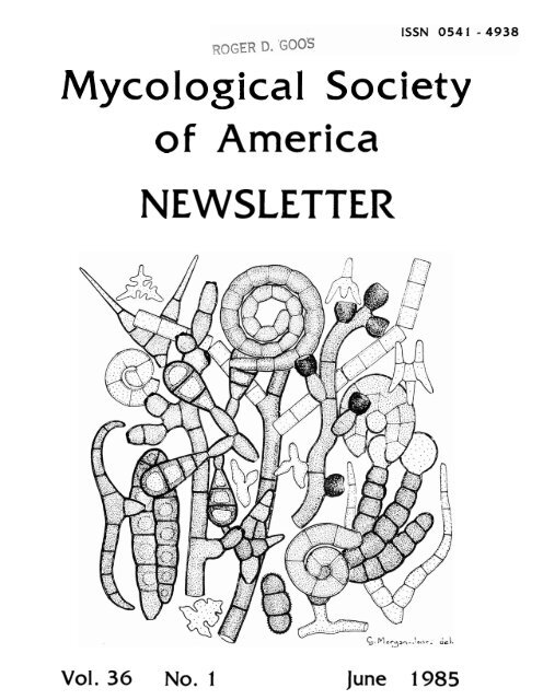
unpublished. Gareth Morgan-Jones prepared the cover "medley". The drawings were done by Jean<br />
J. Sang1 ier via J. W. Bennett (pg. 9), Christopher Best (pg. 12), and Yves Renaud (back cover).<br />
With permission from David Minter, the poetry (pgs. 9 & 15) is reprinted from the BMS Foray<br />
Programne, April <strong>1985</strong>. Finally, we thank Royal1 T. Moore for the submission <strong>of</strong> numerous<br />
"my~010gi~m~" (one-1 iners), some <strong>of</strong> which grace these pages.<br />
In order to conserve space (and reduce cost), readers are encouraged to use the MSA<br />
Di rectory for addresses where thei r response is requested. Mycol ogi sts, however, seem almost<br />
nomadic, sometimes making this approach difficult. Therefore, in this issue we are trying a new<br />
partial solution this problem (see pg. 60). Please make the included changes in your Directory<br />
as they will not appear in future Newsletter issues.<br />
Lastly, I wish to acknowledge the able and pleasant assistance <strong>of</strong> Linda Neuman who typed<br />
most <strong>of</strong> this issue and helped master our new letter qua1 ity printer. The ever-cooperative labor<br />
provided at mailing time by the SIU-C Mycology graduate students is also appreciated.
GENERAL ANNOUNCEMENTS<br />
ATTENTION BRITISH MEMBERS<br />
Dave Minter reminds MSA members in sterling areas that they can avoid bank charges in<br />
converting their MSA subscription to dollars by sending subscriptions in sterling to him at<br />
C.M.I., Ferry Lane, Kew, Surrey, TW9 3AF, U. K. Calculate the value <strong>of</strong> your subscripion using<br />
the dollar/sterling rate in the newspaper <strong>of</strong> the day you post the subscription, and please make<br />
sure your 1986 subscription arrives at the C.Y.I. by 1 January 1986.<br />
MSA PLACEMENT SERVICE<br />
Prospective graduates in mycology seeking employment are urged to register with the MSA<br />
Placement Service by completing the Employee Data Form published elsewhere in this issue <strong>of</strong> the<br />
MSA Newsletter. Data on prospective employees are provided to potential employers where thei r<br />
qualifications meet job requirements. Likewise, persons seeking employment are provided with a<br />
printout <strong>of</strong> vacant positions that match their training. Inqui ries regarding the service should<br />
be addressed to either Gareth Morgan-Jones, Auburn University, or Me1 vin S. Fuller, University<br />
<strong>of</strong> Georgia.<br />
TEACHING CULTURES<br />
Those who teach mycology should be aware that an array <strong>of</strong> identified fungal cultures are<br />
available for teaching purposes from the <strong>America</strong>n Type Culture Collection for $12 a piece. A<br />
catalog <strong>of</strong> these cultures and their uses can be obtained free <strong>of</strong> charge from the ATCC.<br />
THE MYCOLOGY GUIDEBOOK<br />
For several years we have been working, albeit rather slowly, toward a revision <strong>of</strong> the<br />
Mycology Guidebook. It seems appropriate now that we actual ly start thinking about the<br />
organization <strong>of</strong> the new edition and its contents. This requires that members <strong>of</strong> the MSA get<br />
involved in terms <strong>of</strong> their ideas and input, as well as writing parts <strong>of</strong> the text. Below are<br />
some questions which are merely guidelines for getting started. Other comments w i l l be<br />
welcomed with enthusiasm!<br />
1. Are you interested in writing part(s) <strong>of</strong> the new edition <strong>of</strong> the Vycology Guidebook?<br />
2. Did you write one or more sections <strong>of</strong> the present edition.?<br />
3. How useful have you found the present Guidebook?<br />
4. Which subject areas should be added to the text?<br />
5. Which, if any, subject areas might be deleted?<br />
6. Do you have any suggestions on the organization <strong>of</strong> the materials included in the text? For<br />
example, is the present organization (i.e.,<br />
the categories <strong>of</strong> General Information,<br />
Taxonomic Groups, Ecological Groups, Fungi as Bi 01 ogical Tools, and the accompanying<br />
Appendices) a good format?<br />
7. It has been suggested that the new edition <strong>of</strong> the Guidebook be published in Spanish as well<br />
as English. Do you agree or disagree with this idea?<br />
Please send comments to these questions as well as other suggestions, etc. to J. Ammirati,<br />
Chair, Mycology Guidebook Committee, Department <strong>of</strong> Botany, KB-15, University <strong>of</strong> Washington,<br />
Seattle, WA 98195.<br />
TO ALL MYCOLOGICAL WRITERS<br />
The MYCOLOGIA MEMOIRS Committee wants book-length manuscripts for review. Contact T. M.<br />
Hammill for details.
CALENDAR OF MEETINGS, FORAYS, AND WORKSHOPS<br />
August <strong>1985</strong><br />
1-4 The Annual Meeting and foray <strong>of</strong> the NORTH AMERICAN MYCOLOGICAL ASSOCIATION<br />
(NAMA) will be hosted by the <strong>Mycological</strong> Association <strong>of</strong> Washington (MAW), and<br />
will be held at Canaan Valley Resort State Park near El kins, West Virginia.<br />
Kent H. McKnight will serve as head foray mycologist with numerous other guest<br />
speakers and workshop leaders from the YSA membership. <strong>Mycological</strong> and other<br />
resort activities will abound (I plan to bring my roller sk6tes--ed.)<br />
Registration: $60.00. Meals $65.00. Room $44-$120.00. Contact Frances Usenik,<br />
2327 49th Street, NW, Washington, DC 20007.<br />
<strong>1985</strong> ANNUAL MEETING OF THE SOCIETY OF INDUSTRIAL MICROBIOLOGY will be held at<br />
the Westin Hotel, in Copley Place, Boston, MA. For more information contact:<br />
Forrest S. Yoy, Morton Thiokol , Inc., Ventron Division 150 9ndover Street,<br />
Danvers, MA 01932; Phone (617) 774-3100 or Stephen W. Queener, Research<br />
Associate, Eli Lilly and Company, Indianapolis, IN 46285; Phone (317) 261-7454.<br />
TENTH ANNUAL NORTHEASTERN MYCOLOGICAL FORAY will be held at the State University<br />
<strong>of</strong> New York, Oneonta, NY. Principal mycologists will be David Malloch,<br />
University <strong>of</strong> Toronto, David Pegler, Royal Botani cal Gardens, Kew England, and<br />
Currie Marr, State University <strong>of</strong> New York. For more information contact: Pat<br />
and Jim Kronick, 1951 Lowell Lane, Merrick, NY 11566.<br />
BMS SON OF TRUFFLE HUNT will be held at Cotswolds. Contact Jane Ingham, 21<br />
Loughmi 11 Road, Pershore, Worcestershi re.<br />
TELLURIDE MUSHROOM CONFERENCE. Foray in forests surrounding Tell uri de, an<br />
historic Colorado mining town. Courses include mushroom identification,<br />
cul ti vation, and ethnomycol ogy. For further information contact: Emanuel<br />
Salzman, P. 0. Box 5503, Denver, CO 80217-5503. Phone: (303) 296-1218.<br />
1-18 MUSHROOM STUDY TOUR OF THE HIMALAYAS. Organized by Gary Linc<strong>of</strong>f, Andrew Weil<br />
and Emanuel Salzman. Foray in the forests surrounding the hill stations, the<br />
beautiful old summer resorts <strong>of</strong> the British colonial days, in the foothills <strong>of</strong><br />
the great Himalayas. For more information contact: Emanuel Salzman, P. O.Box<br />
5503, Denver, CO 80217-5503. Phone: (303) 296-1218.<br />
BMS AUTUMN FORAY will be held at Chester.<br />
Contact Dave Minter for details.<br />
THE A. H. SMITH FORAY will be held near Baraboo, Wisconsin. Contact Jan Phelps:<br />
UWC-Baraboo, 1006 Connie Road, Baraboo, W I 53913.<br />
The Westwater Inn, Olympia, Washington, will be the site <strong>of</strong> the WESTERN<br />
INTERNATIONAL FOREST DISEASE WORK CONFERENCE. For particulars contact: Ken<br />
Russel 1 , Dept . o f Natural Resources, Di vision <strong>of</strong> Pri vate Forestry and<br />
Recreation, MQ-11, Olympia, WA 98504; (206) 545-0953; or Walt Thies, USDA<br />
Forest Service, Forestry Sciences Laboratory, 3200 Jefferson Way, Corvall is, OR<br />
97331; (503) 757-4396.<br />
32ND CHARLES HORTON PECK ANNUAL MYCOLOGICAL FORAY will be held in the Arnot<br />
Teaching and Research Forest <strong>of</strong> Cornell University, located in Van Etten, New<br />
York. For more information and/or reservation forms contact: R. P. Korf, PECK<br />
FORAY COORDINATOR, Plant Path01 ogy, Cornel 1 University, Ithaca, NY 14853.
4<br />
October - <strong>1985</strong><br />
BMS MINI-FORAY IN SPAIN (Barcelona, Catalonia, and Monseny). Yrite David Minter<br />
or Henry Descal s, Department de Botani ca, Facul tat de Bi 01 ogi cas, Uni versi tat de<br />
Barcelona, Diagonal 645, Rarcel ona 08028, Spain.<br />
THE OREGON MYCOLOGICAL SOCIETY FALL FORAY will be held along the northern Oregon<br />
Coast. Guest mycologist will be Joseph Ammirati. For information on the<br />
location contact J. Preston Alexander, Rt. 1, Box 158, Forest Grove, OR 97116.<br />
November <strong>1985</strong><br />
23<br />
April 1986<br />
BMS Meeting on TOXIC FUNGI at ~odrell' Laboratories, Kew.<br />
to learn more.<br />
Contact David Minter<br />
7-10<br />
THE HKITISH MYCOLOGICAL SOCIETY GEhERAL MEETING in Bristol. Topic:<br />
"Evolutionary Biology <strong>of</strong> the Fungi ." Learn more from David Minter.<br />
23-27<br />
BMS SPECIALIST WORKSHOP ON ASCOMYCETES will occur at CMI.<br />
Contact David Minter.<br />
May - 1986<br />
3-4<br />
MID-ATLANTIC STATES MYCOLOGY CONFERENCE is tentatively set at Towson State<br />
College, Towson, Maryland. Interested persons should contact Jerry Motta, Dept.<br />
<strong>of</strong> Botany, University <strong>of</strong> Maryland, College Park, MD 20742.<br />
BMS ANNUAL SPRING FORAY (jointly held with the Botanical <strong>Society</strong> <strong>of</strong> Edinburgh)<br />
wi 11 be in Gal loway, based at Newton Stewart. Write Alan Bennell , Royal Botanic<br />
Garden, Edinburgh.<br />
August 1986<br />
30-Sept. 4<br />
FOURTH INTERNATIONAL FUNGAL SPORE SYMPOSIUM wi 11 be he1 d in Scot1 and (Sti rl i ng<br />
University). The host wi 11 be John Smith, University <strong>of</strong> Strathclyde, Glasgow,<br />
Scot 1 and.<br />
FORTHCOMING COURSES<br />
David Minter and Paul Cannon wi 11 conduct an international course on ASCOPIYCETE TAXONOMY,<br />
7-18 October <strong>1985</strong>, at the Universidad Austral de Chi le, Valdivia, Chile. Further information<br />
is avai 1 able from Dr. H. Peredo, Instituto Silvicul tura, Uni versidad Austral, Casi 11 a 567,<br />
Valdivia, Chile.<br />
MUSHROOM TOXICOLOGY will be <strong>of</strong>fered by N. S. Weber & K. Cochran, Sept. 15-21 at Dillman's<br />
Sand Lake Lodge, Lac Du Flambeau, W I 54538.<br />
N. S. Weber will teach MUSHROOMS OF SLEEPING BEAK DUNES, July 22-26, at the Leelanau<br />
Center for Education, Glen Arbor, MI 49636.<br />
WOOD DETERIORATION, Fall Quarter <strong>of</strong> <strong>1985</strong>, (3 cr.). by Elmer L. Schmidt, Department Forest<br />
Products, University <strong>of</strong> Minnesota, St. Paul, MN 55108<br />
A MUSHRUMF'US - AN MSA FORAY
NEW MYCOLOGICAL RESEARCH<br />
G. C. ADAMS: Genetics <strong>of</strong> virulence and hypovirulence <strong>of</strong> Leucostoma persoonii and -- L. cincta<br />
(anamorphs: Cytospora spp. ).<br />
J. P. ALEXANDER: The effects <strong>of</strong> endogenous growth regulators on the growth and differentiation<br />
<strong>of</strong> a range <strong>of</strong> wild fleshy fungi.<br />
W. C. HAKEWELL: Studies on color guides, color standards, and color dictionaries.<br />
P. R. BECKJORD: Survey <strong>of</strong> the ectomycorrhizal epigeous fungi <strong>of</strong> oaks and conifers on surface<br />
mines and - in forests <strong>of</strong> western Mary1 and and factors for their ecology, sociology, and<br />
phaenol ogy .<br />
T. SEGAN: The genus Amanita in southern Illinois.<br />
S. E. CARPENTER: Revision <strong>of</strong> the ~hialeoideae and Ombrophiloideae (Helotiales, Leotiaceae).<br />
M. A. CASTELLANO: Monograph <strong>of</strong> Hysterangium.<br />
M.-M. CHEN: Epidemiology <strong>of</strong> white pine blister rust <strong>of</strong> sugar pine and the study <strong>of</strong> resistance<br />
<strong>of</strong> Pinus radiata to western gall rust: identification <strong>of</strong> fungal pathogens; ecology <strong>of</strong> and<br />
resistance to tree rusts; and edible fungi <strong>of</strong> China.<br />
P. K. DUBLISH: Aeromycological studies <strong>of</strong> a stud-farm.<br />
D. H. PFISTER: An inventory <strong>of</strong> the <strong>Mycological</strong> Literature from 1753 to 1821 with Jean Boise as<br />
Research Bibliographer.<br />
G. C. YAYE (with D. H. Pfister & Jean 8oise): <strong>Mycological</strong> Literature.<br />
R. KENDRICK: Pathogenicity <strong>of</strong> Hyphomycetes to Spruce Budworm; model ing the impact <strong>of</strong><br />
herbicides on pond ecosystems; morphology and taxonomy <strong>of</strong> VAY fungi ; and use <strong>of</strong><br />
biotechnology and a dual fungal culture to produce lipids from cellulosic wastes.<br />
R. W. YERRIGAN: Relationships in Agaricus (sub) section Hortenses.<br />
R. KOFFMAN: Studies on Aspergi 11 us clavatus and Psi locybe tampanensi s.<br />
R. K. S. KUSHWAHA: Biology <strong>of</strong> Chrysosporium and allied fungi from soil with special reference<br />
to their morphology and taxonomy.<br />
G. LIM: Fungal enzymes; production <strong>of</strong> aroma chemicals by fungi.<br />
P. D. MILLNER: Interactions among saprophytic root-colonizing bacteria and fungi, and VA<br />
mycorrhi zae that affect plant nutrition; protein and enzyme analysis <strong>of</strong> VA mycorrhizae and<br />
Endogone as related to growth, colonization, and nutrient translocation.<br />
G. M. MUELLER (with Stephen Rehner): DNA-DNA hybridization as a means for studying<br />
phylogeneti c re1 ati onshi ps among species <strong>of</strong> Laccari a.<br />
M. E. NOOKDELOOS: Taxonomy and ecology <strong>of</strong> Agarics from Boreal and Arctic regions (esp.<br />
Entolomataceae, Tricholomataceae). Entoloma sensu lato in eastern Canada and northeastern<br />
USA.<br />
S. L. PEELE: New compounds for anti-tumor and anti-cancer activity from mushrooms.<br />
S. A. REHNER: Taxonomy <strong>of</strong> A roc be sect. Pediadeae and a phylogenetic analysis (based on<br />
mol ecul ar approaches) +b$-<br />
o rocybe sect. Pedi adeae including comparisons to selected taxa<br />
within the Bolbiticeae.
E. L. SCHMIDT: Shiitake yields on red and bur oak, effects <strong>of</strong> white rot fungi on bonding<br />
systems <strong>of</strong> aspen waferboard, and the influence <strong>of</strong> aliphatic acids on spore germination <strong>of</strong><br />
wood decay fungi.<br />
M. SPEAR: Media and techniques for large-scale production <strong>of</strong> ectomycorrhizal inoculum.<br />
W. J. SUNDBERG (with Raafat Mohamnadkhani ): lJl trastructural development and hi stochemi stry <strong>of</strong><br />
clamp connection formation in Cyathus 01 la. -<br />
3. M. TRAPPE: Hypogeous fungi and their mycorrhizae--comparison <strong>of</strong> Spain with Oregon and<br />
Cal i fornia (grant from I1.S.-Spain Cooperative Research Awards) ; mycorrhiza-Rhizobium<br />
interactions in tropical woody legumes (Indo-U.S. Science & Technology Initiative Awards).<br />
R. C. ULLRICH: The following are studying the aspects <strong>of</strong> biology in Schizophyllum in Ull rich's<br />
Lab; Eunice Fruel i ger--0rotidine-5 ' -monophosphate decarboxylase and its gene; Brent<br />
Suckner--ri bosomal RNA genes ; Lisa Phel ps--cytochrome oxidase suhuni t I I mi tochondri a1<br />
gene from; Charles Specht--mappi ng mi tochondri a1 genes; and A1 fredo Munoz--tryptophan<br />
brosynthesi s.<br />
D. WEBER (with Vic Bundersoa): Development <strong>of</strong> an interactive video disc for a visual<br />
identification key to mushrooms (Exxon Educational Foundation Grant).<br />
W. YUN: Taxonomy <strong>of</strong> hypogeous fungi in China.<br />
BASIDIOMYCETES<br />
J. P. Alexander can provide Pleurotus ostreatus (wild types from the Pacific Northwest), and<br />
other Pleurotus sp., and a1 so cul-calvatia booniana (Pacific Northwest).<br />
M. A. Caste1 lano will send Hysterangium specimens.<br />
M.-M.<br />
Chen has stored spores <strong>of</strong> Endocronartium (western gal 1 rust).<br />
P. K. Dub1 ish <strong>of</strong>fers Phyllacteria dal bergiae on leaves <strong>of</strong> Dal bergra sisso (preserved material ).<br />
S. L. Peele has dried specimens, cultures, and spores f rom4mani ta phal loides, A. muscari a,<br />
Pleurotus sapidus as well as many species <strong>of</strong> Psilocybe and Panacolus. sea for free<br />
i nformation or call Florida Mycology ~esearch--904) 478-3912.<br />
R. E.<br />
I<br />
Tulloss <strong>of</strong>fers specimens <strong>of</strong> Amanita spp. from the pine Barrens <strong>of</strong> New Jersey (largely<br />
section Lepi dell a).<br />
MISCELLANEOUS<br />
K. Diebolt has cultures <strong>of</strong> ectomycorrhizal fungi--send for a list.<br />
A MUSHRUMOR - A HIiVT OF ANOTHER NAME CHANGE FOR THE CO!.!MERCIAL MUSHROOM
FUNGI WANTED<br />
ACRASIOVYCETES<br />
R. L. Blanton: Cultures <strong>of</strong> Copromyxa protea.<br />
MYXOMYCETES<br />
K. L. Rraun: Myxomycete specimens from Mexico.<br />
T. W. Gaither: Collections <strong>of</strong> any Didymium species.<br />
H. W. Kel ler: Myxomycetes--speci es <strong>of</strong> the genera Peri chaena and Li cea; corticol ous myxomycetes<br />
from 1 i vi ng trees and vines; myxomycetes from Mexico; specimens <strong>of</strong> Licea fimicol a (common<br />
on dung from herbivorous animal s--cow, bison, horse).<br />
S. L. Stephenson : Myxomycetes, especially col lections from western North <strong>America</strong>.<br />
OOMYCETES<br />
H. H. Ho: Phytophthora japonica (P. - oryzeae), and Phytophthora areceae (A1 type).<br />
ZYGOYYCETES<br />
B. Yendrick (with M. Srundrett): Wishes to exchange pot cultures or spores <strong>of</strong> VAM fungi.<br />
J. M. Trappe: Specimens <strong>of</strong> Endogonaceae.<br />
ASCOMYCETES<br />
G. C. 4dams: Recent collections <strong>of</strong> Leucostoma and Valsa with host identifications.<br />
S. E. Carpenter: Cultures and specimens <strong>of</strong> any Phial eoi deae and Ombrophi 1 oideae (He1 oti ales,<br />
Leot i aceae ).<br />
K. Di ebol t : Cultures <strong>of</strong> Wynnea ameri cana .<br />
K. Esser: The perfect stage <strong>of</strong> the Ascomycete Cochliobolus lunatus.<br />
J. H. Hai nes: Specimens <strong>of</strong> Hyal oscyphaceae.<br />
M. Harrington: Recently collected, ai r-dried specimens <strong>of</strong> Sarcoscypha to culture. Please ship<br />
. . by overnight delivery system, and they shall gladly reimburse costs. Send c/o L. R.<br />
Batra, Rm. 313, Bldg. OllA, USDA, Re1 tsvi 1 le, MD 20705. Telephone 301-344-2317 (call<br />
collect) for any needed information on how to collect or ship these fungi.<br />
T. Iturri aga: Cultures or specimens or both <strong>of</strong> Strossmayeria (Helotiales) and its anamorph<br />
Pseudospi ropes.<br />
L. M. Kohn: Cultures and specimens <strong>of</strong> sclerotium-forming Ascomycetes.<br />
H. J. Kronenberg: Cordyceps si nensi s and Cordyceps ci cadae<br />
J. M. Trappe: Specimens <strong>of</strong> hypogeous Ascomycetes.
W. Zhuang: Discomycetes (with pigmented apothecia) on fungi.<br />
BASIDIOMYCETES<br />
J. P. Alexander: Cultures <strong>of</strong> Ramaria botrytis, Sparassis crispa, 2. radicata, HerIcium<br />
abieti s, Fistul ina hepatica, Vol variel la bombyci na, and Agari cus augustus.<br />
J. Ammi rati : Corti nari us, subgenera Dermocybe, Leprocybe, and Yyxaci um with notes and/or<br />
kodachromes. Yelanotus with note's and/or kodachromes and spore deposits for culture work.<br />
0. Bermudes: Cultures <strong>of</strong> Amanita spp. He would also like cultures <strong>of</strong> known ectomycorrhiza<br />
formers and dried specimens <strong>of</strong> amanitin- and phalloidin-containing mushrooms. He will pay<br />
postage.<br />
H. H. Surdsall : Armillaria mellea collections with complete notes as to field characters,<br />
suspected pathogenicity, and host; cultures, single spore isolates and any other<br />
information concerning the col lections.<br />
M. Caste1 lano: Cultures or specimens <strong>of</strong> Hysterangium (Phal laceae).<br />
K. Diebol t: Cultures <strong>of</strong> Gyrodon merul ioides and Sui 1 lus borinus.<br />
M. F. Doyle: Pacific Island agarics (notes preferred and/or photos, but he's a realist).<br />
R. E. Halling: Collybia and Marasmiellus. Notes on color and odor always helpful.<br />
R. W. Kerrigan: Cultures/spore prints <strong>of</strong> wild Agaricus bisporus and its close relatives.<br />
Vouchers, notes, and kodachromes appreciated.<br />
R. K<strong>of</strong>fman: Gymnopilus spectabilis.<br />
H. J. Kronenberg: Ganoderma lucidum, and Poria -- cocos.<br />
A. S. Yethven: Collections <strong>of</strong> Clavariadelphus from North <strong>America</strong> with color notes and spore<br />
deposits, and cultures <strong>of</strong> Clavariadelphus from Rorth Ameri ca.<br />
H. P. Mol itoris: Cultures <strong>of</strong> Yelanotaenium ruppiae (a marine basidiomycete, Usti laginales).<br />
M. E. Noordeloos: Specimens <strong>of</strong> Entoloma (sensu lato incl . Leptonia; Nolanea: Pouzarel la,<br />
Ecci 1 ia; A1 boleptonia). Notes and slides on fresh material are necessary!<br />
S. L. Peele: Cultures or spores <strong>of</strong> Psilocybe zapotecorum.<br />
J. Pfi ster: Recent exsiccatae and spore prints <strong>of</strong> Me1 anoleuca gramnopodia, - M. graminicola, - M.<br />
verrucipes, - M. subal pina, and - M. subbrevipes for enzymatic studies.<br />
D. Prusso: Specimens <strong>of</strong> Tulostoma from North <strong>America</strong> including collection data.<br />
S. Rehner: Cultures and/or spore prints plus voucher specimens (notes & photos; if possible)<br />
<strong>of</strong> Agrocybe, Bol bi tius, Conocybe, Gastrocybe (especially Agcocybe pediades, A.<br />
subpediades, 4. semiorbiaclaris, A. retigera, A. thinlenta, - A. arval is, - A.<br />
cyl indracalaegeri ta, Gastrocybe lareri tia, C o n o c y b m .<br />
M. Spear: Cultures <strong>of</strong> Agaricus subrufesceus and other unusual Agaricus spp.<br />
W. J. Sundberg: Lepiota spp. (sensu lato; notes and/or photographs helpful ).<br />
J. M. Trappe: Specimens <strong>of</strong> hypogeous Basidiomycetes.
R. E. Tulloss: We1 1 -documented collections <strong>of</strong> Amani t a spp. with slides if possible--please<br />
inquire, first.<br />
DEUTEROMYCETES<br />
W. D. Jennsen : Tri choderma vi ri de .<br />
R. K<strong>of</strong>fman : Aspergi 11 us cl avatus.<br />
R. K. S. Yushwaha: Cultures <strong>of</strong> Chrysosporium, Arthroderma, and Halbranchea.<br />
J. Y. McPartland: Cultures <strong>of</strong> any race (1 thru 6) <strong>of</strong> Fusarium oxysporum F. sp. vasinfectum.<br />
M. Wingfiel d: Leptographi um spp., Verti ci cl adiel 1 a spp., and Phial ocephal a spp.<br />
MISCELLANEOUS<br />
G. Guzman: Duplicates for the Herbarium <strong>of</strong> INIREB (XAL).<br />
W. Lanier: Cultures <strong>of</strong> any fungus which parasitizes pest insects or nematodes. Contact him at<br />
BIOSIS, 3788 Fabian Way, Palo Alto, CA 94303.<br />
R. E. Scott: Cultures <strong>of</strong> psychrophil ic fungi. Please contact him by letter with culturing<br />
conditions, etc. before sending (See Changes <strong>of</strong> Address).<br />
J. F. White & Garry T. Cole: Endophytic fungi from grasses <strong>of</strong> North <strong>America</strong>.<br />
To An Unwanted Ascomycete<br />
A fungus by a spore trap grew,<br />
A di scomycete fresh and new.<br />
And, heedless <strong>of</strong> its likely fate<br />
It soon began to sporulate.<br />
Its spores, released, flew in a trice<br />
Into my spore trap's orifice.<br />
Ah, waste! Its chosen situation<br />
Has spoiled all chance <strong>of</strong> propagation,<br />
And oh! My whole week's record messed<br />
With spores from this unwanted guest.<br />
Fool i sh Pezi za ! Better hide ,<br />
Or 1'11 deploy the fungicide!<br />
---Fel ici ty Jackson
10<br />
IDENTIFICATIONS<br />
The following are willing to identify the taxa specified.<br />
MYXOMYCETES<br />
H. W. Kel ler: Myxomycetes--species <strong>of</strong> Peri chaena and Licea, and corticolous myxomycetes.<br />
ZYGOMYCETES<br />
S. M. Berch: Vesi cular-arbuscul ar mycorrhi zal fungi ,. Endogonaceae<br />
J. M. Trappe: Endogonaceae.<br />
ASCOMYCETES<br />
S. E. Carpenter: Leoti aceae (He1 o ti ales).<br />
J. H. Haines: Hyaloscyphaceae.<br />
A. Y. Rossman: All members <strong>of</strong> the Hypocriales including Nectria and Gibberella as specimens or<br />
cultures forming ascocarps. Fresh specimens are preferred.<br />
L. J. Spielman: Val sa, Leucostoma, and the anamorphs in Cytospora.<br />
J. M. Trappe: Hypogeous Ascomycetes.<br />
BASIDIOMYCETES<br />
J. P. Alexander: Calvatia sp., and Pleurotus sp.<br />
M. A. Caste1 lano: Hysterangium<br />
G. P. Chamuris: Stereum S.S. or s.1.; temperate or tropical.<br />
M.-M.<br />
Chen : Cronartium, Coleosporium, Melampsora, Chrysomyxa; Lenzites, and Daedalea.<br />
R. E. Halling: Collybia and Marasmiellus.<br />
M. E. Noordeloos: Entoloma sensu lato (only possible with notes and slides).<br />
J. M. Trappe: Hypogeous Basidiomycetes.<br />
R. E. Tulloss: Amanita (Please inquire, first).<br />
W. Yun: Me1 anogaster<br />
DEUTEROMYCETES<br />
M.-M.<br />
Chen: Cytospora<br />
M. Wingfield: Leptographium spp.<br />
'<br />
"What's happened to the truffle hunterslr' rrTheyrve gone underground. "
PUBLICATIONS AVAI LABLE--FOR GIVE-AWAY, SALE, OR EXCHANGE<br />
11<br />
Gaston Guzman has the following for exchange or sale: SULLETIN OF THE MEXICAN SOCIETY OF<br />
MYCOLOGY, Vol . 19 (1984), 356 pages and 16 color plates. $15 US (back numbers are a1 so<br />
avai 1 able) .<br />
Machiel E. Noordeloos has reprints on Entoloma for sale or exchange. Send for a list.<br />
Joachim Schl iemann has available: Fries' ICONES SELECTAE HYMENOMYCETUM, 1/11; Persoon Is<br />
SYNOPSIS METHODICA FUNGOORUM 111 I ; Vai 1 lant Is BOTANICON PARISIENSE ; and a 1 arger number <strong>of</strong><br />
other duplicate titles. Send for list or specify desiderata.<br />
Christian Vol bracht has a list <strong>of</strong> duplicates from his mycological collection.<br />
Harold W. Keller has 4. E. Nannenga-Bremekamp's DE NEDERLANDSE NYXOYYCETEN, 1974, 2nd<br />
edition (with supplement).<br />
Richard Rlanton has for sale: AMERICAN JOURNAL OF ROTANY, Vols. 60 (1973) through 70<br />
(1983) for $4/volume plus postage. Also, JOURNAL OF PROTOZOOLOGY, Vols. 23 (1976) through 31<br />
(1984) for $51~01 ume plus postage.<br />
Mo-Mei Chen has for sale PRELIMINARY ANALYSIS OF THE RUST FLORA IN THE FOREST OF TIBET<br />
(abstract in Enqlish); THE GUIDE BOOK ON LABORATORY IDENTIFICATION OF PORE FUNGI (in Chinese);<br />
A NEW GENUS, ST~LBECHRYSOMYXA CHEN GEN NOV. <strong>of</strong> Chrysomyxaceae on Rhododendron, and THE INDEX.OF<br />
TREE DISEASES OF CHINA.<br />
Hi romi tsu Hagiwara wants to exchange the MYXOMYCETOUS WORLD by Hagiwara & Izawa, including<br />
ca. 80 excel lent color photographs with Japanese explanations, for any book(s) or reprint(s) on<br />
Myxomycetes .<br />
Donald T. Kowal ski has for sale originals <strong>of</strong> Lister's A MONOGRAPH OF THE MYCETOZOA, Vols.<br />
1, 2, & 3 for $350; and April 1926 NATIONAL GEOGRAPHIC (Watercolors <strong>of</strong> Slime Molds) for $10.<br />
Robert E. Macho1 has for sale MYCOTAXON, Vols. 1-20 and some old mushroom books--send for<br />
list.<br />
Stephen L. Peele has available one free issue <strong>of</strong> THE MUSHROOM CULTURE, The Journal <strong>of</strong><br />
Mushroom Cultivation (see Changes <strong>of</strong> Address).<br />
Richard W.<br />
Kerri gan has free information on easy classroom cultivation <strong>of</strong> Pleurotus.<br />
Philip A. Orpurt has for sale MYCOLOGIA, Vols. 41-64 (1949-1972) bound (Ch. brown) $8 each<br />
($192); and Vol s. 65-75 (1973-1983) unbound, $5 each ($55).<br />
Walter J. Sundberg will sell Sundberg & Richardson, MUSHROOMS AND OTHER FLESHY FUNGI OF<br />
LAND BETWEEN THE LAKES, 64 p., 98 color photos, 1980. Prepay $3.65 (includes postage and<br />
padded mai 1 i ng envelope).<br />
Jenifer Huang McBeath has a reprint for give away entitled SYYPTOMOLOGY ON SPRUCE TREES<br />
AND SPORE CHARACTERISTICS OF A BUD RUST PATHOGEN, Phytopathology 74:456-461, 1984.<br />
Dieter Schierenberg has special lists <strong>of</strong> books in Botany and Mycology.<br />
Prinsengracht 485-487, 1016 HP, Amsterdam, Holland.<br />
Write him at<br />
Rodham E. Tulloss has, for the cost <strong>of</strong> copying & postage, a BIBLIOGRAPHY & INDEX TO NORTH<br />
AMERICAN LITERATURE ON AMANITA with over 300 references, approx. 250 indexed, over 200 taxa.<br />
Herbert N. Humphrey has for sale a Periodical Library, chiefly mycological, also plant<br />
path01 ogical and botanical journal s dating from 1872. Examples include: ANNALES MYCOLOGICI ,<br />
Vols. 1-36; BULLETIN DE LA SOCIETE MYCOLOGIQUE DE FRANCE, Vols. 7, 11, 29, 35-55;<br />
PHYTOPATHOLOGY, Vol s. 1-59; AMERICAN JOURNAL OF BOTANY, Vol s. 16-23; and many more. For a<br />
complete list, write to Herbert N. Humphrey, 4040 Maybelle Avenue, Oakland, CA 94619.
Bryce Kendrick is <strong>of</strong>fering A YOUNG PERSONS' COLOURING HOOK OF FUNGI, THE FIFTH KINGDOM<br />
(avai 1 able this summer), THE ENCYCLOPEDIA OF MUSHROOMS, and several references on<br />
Deuteromycetes. Write him for a list.<br />
NEW BOOKS BY MSA MEMBERS<br />
The fol 1 owing announcements were recei ved in response to the MSA Newsletter questionnai re:<br />
Alan Bessette. <strong>1985</strong>. GUIDE TO SOME EDIBLE AND POISONOUS FUNGI OF NEW YORK. 41 color<br />
photographs and descriptions <strong>of</strong> fungi, many <strong>of</strong> which are found throughout the United<br />
States. A1 though designed for the novice, experienced collectors will also appreciate the<br />
color qua1 ity and diversity <strong>of</strong> specimens. Cost is $3.95 including postage and handl ing.<br />
Available from A1 an Bessette, Utica College <strong>of</strong> Syracuse Uni versity, 1600 Burrstone Road,<br />
Utica, NY 13502. Make checks payable to Otica College <strong>of</strong> Syracuse University.<br />
J. Cramer, has just received the manuscript for the fourth fully revised edition <strong>of</strong> Rolf<br />
Singer's THE AGAKICULES. Typesetting has been started, and publication is scheduled for<br />
winter <strong>1985</strong>-86.<br />
Nancy S. Weber & Alexander H. Smith. Photographs by Dan Guravich. <strong>1985</strong>. A FIELD GUIDE TO<br />
SOUTHERN MUSHROOMS. Avai lable from Uni veri si ty <strong>of</strong> Michigan Press, Ann Arbor, 48109.<br />
$16.50 less 10% pr<strong>of</strong>essional discount (+ 4% sales tax in MI), + $1.00 handl ing for first<br />
copy, $.25 for each additional copy.<br />
Kenneth B. Raper. 1984. THE DICTYOSTELIDS. Princeton University Press. 453 pp.,<br />
illustrated. $75.00.<br />
Joost A. Stalpers. 1983. A REVISION OF THE GENUS SPOROTRICHUM, Studies in Mycology No. 24.<br />
Centraal bureau voor Schimnelcultures, P. 0. Box 273, 3740 AG Baarn, The Netherlands. 105<br />
pp. hfl. 25.<br />
M. A. Schipper and Joost A. Stalpers. 1984. A REVISION OF THE GENUS KHIZOPUS, Studies in<br />
Mycology No. 25. Centraalbureau voor Schimmelcultures, P. 0. 273, 3740 AG Baarn, The<br />
Nether1 ands. 34pp. hf 1. 10.<br />
Randy Yolina. <strong>1985</strong>. PROCEEDINGS OF THE 6TH NORTH AMERICAN CONFERENCE ON MYCORRHIZAE.<br />
Published by the Forest Research Laboratory, Oregon State University. 471pp. $20.00 plus<br />
$2.25 for postage and handl ing. To order, send check payable to OSU College <strong>of</strong> Forestry,<br />
to Col lege <strong>of</strong> Forestry, Oregon State Uni versi ty, Corvall is, OR 97331-5704.
P. K. Dub1 ish needs pub1 ications on aeromycol ogical studies.<br />
PUBLICATIONS WANTED 13<br />
J. Schl iemann would 1 i ke important old books on mushrooms, iconographies, rare titles; he<br />
is also interested in all give-away, sale, or exchange lists.<br />
E. W. Smith is in need <strong>of</strong> the Index <strong>of</strong> Plant Diseases (USDA Handbook 165).<br />
C. Volbracht is looking for any books on Mycology printed before 1900.<br />
D. Bermudes needs Charles David Badham's ESCULENT FUNGUSES OF ENGLAND 1847.<br />
R. Critchfield, Sr. wants any reprints on identification, taxonomy, morphology, and<br />
ecology <strong>of</strong> myxomycetes, and is willing to reimburse for reprints and/or postage.<br />
R. K<strong>of</strong>fman would like to obtain a copy <strong>of</strong> THE GENUS ASPERGILLUS by Raper & Fennell.<br />
R. Y. S. Kushwaha needs publications on taxonomy, ecology <strong>of</strong> Chrysosporium and members <strong>of</strong><br />
Gymnoascaceae.<br />
R. E. Macho1 wants old mushroom books, including incomplete, very early works, if the<br />
parts on mushrooms are intact.<br />
R. E. Scott would like reprints and any information on microscopic psychrophilic fungi,<br />
(e.g.) culture conditions, enzyme purifications, and secondary products (See Changes <strong>of</strong><br />
Address ).<br />
R. W. Yerri gan needs literature on Agaricus and old spawn catalogs.<br />
S. L. Stephenson would like reprints on Myxomycetes.<br />
J. P. Alexander would like THE FUNGI by Gaumann; Wolf & Wolf's THE FUNGI, Academic Press;<br />
PUFFBALLS & THEIR ALLIES..by A. Smith; and A NEW MONOGRAPH ON PLEUROTUS by Yiller (?).<br />
R. L. Tulloss needs both volumes <strong>of</strong> C. H. Kauffman's AGARICACEAE OF MICHIGAN.<br />
T M. Hammi 11 would 1 i ke the CANADIAN JOURNAL OF BOTANY, 1981, Vol . 59; and Gaumann 's THE<br />
FUNGI: A DESCRIPTION OF THEIR MORPHOLOGICAL FEATURES AND EVOLUTIONARY DEVELOPMENT.<br />
M. F. Doyle would like reprints, etc. on insular mycology and long-distance fungal<br />
dispersal.<br />
Gui 1 lermo Rodri guez-Scherzer wishes reprints or books (pri nci pal ly on Polyporaceae sensu<br />
lato) free or in exchange for mexican fungi (Ascomycetes and Basidiomycetes). Contact him at<br />
Herbario Metropol itano (UAMIZ), Departamento de Biologia, Uni versidad Autonoma<br />
Metropol - i tana-Iztapal apa, Apartado Postal 47-017, Mexico, 0. F. 07800.<br />
T-shirts, with this design<br />
in white on deep Fusarium purple,<br />
are available from:<br />
Mycoproducts, P. 0. Box 3050<br />
P. o. Box 3050<br />
Durham, NC 27705-1050
MYCOLOGICAL SOCIETY OF AMERICA<br />
<strong>1985</strong> ANNUAL MEETING PROGRAM<br />
University <strong>of</strong> Florida, Gainesvil le, Florida<br />
Saturday, August 10<br />
All Day:<br />
Foray to San Felasco Hammock State Preserve and University <strong>of</strong> Florida's<br />
Horticultural Farm, Alachua County.<br />
Morni ng: Yi ni Symposium. Recent Advances in Cryotechnol ogy<br />
Afternoon: Workshop. Recent Advances in Cryotechnol ogy<br />
Sunday, August 11<br />
Morni nglEarly<br />
Afternoon:<br />
Late Afternoon<br />
and Evening:<br />
All Day:<br />
Foray to Lake Yize, Austin Cary Memorial Forest, Alachua Co.<br />
Foray to Cedar Key on Florida's Gulf Coast isightseeing and dinner included)<br />
Meeting <strong>of</strong> the MSA Council<br />
Monday, August 12<br />
Morning: Session 1. Contributed Papers. Taxonomy<br />
Session 2. Symposium. Factors Effecting Morphogenesis in Achlya<br />
Session 3. Posters. Ecol ogy, Morphol ogy , and Taxonomy<br />
Afternoon: Session 3. (continued)<br />
Session 4. Contributed Papers. Siochemistry and Physiology<br />
Session 5. Contributed Papers. Ecology<br />
Session 6. Symposium. Recent Trends in the Study <strong>of</strong><br />
Heterobasidi omycetes<br />
Evening:<br />
AIBS Plenary Session<br />
Tuesday, August 13<br />
Morning: Session 7. Contributed Papers. Morphology and Taxonomy<br />
Session 8. Symposium. Recent Advances in the Biology <strong>of</strong> the Cultivated<br />
Mushroom<br />
Session 9. Posters. Biochemistry, Cytology, Genetics, Medical Mycology,<br />
Physiology and Ul trastructure<br />
Afternoon: Session 9. (continued)<br />
Session 10. Contributed Papers. Cytology and Ul trastructure<br />
Session 11. Contributed Papers. Genetics and Taxonomy<br />
Session 12. .Symposi urn. Beta-Gl ucans: Thei r Biosynthesi s and Degradation<br />
Evening: Session 13. Nomenclature Open House and Discussion<br />
Wednesday, - August 14<br />
Morni ng :<br />
MSA Breakfast and Business Meeting<br />
Presidential Address. Henry C. Aldrich. From Taxonomy to<br />
Biochemistry - The Odyssey <strong>of</strong> a Myxomycetologist
Afternoon : Annual Lecture. Franz Oberwi nkler. Evolutionary Trends in Basidi omycetes.<br />
Session 14: Contributed Papers. Biochemistry and Physiology<br />
Session 15: Contributed Papers. Genetics, Taxonomy, and Ul trastructure<br />
15<br />
Evening:<br />
Awards Presentat ion and Soci a1<br />
Thursday, August 15<br />
Morning: Session 16: Contributed Papers. Cytology and Ul trastructure<br />
Session 17. Symposium. Observations on the Macromycetes <strong>of</strong> the Deep South<br />
and Gulf Coast Regions<br />
MSA MEETING NOTES<br />
Gerald Benny wants to make all attending this year's MSA Annual Meeting in Gainesville<br />
aware <strong>of</strong> the following points.<br />
It may be possible to arrange for (a) .pick up at the airport or bus station if using these<br />
modes <strong>of</strong> transportation and (b) some pre- or post-meeting field excursions to nearby collecting<br />
areas. Write or call Gerald Benny in advance for arrangements.<br />
For those arriving for field trips, appetites can be serviced at the Snack Bar on the<br />
second floor <strong>of</strong> the Reitz Union or <strong>of</strong>f campus on University Avenue (just north <strong>of</strong> the campus).<br />
Several places, which can be reached by car, are a1 so located on Archer Road (south <strong>of</strong> the<br />
campus ) .<br />
Bartram Room B-22, which wi 11 be used for specimen study, identification, display, etc.,<br />
will be open Saturday and Sunday evenings and Sunday during the day by arrangement. Jim<br />
Kimbrough's room (Bartram 218, West Wing) houses the Mycology Library and will serve as a<br />
resource room.<br />
ABSTRACTS<br />
Abstracts <strong>of</strong> the papers scheduled for presentation at the <strong>1985</strong> MSA Annual Meeting are<br />
included a1 phabetical ly by author on the following pages.<br />
Truffle Hunting<br />
The truffle's an elusive chap,<br />
he doesn't have a sti pe or cap!<br />
He doesn't have a gill or pore,<br />
or veil or ring or volva or.. .<br />
skeletal trama, what a miser!<br />
Yet still he makes good mycorrhiza!<br />
And if you open him then ee-bah<br />
gum! He's got a 1 ovely gleba!<br />
With every tree he will not grow,<br />
when Autumn comes, no flashy show.<br />
A modest fungus, that's for sure,<br />
all curled up in the forest floor.<br />
4 we1 1 -trained dog or truffle pig<br />
(they say) will scent him out and dig<br />
him up, though with his habits mild<br />
I s till prefer the truffle child!<br />
Black, white, or brown our truffles be,<br />
too small and soily for your tea.<br />
So gastronomes must go abroad,<br />
to eat the truffle perigord.<br />
But to the beech woods take your rake<br />
and clear the leaves and start to<br />
scrape:<br />
they're fun to fine in their own way,<br />
so come on, join our next foray!<br />
---Jane Ingham
Adarns, G. M. W., see Rlackwell, M.<br />
G.R. ALlAGA and J. POMMERVILLE. The Department <strong>of</strong><br />
B~ology, Texas A&M IJnt versity, College Station, TX<br />
77843. Characterizat~on <strong>of</strong> the ktnetosomal region in the<br />
zoospores <strong>of</strong> Allomyces macrogynus.<br />
The aquatic fungus, Allomyces macrogynus, possesses<br />
unlflagllate zoospores with a slngle functional kinetosome<br />
(basal body) closely associated with the nucleus, the<br />
flagellar rootlet, and the basal mitochondr~on. This study<br />
was undertaken in an effort to determine more precisely<br />
the structure, orientation, and cornposition <strong>of</strong> the rootlet.<br />
Electron micrographs show that the rootlet is located next<br />
to that portion <strong>of</strong> the klnetosome which contains the<br />
characteristlc cartwheel structure. In longitudinally and<br />
transversely sectioned zoospores, the rootlet is composed<br />
<strong>of</strong> three electron dense bands, each separated by loosely<br />
packed fibrillar material. The outer band <strong>of</strong> the rootlet is<br />
adjacent to the basal mttochondrion whlch partially<br />
surrounds the rootlet while the inner band is linked to the<br />
kinetosome by short repeating extensions. The posterior<br />
portion <strong>of</strong> the nucleus is closely associated with the<br />
klnetosome forming an extension Into the most proximal<br />
region. Zoospores were osmotically lysed or mechanically<br />
disrupted in order to isolate the kinetosome fraction by<br />
density gradient centrif ugatlon. L~ght and electron<br />
microscopy <strong>of</strong> thls fraction showed that the kinetosome<br />
was separated w~th the nucleus, nuclear cap,<br />
cap-associated mitochondria, basal m~tochondrion, and<br />
flagellar rootlet, indicating that these organelles are<br />
associated with the kinetosome. Two-dtmensional<br />
polyacrylamide gel electrophores~s was performed on the<br />
kinetosomal fractions in order to characterize the proteins<br />
present. Further puriftcatlon <strong>of</strong> the kinetosome and the<br />
associated rootlet IS needed in order to determine the<br />
molecular composition <strong>of</strong> the structures.<br />
Arnerson, H. V., see Gray, 0. J.<br />
Arnrnirati , J. F., see Muel ler, G. M., et. a1 .<br />
Anderson, J. R., see Hintz, W. E., et. al.<br />
Anderson, J. R., see Meyer, R. J., et. al.<br />
J.B. ANDERSON. Mushroom Research Group, Erindale<br />
College, University <strong>of</strong> Toronto, Mississauga,<br />
Ontario, Canada L5L 1C6. Breeding behavior <strong>of</strong><br />
Agaricus species.<br />
Cultivated strains <strong>of</strong> Agaricus bisporus are, for the<br />
most part, genetically uniform. One goal <strong>of</strong> our research<br />
is to use wild populations <strong>of</strong> Agaricus as a<br />
source <strong>of</strong> genetic variability which can be transferred<br />
into the background <strong>of</strong> A. bisporus by forced somatic<br />
hybridization. From the increased range <strong>of</strong> inherited<br />
variability in hybrid cells or their derivatives,<br />
strains improved with respect to yield, shelf<br />
life, temperature optima for growth and fruiting, or<br />
any other parameter could be selected. Information<br />
on the mating systems <strong>of</strong> wild Agaricus spp. is a prerequisite<br />
to any breeding program. We have found,<br />
that, consistent with earlier reports, A. bitorquis<br />
is unifactorially heterothallic, with multiple<br />
alleles at the mating-type locus. Although nuclear<br />
migration was previously unknown in Agaricus, we<br />
found a strain <strong>of</strong> A. bitorquis whose nuclei apparently<br />
migrate within the opposing mycelium <strong>of</strong> some compatible<br />
mates. A. vaporarius, which is very closely<br />
related to A. bisporus, was also unifactorially<br />
hrterothallic. Further, A. bisporus, -and A.<br />
bitorquis, and A. vaporariuswere intersterile wiTh<br />
one another. The mating systems <strong>of</strong> several other<br />
species, A. silvicola, A. campestris. A. placomyces,<br />
and A. arvensis -yere not clear. I w~li describe the<br />
use <strong>of</strong> auxotrophic and drug-resistance aucations as<br />
markers for selec~ion <strong>of</strong> interspecies h~!terokoryons<br />
in pairings <strong>of</strong> intact, living mycelia ~nd in fusions<br />
<strong>of</strong> protoplasts and I will discuss the DKOSDeCtS<br />
. .<br />
<strong>of</strong><br />
- -<br />
overcoming intersterility barriers in Agaricus for<br />
breeding purposes.<br />
Anderson, R. C., see Liberta, A. E.<br />
Antonopoulos, A. A., see Wene, E. G.<br />
A.A. ANTONOPOULOS and E.G. WENE, Argonne National Laboratory,<br />
Energy and Environmental Systems, 9700 South<br />
Cass Ave., Argonne, IL 60439. Mutagenesis studies on<br />
Fusariwn oxyspom isolates.<br />
Selected Ftlsariwn strains have been studied to determine<br />
their potential for ethanol production from the<br />
decomposition and fermentation <strong>of</strong> biomass. In addition<br />
to screening strains isolated from natural habitats,<br />
new strains have been developed through W-<br />
irradiation <strong>of</strong> microconidia. In several cases W-<br />
mutants were more effective glucose and xylose fermenters<br />
and cellulase enzyme producers than the parental<br />
strains. Methodology and the results <strong>of</strong> mutagenesis<br />
efforts will be discussed.<br />
Arnott, H. J., see Whitney, K. D.<br />
Austin, W. L., see Wilfred, A., et. a1 .<br />
C. W. BACON and D. M. EINTON. Toxicology and<br />
Biological Constituents Research Unit, Russell<br />
Research Center, USDAIARS, Athens, GA 30613.<br />
Efficacy <strong>of</strong> Iodonitrotetrazolium violet for<br />
determining endophyte infected tall fescue seeds.<br />
A rapid and simple spectrophotometric method <strong>of</strong><br />
measuring the infection and viability <strong>of</strong> the fungal<br />
endophyte, Acremonium sp., in seed <strong>of</strong> tall fescue is<br />
presented and partially characterized. The assay is<br />
based on the reduction <strong>of</strong> a tetrazolium salt,<br />
2-(p-iodopheny1)-3-(p-nitropheny1)-5-phenyl<br />
tetrazolium chloride (INTI, by whole seed in the<br />
presence <strong>of</strong> nitrogen and Triton X-100. The method<br />
depends upon dehydrogenase enzyme activity to reduce<br />
the colorless INT into a violet-red compound<br />
(formazan) which is made water soluble by the Triton<br />
X-100. The procedure can be completed in a 24 to 48<br />
h period; the INT-formazan product is measured at<br />
490 nm, and the infection status <strong>of</strong> the seed lot<br />
assessed. The simplicity and rapidity <strong>of</strong> this<br />
method have many advantages over previously used<br />
methods (Eliza, seed growth, and microscopy) that<br />
are either complex, long andlor cannot distinguish<br />
living from dead fungi in seed. Several inhibitors<br />
and substrates <strong>of</strong> the electron transport system were<br />
used to determine the site <strong>of</strong> INT reduction in<br />
noninfected seed and contrasted with the apparent<br />
absence under anaerobic conditions in infected seed.<br />
E. R. BADHAM. Carolina Fungi, Inc. 2736<br />
Lakeview Dr., Raleigh, NC 27609. The influence <strong>of</strong><br />
humidity upon transpiration and growth in the mushroom<br />
Psilocybe cubensis.<br />
The influence <strong>of</strong> humidity upon individual<br />
basidiocarps <strong>of</strong> Psilocybe cubensis was studied using<br />
an environmentally controlled wind tunnel and a computer<br />
program which helped to model growth and
development. Regression models were developed which<br />
were able to explain 77% <strong>of</strong> the variation in the<br />
transpiration rate and 68% <strong>of</strong> the variation in growth<br />
rate. Transpiration and growth <strong>of</strong> this mushroom were<br />
significantly correlated with the humidity <strong>of</strong> the air.<br />
The fastest growth and the lowest transpiration<br />
occurred at the highest humidities. No inhibition <strong>of</strong><br />
growth was detected at 0 pascals VDD (100% RH).<br />
Misting accelerated growth and transpiration while<br />
light had no effect. Although humidity was a very<br />
important factor influencing transpiration and growth,<br />
the size and shape <strong>of</strong> the mushroom were also important<br />
in water relations. The final water content <strong>of</strong><br />
basidiocarps with thin stipes or those with large<br />
surface area/volume ratios was significantly lower<br />
than that <strong>of</strong> thick stiped mushrooms or those with<br />
small surface area/volume ratios even though humidity<br />
was equal. Growth rates under conditions which<br />
promoted the highest levels <strong>of</strong> hydration <strong>of</strong> the<br />
basidiocarp were rapid (up to estimated 4% increase<br />
in dry weight per hour).<br />
Barro, S. C., see Dunn, P. H., et. al.<br />
Barstow, W. E., see Freshour, G. D., et. al.<br />
- W.E. - BARSTOW, W.L. LINGLE and G.D. FRESHOUR.<br />
Botany Department, The Uni versi ty <strong>of</strong><br />
Georgia, Athens, GA 30602. The association<br />
<strong>of</strong> tubular and rough ER with gamma particle<br />
proteins in Blastocladiella emersonii ,<br />
Blastocladiella bri tannica, m a r i a<br />
anguillulae.<br />
The presporulation coenocytic thallus <strong>of</strong> B.<br />
emersonii, E.britannica and<br />
characterized by the<br />
~~~<br />
bundles' <strong>of</strong> tubular elements which ramify<br />
through the cytoplasm. The individual<br />
tubules are composed <strong>of</strong> a single unit<br />
membrane averaging 8.7 nm in thickness. At<br />
this time each individual tubule is<br />
surrounded by an 80 to 100 nm ribosome-free<br />
zone. When the tubules occur in bundles<br />
there is a regular 100 to 150 nm center to<br />
center spacing which results in numerous<br />
hexagonal arrays as observed in cross<br />
section. During the early stages <strong>of</strong><br />
zoosporangium formation the bundles <strong>of</strong><br />
tubules lose their ordered re1 ationship to<br />
each other and the previously clearly<br />
defined ri bosome-f ree area around each<br />
tubule becomes less distinct. At this time<br />
there is a rapid increase in rough ER and<br />
frequent connections are found between the<br />
tubules and cisternae <strong>of</strong> rough EH. The<br />
cisternae <strong>of</strong> rough ER contain the protein<br />
precursors <strong>of</strong> gamma particles. By the time<br />
<strong>of</strong> papilla formation the tubules had<br />
disappeared. We assume that the tubule<br />
membranes had been converted to rough EK.<br />
L. R. BATRA. U. S. Dept. Agriculture, Beltsville<br />
Agricultural Research Center, Beltsville, Maryland<br />
20705. Deceit and corruption: Ericaceae blights<br />
caused by Monilinia spp. mimic host flowers and<br />
exploit pollinators as vectors.<br />
A group <strong>of</strong> Monilinia spp. invades diverse Ericaceae<br />
(1). One species,, M. vaccinii-corymbosi (Reade)<br />
Honey is polytrophic on Vaccinium corymbosum L. and<br />
its several related species. In contrast, most other<br />
Monilinia spp. on the Ericaceae are rather species<br />
specific. They cause' a blight <strong>of</strong> nascent spring<br />
17<br />
shoots and mummification <strong>of</strong> fruit, e.g. the "mummy<br />
berry" stage in blueberries, cranberries (Vaccinium)<br />
and huckelberries (Gaylussacia). Recently we reported<br />
a new and unusual mechanism for dispersal <strong>of</strong> these<br />
pathogens where infected shoots, including leaves<br />
mimic flowers (2). The pathogen elicits behavior <strong>of</strong><br />
particular insect pollinators so that they are<br />
attracted to infected parts where they lick the<br />
mantle <strong>of</strong> conidia, and become contaminated with<br />
spores. When these insects subsequently visit host<br />
flowers, the conidia are deposited on their stigmas,<br />
resulting in infected ovaries; and other floral<br />
parts, to be further dispersed. Here I discuss<br />
significance <strong>of</strong> comingling <strong>of</strong> inocula <strong>of</strong> several<br />
sympatric Monilinia spp.: M. azaleae Honey, M.<br />
baccarum (SchrBt.) Whet. , g. megalospora (Wor.)<br />
Whet., fi. oxycocci (Wor.) Honey, M. polycodii<br />
(Reade) Honey, 1. vaccinii-corymbosi and others.<br />
At least one pair <strong>of</strong> Monilinia spp. hybridizes.<br />
1. Batra, L. R. 1983. Mycologia 75: 131-152.<br />
2. Batra, L. R. and S. W. T. Batra. <strong>1985</strong>.<br />
Science 226 (May issue).<br />
Beattie, S. W., see Hammill, T. M., et. al., a.<br />
Beattie, S. W., see Hammill, T. Y., et. al., b.<br />
- G. - L. BENNY J. L. .GIBSON, and J. W. KIMBROUGH.<br />
-9<br />
Department <strong>of</strong> Botany, University <strong>of</strong> Florida,<br />
Gainesville, FL 32611.<br />
Cytochemical observations on the merosporangia,<br />
merosporangiospores, and pseudophial ides <strong>of</strong><br />
Linderina pennispora.<br />
Young Linderina pennispora merosporangiospores<br />
have several discernible layers and spines<br />
embedded in their walls. Fewer layers are<br />
produced in the walls <strong>of</strong> the subtending<br />
pseudophialides, but the septum and an associated<br />
lobate structure (the abscission vacuole or<br />
labyrinthiform organelle) are chemically different<br />
than the other walls. Cytochemical studies<br />
revealed the presence <strong>of</strong> a glucose- or<br />
mannose-containing cell coat on the merosporangia.<br />
The wall <strong>of</strong> the merosporangium and<br />
merosporangiospore, and the neck <strong>of</strong> the<br />
pseudophialide are chemically different from the<br />
remainder <strong>of</strong> the wall <strong>of</strong> the pseudophialide or the<br />
labyrinthiform organelle. Polysaccharides appear<br />
to be present in the wall <strong>of</strong> the merosporangium,<br />
the spore spines, and the lenticular cavity.<br />
D. A. BETTERLEY. Spawn Mate, Inc., 555 North<br />
First St., San Jose, CA 95112.<br />
Biological control <strong>of</strong> pathogens <strong>of</strong><br />
cultivated mushrooms (Agaricus spp.).<br />
This paper serves as a brief review <strong>of</strong> biological<br />
control attempts in mushroom cultivation<br />
and discussion <strong>of</strong> recent progress in<br />
the isolation and introduction <strong>of</strong> microbial<br />
antagonists to reduce disease incidence. In<br />
most cases, the antagonist must colonize the<br />
compost and/or casing materials in the<br />
presence <strong>of</strong> pesticides or conditions used to<br />
control other pathogens.<br />
Sciarid flies (Lycoriella spp.:Diptera)<br />
have been effectively controlled in small<br />
scale trials using several systems, among<br />
them isolates <strong>of</strong> Erynia montana (Entomophthorales),<br />
nematodes parasitic to sciarid<br />
larvae, and Bacillus thuringiensis.
Nematodes, both parasitic and saprophytic,<br />
can cause yield reductions. Arthrobotrys<br />
species can be effective, although proper<br />
sanitation is a simpler remedy. Work is<br />
beginning on the control <strong>of</strong> Verticillium<br />
disease and bacterial mummy disease with the<br />
use <strong>of</strong> bacterial antagonists. The greatest<br />
success to date has been the control <strong>of</strong><br />
bacterial blotch disease with antagonistic<br />
strains <strong>of</strong> Pseudomonas fluorescens selected<br />
by Dr. Peter Fahy in Australia. Cooperative<br />
trials are underway in the U.S. for disease<br />
control in mushroom houses and also for<br />
increased postharvest quality <strong>of</strong> mushrooms.<br />
6. E. & H. V. T. COTiER. Department <strong>of</strong> Biology,<br />
Virginia Polytechnic Institute and State University,<br />
Blacksburg, VA 24061, U. S. A.<br />
Can sporocarp pattern measure the spatial pattern <strong>of</strong><br />
the vegetative mycelium? - A test using the bolete,<br />
Boletinellus merulioida.<br />
Ecological studies <strong>of</strong> higher fungi have depended on<br />
sporocarps as a measure <strong>of</strong> fungal presence,<br />
productivity, or dominance. A major criticism <strong>of</strong> these<br />
studies has been that the relationship <strong>of</strong> the<br />
sporocarps to the vegetative mycelium is unknown. The<br />
accuracy <strong>of</strong> a sporocarp-determined distribution pattern<br />
was tested by using sclerotia as a marker <strong>of</strong> the<br />
vegetative mycelium <strong>of</strong> Boletinellus rerulioidea. The<br />
spatial pattern <strong>of</strong> the vegetative mycelium was<br />
determined in 64 2 x 2 q contiguous quadrats and was<br />
compared to the spatial pattern <strong>of</strong> sporocarps produced<br />
in the quadrats over a 4-year period. Year-to-year<br />
sporocarp frequency in the quadrats varied greatly, and<br />
and sporocarp frequency for a single year was an<br />
unreliable indication <strong>of</strong> the pattern <strong>of</strong> the vegetative<br />
mycelium. However, cumulative sporocarp frequency over<br />
the 4-year period provided a good estimate <strong>of</strong> the<br />
spatial pattern <strong>of</strong> the mycelium. Sporocarp and<br />
sclerotial densities were centered around and declined<br />
outward from Fraxinus americam trees. In a nearby,<br />
second set <strong>of</strong> contiguous quadrats, no sporocarps were<br />
observed over the four years; neither were sclerotia<br />
present in the plot despite the presence <strong>of</strong> E.<br />
emeric-. The relationship between B. peruliow and<br />
E. meric- is also discussed.<br />
MEREDITH BLACKWELL, ANTHONY J. KINNEY, PAUL T.<br />
RADFORD, and CATHERINE M. DUGAS. Department <strong>of</strong><br />
Botany, Louisiana State University, Baton Rouge,<br />
Louisiana 70803, and R. L. GILBERTSON. Department<br />
<strong>of</strong> Plant Pathology, University <strong>of</strong> Arizona, Tucson,<br />
Arizona 85721. The chemical basis <strong>of</strong> Melzer's<br />
reaction.<br />
For the last hundred years mycologists have used<br />
Melzer's reaction as a taxonomic character. Two<br />
previous studies attribute the blue staining<br />
(amyloid) reaction to the presence <strong>of</strong> amylose in<br />
five species <strong>of</strong> Basidiomycetes. We have verified<br />
the presence <strong>of</strong> amylose and a correlated amyloid<br />
reaction in additional species <strong>of</strong> Basidiomycetes<br />
and in an ascomycete. Amylose and amylopectin were<br />
not detected in species which showed a red<br />
(dextrinoid) reaction. Species with a dextrinoid<br />
reaction did appear to have high concentrations <strong>of</strong><br />
certain quarternary ammonium compounds (QACs),<br />
which are known to form a red periodide complex<br />
with KI The QACs were measured quantitatively,<br />
after &A extraction <strong>of</strong> the fungus, by a<br />
spectrophotometric technique. They were tentatively<br />
identified as choline and glycine betaine by a<br />
combination <strong>of</strong> ion-exchange chromatography and<br />
2-dimensional thin layer chromatography. Con-<br />
firmation <strong>of</strong> this identification is at present<br />
being sought utilizing mass spectrometry. It is<br />
interesting to note that QAC accumulation,<br />
especially accumulation <strong>of</strong> glycine betaine, has<br />
been associated with salt and water stresses in<br />
angiosperms. QACs could be <strong>of</strong> ecological<br />
significance in fungi.<br />
MEREDITH BLACKWELL and G. MIKE W. ADAMS.<br />
Department <strong>of</strong> Botany, Louisiana State University,<br />
Baton Rouge, Louisiana 70803. The exudate <strong>of</strong><br />
Inonotus dryadeus.<br />
Inonotus dryadeus is a common root parasite <strong>of</strong><br />
hardwoods in the Gulf Coast region. Basidiocarp<br />
development is characterized by the transport <strong>of</strong> a<br />
watery exudate to the maturing basidiocarp surface.<br />
As much as 5 ml <strong>of</strong> exudate can be transported in 12<br />
hrs. While the mechanism <strong>of</strong> transport is<br />
uncertain, it is known that the exudate moves in<br />
thick-walled hyphae which lack cytoplasm and<br />
contain few septa; these hyphae end 1-2 mm from the<br />
basidiocarp surface. Cytoplasm is restricted to<br />
thin-walled hyphae which extend to the margin <strong>of</strong><br />
the basidiocarp. Similar transport hyphae are also<br />
present in Laetiporus sulphureus, d. persicinus,<br />
and other polypores which produce exudates.<br />
Exudate <strong>of</strong> i. dtiadeus from both living and dead<br />
trees <strong>of</strong> different species in characterized by low<br />
pH, high organic salt content, several free amino<br />
acids in low concentrations, and a variety <strong>of</strong><br />
unidentified proteins. The unconcentrated exudate<br />
has antibiotic activity at room temperature against<br />
all gram positive bacteria tested as well as<br />
Pseudomonas aeru~inosa. Activity is confined to a<br />
heat stable, low (
JEAN R. BOISE. The New York Botanical Garden,<br />
Bronx, NY 10458. A traditional fungal taxonomist<br />
tries cladistics.<br />
Resting aporea were eeparate, not organized into a<br />
cyetoeorue: they were ruet brown in color, amooth<br />
walled, and were 4-4.6 pa in diameter.<br />
19<br />
Cladistic methodology is employed to avail a fresh<br />
perspective on the taxonomy <strong>of</strong> the melanommataceous<br />
species named in Trematosphaeria. Congruence<br />
testing supports disposition <strong>of</strong> the taxa in a manner<br />
similar to that arrived at by traditional analysis.<br />
Outgroup comparison, using the Lophiostomataceae,<br />
provides evidence to reject the proposal that<br />
Trematosphaeria is a synonym <strong>of</strong> Melanomma, but more<br />
detailed statements about species placement are<br />
severely limited by the lack <strong>of</strong> studies on intrafamilial<br />
relationships in the outgroup and by the<br />
high degree <strong>of</strong> parallelism that is accepted in the<br />
taxonomy <strong>of</strong> the two families.<br />
Boucias, 0. G., see Pendland, J. C.<br />
S.M. BOYETCHKO and J.P. Tewari. Department <strong>of</strong> Plant<br />
Science, University <strong>of</strong> Alberta, Edmonton, Alberta,<br />
Canada T6G 2P5. A new Glomus with dimorphic<br />
chlamydospores.<br />
A previously unknown species <strong>of</strong> Glomus mycorrhizal<br />
with barley in Alberta was isolated.The proposed<br />
new species, Glomus dimorphicum sp. nov. is<br />
dimorphic, poTETTng both single (130 - 300 pm in<br />
diameter) and grouped chlamydospores (50 - 130 pm in<br />
diameter). The young single spores possess three<br />
wall layers: an outer hyaline wall layer which<br />
eventually sloughs <strong>of</strong>f, a middle laminated layer and<br />
a thin inner wall layer. Older spores <strong>of</strong> the single<br />
type lack the outer hyaline wall layer. The spores<br />
<strong>of</strong> the grouped type form discrete radiate<br />
clusters. The wall layers in these grouped spores<br />
are similar to those <strong>of</strong> the older single spore in<br />
having two wall layers. The spores <strong>of</strong>ten have soil<br />
particles agglutinated to their wall surfaces which<br />
makes study <strong>of</strong> the spore surface difficult. X-ray<br />
energy dispersive analyses facilitated the<br />
identification <strong>of</strong> the relatively clean areas <strong>of</strong> the<br />
outer hyaline wall layer. Examination <strong>of</strong> the roots<br />
<strong>of</strong> barley has revealed extensive hyphal<br />
colonization, very few vesicles and no arbuscul es.<br />
J. P. BRASELTON. Department <strong>of</strong> Botany, Ohio<br />
Univereity, Athene, OH 45701.<br />
Ultraatructure <strong>of</strong> Plaemodio~hora di~lantherae, A<br />
Plaamodiophoromycete Parasite on the Shoal Graaa.<br />
- ---<br />
Halodule wriahtii<br />
Plaemodio~hora di~lantherae (Ferdinandaen et<br />
Winge) Cook infected shoal graee, Halodule<br />
wriahtii Aecheraon, collected from the Indian<br />
River in Florida. In actively growing ehoota, color.<br />
<strong>of</strong> galla were white to light creaa near the shoot apex<br />
but light to dark brown beginning three to five nodea<br />
from the apex. The paraeite wae contained within the<br />
inner cortex, infected celle <strong>of</strong> which were enlarged to<br />
ee much aa 250 pm in maximum diameter. All etagee <strong>of</strong><br />
development appeared to be <strong>of</strong> the cyetogenoua phaae:<br />
no eporangie were obaerved. Ultraatructurally P.<br />
di~lantherae had eeveral feature8 coneietent with<br />
cystogenoue plasmodia <strong>of</strong> other Plaemodiophoromycetee:<br />
Plaemodia occurred within cytoplaaa <strong>of</strong> hoet celle,<br />
centrioles were paired end-to-end, and aynaptonemal<br />
coaplexee (SCe) occurred in nuclei <strong>of</strong> transitional<br />
plasmodia. The haploid chromosome number a8<br />
determined by counting the SCa wae nine, the loweet<br />
haploid number reported for Plaaaodiophoromycetee.<br />
Brewer, C., see Iturriaga, T.<br />
S.A. BRUNT and J.C. SILVER. Department <strong>of</strong><br />
Microbiology. University <strong>of</strong> Toronto, West Hill,<br />
Ontario, Canada, MlClA4. The effect <strong>of</strong> the steroid<br />
hormone antheridiol on secreted proteins in the fungus<br />
Achlya ambisexualis.<br />
The steroid hormone antheridiol induced changes in<br />
proteins in the secreted fraction <strong>of</strong> the fungus<br />
Achlya 'ambisexualis. When the 35~-labeled proteins<br />
from control and hormone-treated cells were analyzed<br />
using SDS polyacrylamide gel electrophoresis and<br />
fluorography both qualitative and quantitative changes<br />
were observed. The most prominent proteins observed<br />
in the secreted protein fraction from control cells<br />
had molecular weights <strong>of</strong> 57,000 (57K), 54,000 (54K)<br />
and 50,000 (50K) respectively. Con A affinity<br />
chromotography and treatment with the enzyme Endoglycosidase<br />
H strongly suggested that the latter two<br />
proteins were glycoproteins. After hormone treatment<br />
the 54K and 50K proteins were barely detectable but<br />
there was nearly a two-fold increase in the<br />
accumulation <strong>of</strong> a doublet <strong>of</strong> prominently labeled<br />
proteins with relative molecular weights <strong>of</strong> 44,400<br />
(44.4K) and 43,000 (43K) respectively. Neither <strong>of</strong><br />
these two proteins bound to a Con A column or were<br />
sensitive to enzymatic cleavage with Endoglycosidase<br />
H. Cleavage <strong>of</strong> the 54K and 50K proteins from control<br />
cells with Endoglycosidase H produced a 45K protein<br />
which migrated close to the 44.4K and 43K protein<br />
doublet from hormone-treated cells. These<br />
observations suggest that the steroid hormone<br />
antheridiol may induce changes in glycoprotein<br />
processing <strong>of</strong> secreted proteins in Achlya.<br />
(Supported by NSERC, Canada)<br />
Bunderson, C. V., see Weber, D. J., et. al.<br />
Sunderson, E. O., see Weber, D. J., et. al.<br />
L. M. CARRIS, D. A. GLAWE, and L. I?. GRAY. Dept. <strong>of</strong><br />
Plant Pathology, and USDA-ARS, Univ. <strong>of</strong> Illinois,<br />
Urbana, IL 61801. Isdlation <strong>of</strong> fungal pathogens <strong>of</strong><br />
soybeans from cysts <strong>of</strong> Heterodera glycines in<br />
Illinois.<br />
Cysts <strong>of</strong> Heterodera glycines, the soybean cyst nematode,<br />
were collected 1983-1984 from soybean fields in<br />
Illinois. Of over 50 species <strong>of</strong> fungi isolated from<br />
cysts, three were identified as known soybean pathogens.<br />
Identification <strong>of</strong> Corynespora cassiicola and<br />
Phialophora gregata, causal agents <strong>of</strong> Target Spot and<br />
Brown Stem Rot diseases, respectively, was confirmed<br />
by demonstrating their pathogenicity on 'Century'<br />
soybeans. Neocosmospora vasinfecta, causal agent <strong>of</strong><br />
Neocosmospora Stem Rot, was identified on the basis<br />
<strong>of</strong> morphological and cultural characters. This is<br />
the first report <strong>of</strong> C. cassiicola and N. vasinfecta<br />
from soybean fields in Illinois.<br />
MICHAEL A. CASTELLANO and R. MOLINA Forestry<br />
Sciences Laboatory, Pacific Northwest Forest and<br />
Range Experiment Station, Corvallis, OR 97331.<br />
Distribution, ecology and mycorrhizal potential <strong>of</strong><br />
some Pacific Northwest fungi.<br />
Mycorrhizae have been synthesized in pure culture
20<br />
for the first time with hypogeous Basidiomycota in<br />
the genera Alpova, Cautieria, Leucogaster,<br />
Leucophleps, Mycolevis, Radiigera and Ascomycotina<br />
in the genus Ceopora. These combinations gave<br />
positive results: Alpova trappei Fogel with<br />
Douglas-f ir, lodgepole pine, and Sitka spruce ;<br />
Gautieria monticola Harkn. and g. crispa Stewart L<br />
Trappe with lodgepole pine and Douglas-fir or Sitka<br />
spruce, respectively; Cautieria otthii Trog. with<br />
lodgepole pine and Sitka spruce; Geopora cooperi<br />
Harkn. and Leucogaster rubescens Zeller h Dodge<br />
only with Douglas-fir; Leucophleps spinispora Fogel<br />
and Mycolevis Sicci~leba Smith with Douglas-fir,<br />
~p ~-<br />
lodgepole pine, ponderosa pine, and western<br />
hemlock; Radiigera fuscoaleba Zeller only with<br />
western hemlock. In addition, Boletus mirabilis<br />
Murr., a lignicolous agaric, and Lycoperdon<br />
pyriforme Pers., a <strong>of</strong>ten lignicolous true puffball,<br />
formed mycorrhizae with western hemlock. The<br />
relationship <strong>of</strong> these results to the ecology <strong>of</strong><br />
these taxa is discussed.<br />
Cavender, J. C., see Hammer C. A.<br />
T.E. CHASE and R.C. ULLRICH. Botany Dept., University<br />
-- <strong>of</strong> Vermont, Burlington, VT 05405. Genetics <strong>of</strong> intersterility<br />
in Heterobasidion annosum.<br />
Our mating studies using a world-wide collection <strong>of</strong><br />
strains <strong>of</strong> this root-rotting basidiomycete provide<br />
data on the genetic system determining intersterile<br />
groups (i.e., biological, species). We have identified<br />
five intersterility (IS) genes. The IS genes and alleles<br />
are designated: VI+/Vl-, Vz+/VZ-, V3+/V3-, St/<br />
S-, and P+/P-. To be interfertile, two homokaryons<br />
must each carry a + allele for the same IS gene (e.g.,<br />
in the case <strong>of</strong> V1+ VZ- V3- S+ P- x Vl- V2- L13+ S+ P-<br />
the two are interfertile by virtue <strong>of</strong> St. Homozygosity<br />
for the positive alleles <strong>of</strong> any IS gene suffices<br />
and negative alleles do not affect the positive interaction.<br />
These IS genes function to determine intersterility<br />
and interfertility independently <strong>of</strong> incompatibility<br />
(i.e., mating type) alleles. Therefore,<br />
within interfertile groups, compatible homokaryons<br />
must be heterozygous for incompatibility alleles<br />
(e.g., A1 x A?). Many <strong>of</strong> the first isolates studied<br />
from nature carry - alleles for all V genes and are<br />
either S+/P- or S-/P+. Consequently, the IS' and 'PI<br />
intersterile groups were recognized first. The V<br />
genes were implicated subsequently when samples were<br />
increased. Our conclusions are based on analyses <strong>of</strong><br />
crosses involving progeny from naturally-occurring<br />
isolates and interfertile crosses. These data provide<br />
the first model for the genetic basis <strong>of</strong> intersterility<br />
and speciation in higher fungi.<br />
Choi, H. T., see Ross, I. K., et. al.<br />
Chung, W. C., see Setliff, E. C.<br />
W. G. CIBULA and C. L. OVREBO. Earth Resources Laboratory,<br />
NASA, NSTL Station, MS 39529 and Matthaei<br />
Botanical Gardens, The University <strong>of</strong> Michigan, Ann<br />
Arbor, MI 48105. Diversity and distribution <strong>of</strong><br />
mycorrhizal basidiomycetes in two Pinus taeda plots<br />
in Mississippi.<br />
Mycorrhizal basidiomycetes were inventoried in 2<br />
plots <strong>of</strong> Pinus taeda on a weekly basis in 1974-1975,<br />
1975-76 and 1983-84. The 120 x 120 foot plots have<br />
25 year old trees at 10 foot spacings, resulting in<br />
144 subplots. The two plots differ with seedlings<br />
<strong>of</strong> one having received a single dose <strong>of</strong> N-P-K<br />
fertilizer at the age <strong>of</strong> one year. The total wood<br />
volume today in the fertilized plot is twice that<br />
<strong>of</strong> the unfertilized plot. A greater diversity <strong>of</strong><br />
fungi and a greater number <strong>of</strong> sporophores occurred in<br />
the fertilized plot during each sampling period.<br />
Collection data from each subplot have been entered<br />
into a computer in such a manner that the spatial<br />
distribution <strong>of</strong> sporophore production for each<br />
species on each collectiondate, along with seasonal<br />
or yearly sums, has been preserved. These sum files<br />
were subjected to various statistical tests, such as<br />
the Pearson r correlation coefficient, for further<br />
analysis. Species showing good spatial correlations<br />
between seasons include Cortinarius semisanguineus,<br />
Suillus decipiens and Tricholoma flavovirens.<br />
Also,.an algorithm has been developed to calculate<br />
the relationship between the species in a specific<br />
subplot with all other species within the subplot.<br />
Differences in aircraft radiometric remote sensor<br />
values between 0.4 & 2.5 pm correlate well with the<br />
differences in mycorrhizal involvement demonstrated<br />
by the observed field data.<br />
W. G. CIBULA, D. P. LEWIS and N. S. WEBER.<br />
m~r/~RL,at'l. Space Tech. Lab., NSTL, MS<br />
39529; Temple-Eastex, Evadale TX 77615;<br />
Herbarium, Univ. <strong>of</strong> Mich., Ann Arbor, MI<br />
48109. Observations on the boletes <strong>of</strong> the<br />
Gulf Coast region.<br />
The diversity <strong>of</strong> boletes in southeastern<br />
North <strong>America</strong> is perhaps the greatest on the<br />
continent. These mycorrhizal fungi include a<br />
number <strong>of</strong> species which are seldom<br />
encountered in other regions <strong>of</strong> North<br />
<strong>America</strong>. Some elements <strong>of</strong> the bolete flora<br />
such as Boletellus ananus occur elsewhere in<br />
tropical regions, particularly the Asian<br />
tropics. Other species such as Boletus<br />
catervatus, described from the Asian tropics,<br />
have been found on the Gulf Coast. Species<br />
diversity in Suillus and Leccinum appears to<br />
be lower in the Coastal Plain than elswhere<br />
in North <strong>America</strong> while that <strong>of</strong> Tylopilus,<br />
Strobilomyces, and Boletus seems to be<br />
greater.<br />
Clark, J., see Hu, F.-S, et. al. -<br />
Clark, J., see Hu, F.-S.<br />
- K.W. OOCHRAN. Departments <strong>of</strong> Epidemiology and <strong>of</strong><br />
~rmacology, University <strong>of</strong> Michigan, Ann Arbor, M I<br />
48109, and NAMA Committee. Cases reported to the<br />
NAMA Mushroan Poisoning Case Registry in 1984.<br />
In 1984 the Registry received 61 reports involving<br />
87 cases, sane involving earlier events. Most cases<br />
(87%) involved mushrcuns ingested for fccd; 8% for<br />
non-nutritional recreation; 5% accidently by<br />
children Eight cases involved unknown s~ecies: 6<br />
cases, mixed species - all Amanita. ~hlo~o~hyllun<br />
mol Mites was the most frequently reported species<br />
&followed<br />
by Annillaria mellea, 6; mnita<br />
pantherina and Collybia acervata, 4; Amanita<br />
muscaria and Leucoagaricus naucinus, 3. TWO fatal<br />
cases involved Amanita bisporigera and Psi locybe<br />
cubensis, the latter anaphylactically. With the<br />
newly reported cases, 2 or more incidents have now<br />
been reported to the Registry for the following<br />
species (syrnptams for all were gastrointestinal and<br />
as noted): Amanita muscaria, 5 -2 with very viscous<br />
mucus, 1 each ha1 lucination and unconscious; A.<br />
virosa 4; millaria mellea, 6; Collybia acervatya<br />
-I
4-a 11 severe GI; Cratere llus cornucopioides, 2;<br />
Galerina autumnalis, 2 - severe GI; Lactarius<br />
chelidonius, 2; Leumagarias naucinus, 4; Lepiota<br />
-<br />
rubrotincta, 2; Phaeolepiota aurea, 2f Pleurotus<br />
ostreatus, 2; Psilocybe plbensis. 2 muscle tremors<br />
and CNS; Russula & 3ental is, 2; Scleroderma<br />
citrinum, 2. More extensive reporting <strong>of</strong> cases,<br />
where pr<strong>of</strong>essiona 1 or amateur mycologists are<br />
involved, is needed.<br />
S. D. COHEN and J. J. MOTTA. Department <strong>of</strong> Botany,<br />
University <strong>of</strong> Maryland, College Park, MD 20742.<br />
Associated protein changes with rhizomorph differentiation<br />
in Armillaria mellea.<br />
Molecular events associated with the differentiation<br />
<strong>of</strong> rhizomorphs on undifferentiated mycelium <strong>of</strong><br />
Armillaria mellea have not been studied previously.<br />
Undifferentiated mycelium is induced to differentiate<br />
rhizomorphs in a minimal medium containing minute<br />
amounts <strong>of</strong> alcohol. This transformation is accomplished<br />
in four distinct stages; induction, initiat<br />
ion, differentiation and rhizomorph elongat ion.<br />
Inhibitor studies with cycloheximide and actinomycin<br />
D indicate that rhizomorph differentiation is regulated<br />
at the transcriptional level. Quantitative<br />
differences exist between induced and non-induced<br />
mycelia with respect to the total amount <strong>of</strong> protein.<br />
Protein extracts from induced and non-induced mycelia<br />
were analyzed by one-dimensional polyacrylamide gel<br />
electrophoresis. The number <strong>of</strong> peptides differed<br />
when induced and non-induced mycelia were coinpared.<br />
Cole. R. J., see Yates, I. E., et. al.<br />
J. C. Cooke Biology Group, University <strong>of</strong> Connecticut<br />
at Avery Point, Groton, CT 06340. VA mycorrhizal<br />
fungi associated with Amnophila breviligulata in a<br />
Connecticut sand dune.<br />
Recent studies on VA fungi associated with Ammophila<br />
breviligulata from marine sand dunes have shown a<br />
positive correlation between the percent cover <strong>of</strong><br />
the dune by Ammophila and VA fungal spores from<br />
rhizosphere samples. Spore density and spore species<br />
have been shown to increase with the age and stability<br />
<strong>of</strong> the dunes. Samples from a dune-beach area <strong>of</strong><br />
Bluff Point State Preserve in southeastern Connecticut<br />
have produced findings that support the results<br />
reported in other studies. The dune is a tombolo <strong>of</strong><br />
medium fine sand and is known as Bluff Point Beach<br />
and Bushy Point Beach. Samples were taken from the<br />
rhizosphere <strong>of</strong> Ammophila plants on three parallel<br />
transects at regular intervals. The transects were<br />
2 meters apart and crossed the dune from the south<br />
facing side on Fisher's Island Sound to the north<br />
facing side on the Poquonock River. A modified wetseiving<br />
procedure that emphasized counts <strong>of</strong> VA spores<br />
<strong>of</strong> Gigaspora larger than 0.27 mm was used. The<br />
results <strong>of</strong> the findings will be presented.<br />
Cooper, M. O., see Ellzey, J. T., et. al.<br />
Cotter, D. 4., see Harris, S. D.<br />
Cotter, H. V. T., see Bills, G. F.<br />
J.M. CURLIN, Department <strong>of</strong> Botany, University <strong>of</strong><br />
California, Davis, California 95616. Ultrastructural<br />
features <strong>of</strong> Microstroma juglandis:<br />
A taxonomic anomaly.<br />
Microstroma juglandis (Heterobasidiomycetes-<br />
Cryptobasidial El-a eaf parasite <strong>of</strong> several<br />
Juglans species. The life cycle includes a<br />
yeast phase but repetitive germination is<br />
unknown. The basidium, which is aseptate, bears<br />
8 or more symnetrical ly attached basidiospores<br />
on short, peg-like structures. In the process<br />
<strong>of</strong> conducting growth studies under various<br />
environmental conditions, it was observed that<br />
some isolates form hyphal strands in culture.<br />
Three fixation techniques have been compared in<br />
the preparation <strong>of</strong> this organism for electron<br />
microscopy, including the technique <strong>of</strong> freeze<br />
substitution. A study <strong>of</strong> the ul trastructrual<br />
features <strong>of</strong> this fragile organism <strong>of</strong>fers new<br />
data concerning the taxonomic position <strong>of</strong> this<br />
obscure pathogen.<br />
KURT R. DAHLRERG. Campbell Institute for<br />
Research and Technology. P.O. Box 311.<br />
Napoleon, OH 13545.<br />
AGARICUS SOMATIC CELL HYBRIDIZATION.<br />
The commercial mushroom fungus Agaricus<br />
bisporus (A. brunnescens) is generally<br />
considered to have limited genetic<br />
diversity. Extensive inbreeding <strong>of</strong><br />
commercial strains as well as the inherent<br />
difficulties associated with secondary<br />
homothallism have led researchers to<br />
believe that the upper l i m i t <strong>of</strong> strain<br />
improvement is being approached.<br />
One alternative to a traditional mushroom<br />
breeding program which may hold promise to<br />
increase genetic diversity and result in<br />
improved mushroom strains is interspecies<br />
hybridization. Recent efforts have<br />
attempted to create interspecies hybrids<br />
through protoplast fusion. Although this<br />
technique has proven to be quite<br />
successful among other fungal groups,<br />
methods to create, fuse, and regenerate<br />
Agaricus protoplasts are inefficient, and<br />
much additional research is needed.<br />
Research efforts by several groups to<br />
achieve successful fusion <strong>of</strong> Agaricus<br />
protoplasts and the advantages <strong>of</strong> using<br />
protoplast methodologies w i l l be reviewed<br />
and compared to previous attempts to<br />
create improved mushroom strains.<br />
Oashek, W. V., see Taylor, R., et. al.<br />
-- 6. DEML, Lehrstuhl Spezielle Botanik, Universitat<br />
Tubingen, Auf der Morgenstelle 1, D-7400 Tubingen,<br />
Fed. Rep. <strong>of</strong> Germany.<br />
Recent trends in the taxonomy <strong>of</strong> the phragmobasidial<br />
Ustilaginales.<br />
For a long time the smuts have been considered as a<br />
more or less uniform group <strong>of</strong> parasitic fungi. But<br />
in truth the term "smut fungi" describes rather a<br />
type <strong>of</strong> infection than a natural group <strong>of</strong> fungi.<br />
Quite recently the phragmobasidial Ustilaginales have<br />
been analized by modern techniques, such as enzymatic<br />
patterns, siderophore formation under low iron conditions,<br />
and sequences <strong>of</strong> 5s ribosomal RNA. As a<br />
result <strong>of</strong> these studies carried out on cultures it
-<br />
can be assumed that even the Ustilaginales s. str.<br />
seem not to represent a homogeneous taxon.<br />
On that basis a number <strong>of</strong> species <strong>of</strong> the genera<br />
Anthracoidea, Far sia, ~icrobotr~um, S hacelotheca<br />
soorisorium. ~- ~<br />
a&laao<br />
~<br />
were restu#kTGTiE;<br />
mbrphologicai characteristics and the germinat ion <strong>of</strong><br />
teliospores.<br />
In consideration <strong>of</strong> the host plants, the site and<br />
morphology <strong>of</strong> the sori, tel iospore formation and<br />
germination, as well as the criteria cited above, the<br />
phragmobasidial Ustilaginales can be divided into at<br />
least three clusters.<br />
1) Genera which parasitize monocotyledonous hosts,<br />
2) genera which parasiteze dicotyledonous hosts -<br />
these groups show an obvious affinity to the<br />
heterobasidiomycetous yeast genera Leucosporidium<br />
and Rhodos oridium -, and<br />
3) Far ria, aPgenus which is unique because <strong>of</strong> the<br />
&on <strong>of</strong> the tel iospores.<br />
Dorner, J. W., see Yates, I. E., et. al.<br />
Dugas, C. M., see Blackwell, M., et. al.<br />
Dunn, P. H., see Durall, 0. M.<br />
P. H. DUNN, S. C. BARRO, AND M. A. POTH. USDA Forest<br />
Service, Pacific Southwest Forest and Range Experiment<br />
Station, Forest Fire Laboratory, 4955 Canyon Crest Dr.,<br />
Riverside, CA 92507. Comparison <strong>of</strong> physiological<br />
methods to measure soil microbial biomass.<br />
Fungi generally account for three-fourths <strong>of</strong> the<br />
microbial biomass in soil. Two <strong>of</strong> the preferred<br />
physiological methods <strong>of</strong> measuring the total microbial<br />
biomass are the fumigation and incubation technique<br />
<strong>of</strong> Jenkinson and Powlson and the glucose addition<br />
technique <strong>of</strong> Anderson and Domsch. The glucose<br />
addition method gives potential biomass for a site<br />
while the fumigation technique gives a measure <strong>of</strong><br />
current biomass. The soil microbial biomass in a<br />
chaparral chronosequence (six separate sites) was<br />
evaluated with both methods using soil from beneath<br />
several Adenostoma fasciculatum (~f) and Ceanothus<br />
greggii (Cg) shrubs at each site. The two methods<br />
indicated similar trends in biomass fluctuation with<br />
stand age. Regression analysis showed that the two<br />
methods were directly related for both shrub species<br />
(Af: r= 0.85, Cg: r= 0.62, combined: r= 0.73).<br />
D. M. and P. H. WNN. Pacific Southwest<br />
Forest and mge Experiment Station, Forest Service,<br />
U. S. Department <strong>of</strong> Agriculture, Forest Fire Laboratory,<br />
4955 Canyon Crest Drive, Riverside, CA 92507.<br />
The use <strong>of</strong> an ultrasonic probe for renwing fungal<br />
spores from California bay ( m a r i a cal ifornica<br />
(H. & A.) Nutt.) leaves.<br />
Root, leaf, and soil washing techniques were developed<br />
to facilitate the isolation <strong>of</strong> fmgi present in the<br />
vegetative state rather than those in a transient<br />
spore state. These techniques have used shaking<br />
and txlbbling mechanics to remwe transient spores,<br />
leaving behind resident fmgi. The resident fmgi<br />
can then be isolated by plating leaf, root, and<br />
soil particles onto growing media. The washing<br />
efficiency <strong>of</strong> these methods has been low. In some<br />
studies, as many as 60 one-minute washings per sanple<br />
were required. In bacteriological research, an<br />
ultrasonic probe has been used to dislodge bacteria<br />
from soil. Ramval efficiency is higher than that<br />
achieved by shaking mechanics. Results <strong>of</strong> washing<br />
cal if ornia bay leaves and subsequent f ungal isolation<br />
confirm previous studies m bacteria. Ultrasonicaticm<br />
was fmd to be more efficient than shaking in the<br />
renwal <strong>of</strong> transient spores from leaves. Results<br />
from sampling waste water for viable transient spores<br />
indicated that the duration <strong>of</strong> washing and the<br />
anperage used with the probe must be adjusted to<br />
aquire maximm remwal efficiency, but at the saw<br />
time avoid injury to the transient spores and<br />
vegetative mycel im.<br />
M.J.DYKSTRA and E.J.NOGA. School <strong>of</strong> Veterinary<br />
Medicine, North Carol ina State University, Raleigh,<br />
NC 27606. A Newly Described Oomycete Disease <strong>of</strong><br />
Fish, Menhaden Ulcerative Mycosi s (MUM).<br />
In the spring, summer, and fall <strong>of</strong> 1984, deep skin<br />
ulcers were noted in a large proportion <strong>of</strong> menhaden<br />
collected from the estuaries <strong>of</strong> North Carolina. Wet<br />
mounts <strong>of</strong> lesion material showed broad, aseptate<br />
hyphae in 54 out <strong>of</strong> 56 lesions. Histopathology<br />
revealed broad, aseptate hyphae in 90% <strong>of</strong> the lesions<br />
to which the fish had mounted an immune response leading<br />
to large granulomas surrounding the hyphae. Lesion<br />
material from 39 fish was placed on nutrient media for<br />
14 hours at room temperature at which time emerging<br />
hyphal tips were removed to fresh media. Twelve <strong>of</strong><br />
the lesions contained Achls or Sa role nia sp., 13<br />
contained imperfect fuF, and 9$kXdk fungi.<br />
In some cases, hyphae were teased from the lesion.<br />
After 14 hours, the growing tips were transferred and<br />
Achly; sp. was subsequently identified. The intense<br />
granu omatous response to the fungus coupled with the<br />
absence <strong>of</strong> any other predominant parasites in the<br />
lesions suggests the deep involvement <strong>of</strong> the fungus<br />
with the disease. The ubiquity <strong>of</strong> the fungal genera<br />
involved further suggests that other environmental<br />
factors are the primary cause <strong>of</strong> the disease with the<br />
fungi being heavily involved in the lethal end-stage<br />
<strong>of</strong> the disease. It is particularly unusual to find<br />
Oomycetes in the high salinities (up to 1.3 ppt) from<br />
which the fish were collected.<br />
J. J. ELLIS. Northern Regional Research Center, ARS,<br />
USDA, Peoria, IL 61604. Species and varieties in the<br />
Rhizopus microsporus group as indicated by their DNA<br />
complementarity.<br />
Deoxyribonucleic acid renaturation studies between<br />
authenticated strains <strong>of</strong> Rhizopus species that f om<br />
short sporangiophores support conclusions that most<br />
<strong>of</strong> those published species should be considered<br />
varieties <strong>of</strong> R. microsporus.<br />
Strains <strong>of</strong> R. chinensis<br />
var . liquefaciens , ;: . pseudochinensis, R . oligosporus,<br />
R. cohnii, R. rhizopodiformis, and R. pygmaeus<br />
gave high nuclear DNA relatedness with strains <strong>of</strong> R.<br />
microsporus and R. chinensis. In contrast, strains<br />
<strong>of</strong> R. tritici and R. niveus showed low relatedness<br />
with R. microsporus and R . chinensis, but much higher<br />
relatedness with strains <strong>of</strong> R . arrhizus . Theref ore,<br />
nuclear DNA complementarity studies are consistent,<br />
for the most part, with conclusions recently based on<br />
morphological observations concerning varieties and<br />
species <strong>of</strong> Rhizopus .<br />
J. T. ELLZEY, M. 0. COOPER and T. H. HAMMONS.<br />
Biological Sciences, University <strong>of</strong> Texas at<br />
El Paso, El Paso, TX. 79968-0519. Ultrastructure<br />
<strong>of</strong> membrane-bounded structures<br />
within hypovirulent strains <strong>of</strong> Endothia<br />
(Cryphonectria) parasitica.<br />
Transmission electron microscopy <strong>of</strong> freezesubstituted<br />
hyphae <strong>of</strong> virulent and hypovirulent<br />
strains <strong>of</strong> Endothia (Cryph0nectria)para-
sitica has revealed the presence <strong>of</strong> aggregates<br />
<strong>of</strong> membrane-bounded structures (30-80nm<br />
in diam) within the hypovirulent and convert<br />
strains (113; 802 and 713). Such particles<br />
were not observed within the virulent strains<br />
(67 and 155). Utilizing a polyethylene glycol<br />
extraction procedure followed by negative<br />
staining with 0.5% uranyl acetate, two subpopulations<br />
<strong>of</strong> spherical particles (21-42 nm<br />
in diam) and (52-92 nm in diam) were obtained<br />
from the hypovirulent and convert strains.<br />
No such particles were isolated from the virulent<br />
strains. Diphenylamine, orcinol and e-<br />
thidium bromide tests are being conducted to<br />
determine if there is an association between<br />
nucleic acids and the membrane-bounded structures<br />
which were obtained from polyethylene<br />
glycol extractions <strong>of</strong> hyphae.<br />
--<br />
G.W. Erdos Dept. Micro. & Cell Sci. & C.M. West<br />
Dept. Anatomy, Univ. <strong>of</strong> Florida, Gainesville, FL<br />
32611. Use <strong>of</strong> monoclonal antibodies for the<br />
localization <strong>of</strong> carbohydrate in Dictyostelium.<br />
Glycoproteins (GP) that bind wheat germ agglutinin<br />
(WGA) are secreted into the medium during<br />
development in liquid. To determine which <strong>of</strong><br />
these became associated with the insoluble<br />
extracel lular matrix during normal development on<br />
a solid substratum, monoclonal antibodies were<br />
raised against cellular WGA binding glycoproteins.<br />
Three clones were chosen which produced anti bodies<br />
against distinct families <strong>of</strong> developmentally<br />
regulated GPs. The epitopes are carbohydrates<br />
since they are pronase resistant and periodate<br />
sensitive. Thin sections <strong>of</strong> 20 hr. culminants<br />
were labelled indirectly using an ammonium sulfate<br />
fraction <strong>of</strong> hybridoma culture supernatant followed<br />
by goat antimouse IgG bound to colloidal gold.<br />
Antibody 81.8 labelled the plasma membrane (PM) &<br />
internal vesicles <strong>of</strong> prespore & prestalk cells &<br />
the PM 81 central vacuole <strong>of</strong> stalk cells. Secreted<br />
epitopes <strong>of</strong> this anti body were incorporated into<br />
the slime sheath. Antibody 83.5 was prespore<br />
specific being localized on the PM & internally.<br />
Secreted GPs <strong>of</strong> this family were incorporated in<br />
the slime sheath & the stalk tube. Antibody 40<br />
labelled both prestalk cells & to a lesser extent<br />
prespore cells. This epitope was localized in the<br />
slime sheath only in the prespore area. Thoughts<br />
on methodologies for the preservation <strong>of</strong><br />
carbohydrate epitopes are also considered.<br />
R. C. EVANS and H. STEMPEN. Biology Department,Rutgers<br />
University, Camden, NJ 08102. Localization <strong>of</strong> protein<br />
in the hyphal sheath <strong>of</strong> Bipolaris maydis race T.<br />
Previous studies have demonstrated that thesheath-like<br />
extension <strong>of</strong> the cell wall found on germ tubes and hyphal<br />
tips <strong>of</strong> Bipolaris maydis race T contains antigenic<br />
material. Experiments utilizing rabbit IgG antiserum<br />
showed the ferritin label concentrated in discrete<br />
patches within the fibrillar matrix <strong>of</strong> the<br />
sheath and more evenly distributed on the outer surface<br />
<strong>of</strong> the hyphal wall proper. When germinated conidia<br />
were treated with papain there was a considerable<br />
reduction in the localization <strong>of</strong> the ferritin label in<br />
the sheath, indicating that the antigenic material is<br />
proteinaceous in nature. When germinated conidia were<br />
reacted with ~ odavue~ protein visualization reagents,<br />
darkly-staining patches could also be observed in the<br />
sheath using light microscopy. Prior treatment with<br />
papain reduced or eliminated this staining, lending<br />
support to the hypothesis that protein is localized in<br />
these regions. Previous studies have also shown that<br />
the sheath reacts with diaminobenzidine and hydrogen<br />
peroxide (DAB/H202) to form a dark product, and experiments<br />
were performed to determine if the protein component<br />
<strong>of</strong> the sheath has peroxidase activity. However,<br />
the DAB/H202 reaction product accumulates primarily in<br />
the fibrillar portion <strong>of</strong> the sheath, and the intensity<br />
<strong>of</strong> the reaction is not diminished by prior treatment<br />
with anti-Bipolaris antiserum nor heat. Thus, the<br />
proteinaceous patches in the sheath are not directly<br />
involved in the DAB/H202 reaction.<br />
D. F. FARR. Mycology Laboratory, Plant Protection<br />
Institute,U.S. Department <strong>of</strong> Agriculture,Beltsville<br />
Agricultural Research Center, Beltsville, Maryland<br />
20705. Ccmputerization <strong>of</strong> the National Fungus<br />
Collections.<br />
Over the last five years the Mycology Laboratory has<br />
successfiill~ implenazted a ~rojecto computerize<br />
the specimen label data in the National Fungus<br />
Collections. Currently about 220,000 specimens <strong>of</strong><br />
rusts, smuts, and polypores have been catalogued.<br />
OLn ex~eriences during the project's developent as<br />
well as our current procedures will be discussed. We<br />
have found that to a large extent the overall management<br />
<strong>of</strong> the project is wre brtant than the types<br />
<strong>of</strong> cquter hardware or s<strong>of</strong>tware. Factors that we<br />
have found critical to the success <strong>of</strong> the project,<br />
such as the addition <strong>of</strong> unique nunbers to each specimen,<br />
will be briefly discussed. Same general characteristics<br />
<strong>of</strong> the kinds and distributions <strong>of</strong> the<br />
specimens in the NFC will be ~resented. In addition<br />
to providing research scientists with listings <strong>of</strong><br />
various groups <strong>of</strong> f q i for their own studies, the<br />
data base is also being utilized in other ways such<br />
as the revision <strong>of</strong> the Index <strong>of</strong> Plant Diseases in<br />
the United States (USDA, Agriculture Handbook No.<br />
165) .<br />
'and 0. K. Miller Jr. Department <strong>of</strong> Biology,<br />
VPI & SU, Blacksburg, Va. 24061. A biosystematic study<br />
<strong>of</strong> the AGROCYBE PRAECOX species complex.<br />
Certain members <strong>of</strong> the genus AGROCYBE (Bolbitiaceae,<br />
Basidiomycetes) are highly polymorphic and form species<br />
complexes. As a result, considerable taxonomic<br />
confusion is associated with these groups <strong>of</strong> species.<br />
The A. PRAECOX species complex is the subject <strong>of</strong> this<br />
study. Biosystematic techniques have been employed to<br />
help define the range and limits <strong>of</strong> variation among<br />
taxa in this complex. This study focuses on mating<br />
compatibility and correlated morphological<br />
characteristics. Mating relationships <strong>of</strong> populations<br />
obtained from three disjunct geographic. regions<br />
(western North <strong>America</strong>, eastern North <strong>America</strong>, and<br />
western Europe) have been examined. Regional mating<br />
relationships were analysed by confronting two<br />
compatible single spore isolates per collection in<br />
every unique combination. Out <strong>of</strong> nine populations<br />
representing w. North <strong>America</strong>, two intersterile mating<br />
compatibility groups were identified. However, only one<br />
mating compatibility group was identified from nine<br />
populations representing e. North <strong>America</strong>. Two<br />
intersterility groups were identified from four<br />
collections representing w. Europe. Mating<br />
relationships among populations from the three<br />
geographic regions and their correlated morphological<br />
characteristics are compared and discussed.
24<br />
L. FREDERICK, IVAN L. ROTlI, and lXETER P E N D ~ ~ .<br />
Howard University, Washington, DC 20059, University <strong>of</strong><br />
Georgia, Athens, GA 30605, and U. S. Forest Service,<br />
Mge Tinter and Wildlife Division, Atlanta, GA 30309<br />
The "peridial platelets" <strong>of</strong> Clast&m<br />
cation.<br />
- a clarifi-<br />
Fuller, N. S., see Freshour, G. 0.. et. al.<br />
J.1:. GEMMA and R.E. KOSKE. Department <strong>of</strong> Botany,<br />
university <strong>of</strong> Rhode Island, Kingston, R. I. 02881.<br />
Seasonal Spore Dormancy in Gigaspor5 gigantea.<br />
Structures that have been described as "peridial<br />
platelets" =present the key diagnostic feature typifying<br />
the myxcmycete genus Clastodenra. Tnese discrete<br />
flattened structures that persist in sporangia<br />
<strong>of</strong> I17embers <strong>of</strong> the genus have been reported to be <strong>of</strong><br />
peridial origin. \Ve have investigated the nature <strong>of</strong><br />
these "peridial" platelets with scanning electron<br />
microscopy and have found them to be ontogenetically<br />
related to the capillitial system instead <strong>of</strong> being<br />
derived from the peridium. Platelets develop from<br />
the dichotcmised ends <strong>of</strong> capillitial branches that<br />
broaden into oval or elongate, flattened, anastarosed<br />
segrrents. They nray have a m t h to slightly pitted<br />
outer surface and smat veined undersurface. me<br />
platelets are relatively thick and the expanded portion<br />
has a clear uninterrupted continuity with the<br />
narm cvlindric ends <strong>of</strong> the ca~illitial branches.<br />
During the early stages <strong>of</strong> sporkgial develomnt the<br />
capillitial system and sporoplasm are enveloped by a<br />
thin &rano& peridium. When the sporangib<br />
matures the peridium disintegrates exposing the<br />
underlying cmillitial platelets and the spore mass.<br />
The only portion <strong>of</strong> the-peridium that persists in a<br />
nature sporangium is a collar-like mant at the top<br />
<strong>of</strong> the stalk.<br />
Frederick, L., see Howell-Major, Y., et. al.<br />
Frederick, L., see Wilfred, A., et. al.<br />
- G.D.FRESHOUR, -<br />
W.E.8ARSTUW and M.S.FULLER.<br />
Botany Department, The University <strong>of</strong><br />
Georgia, Athens, GA 30602. The<br />
ultrastructure <strong>of</strong> mitosis in Rhizidiomyces<br />
apophysatus.<br />
Serial sections <strong>of</strong> the early stages <strong>of</strong><br />
germination and <strong>of</strong> sporogenesi s in R.<br />
a o h satus revealed the following features<br />
(I) the angle between the<br />
centrioles <strong>of</strong> prophase nuclei is ca. 135'<br />
(2) the nuclear envelope persists and is<br />
fenestrated at the poles (3) the nucleolus<br />
disperses into a pocket to the sides <strong>of</strong> the<br />
metaphase chromosomes and is not seen at<br />
later stages (4) Golgi membranes and<br />
vesicles are abundant adjacent to the<br />
centrioles and polar fenestrae at prophase<br />
and metaphase (5) during metaphase vesicles<br />
move into the spindle apparatus and are<br />
found on either side <strong>of</strong> the metaphase<br />
chromosomes. At anaphase these i n t ranucl ear<br />
vesicles move ahead <strong>of</strong> the advancing<br />
chromosomes (6) perinuclear ER and<br />
microbodies surround the nuclei during<br />
anaphase and telophase (7) during telophase<br />
the daughter nuclei are formed by the<br />
addition <strong>of</strong> new envelope to existing<br />
membranes (8) the mid-region <strong>of</strong> the original<br />
nucleus is excluded at telophase. The<br />
morphological events <strong>of</strong> mitosis in K.<br />
apophysatus supports the taxonomic<br />
separation <strong>of</strong> the Hyphochytriomycota from<br />
the Oomycota and Chytridiomycota.<br />
Freshour, G. D., see Rarstow, W. E., et. al.<br />
Monthly field collections <strong>of</strong> Gigaspora gigantea<br />
demonstrated seasonal variation in spore number and<br />
percent spore germination. An increase in spore<br />
number in September was correlated with a reduction<br />
in percent germination that continued over a 2 month,<br />
period. Germination <strong>of</strong> spores on sand plates increased<br />
from 4.2% in September to 945 in December. This<br />
agrees with previously reported evidence for a spore<br />
dormancy period that was found for other vesicular<br />
arbuscular mycorrhizal (VAM) species grown in pot<br />
culture.<br />
R. V. GESSNER, R. I\'. SCIIUL:, :lnd bl. :A. RObLXYO.<br />
Department <strong>of</strong> Bio1ogic:ll Sciences, licstcrn I1 1 ino is<br />
University, blacomb, IL blJS5. Popul:~t ion gcnct ic<br />
analysis <strong>of</strong> blorchel la spp.<br />
Starch gel electrophoresis was utilized to investigate<br />
inheritance patterns and the population<br />
structure <strong>of</strong> blorcllella spp. from west centrill<br />
I1 1 inois. Twelve enzymes encoded by 16 presumpr ive<br />
loci were analyzed from tissue homogenotes <strong>of</strong> broth<br />
grown mycelium and field collected ascocarps. Data<br />
were analyzed to determine the extent <strong>of</strong> intrn- and<br />
inter-specific variation within and among populations.<br />
In order to determine the potential for gene flow<br />
between individuals, single iiscospore cultures from<br />
individual ascocnrps <strong>of</strong><br />
-<br />
M.<br />
--<br />
del iciosa were studied<br />
and preliminary data silggcst that the a1 1 eles<br />
segregate in a Hendel inn manner. Elect romorph<br />
differences were greater between M. ~leliciosa and<br />
-!I.<br />
csculenta than among individual isolates <strong>of</strong><br />
either species. These data support the division <strong>of</strong><br />
these taxa into separate species.<br />
- J. L. GIBSON. Department <strong>of</strong> Botany, University <strong>of</strong><br />
FloZdGainesvil le, FL 32611.<br />
Light and Electron Microscopy <strong>of</strong> Endogone<br />
pisiformis.<br />
Sporocarpic tissues, including zygosporangia and<br />
zygospores, <strong>of</strong> Endogone pisiformis were examined<br />
with both light and electron microscopy. The<br />
sporocarps are covered by a peridium <strong>of</strong><br />
thick-walled, branched, tapering hyphae that stain<br />
reddish-brown in Melzer's reagent. The<br />
zygosporangia develop from the fusion <strong>of</strong> paired,<br />
apposed gametangia. A primary zygosporangium is<br />
disrupted by an expanding secondary<br />
zygosporangium, within which the zygospore<br />
develops. Zygosporangial and zygospore walls<br />
contrast in staining properties with both light<br />
and electron microscopy. Gametangial septa are<br />
mult iperforate and glebal hyphae possess<br />
infrequent septa with single, central<br />
perforations. The primary constituents <strong>of</strong> the<br />
zygospore cytoplasm are two types <strong>of</strong> lipid globule<br />
that differ in their staining properties.<br />
-- J. L. GIBSON and J. W. Kimbrough. Department <strong>of</strong><br />
Botany, University <strong>of</strong> Florida, Gainesville, FL<br />
32611.<br />
Ultrastructure and development <strong>of</strong> Glaziella<br />
aurantiaca.<br />
Glaziella aurantiaca is a fungus that has been
collected infrequently in lowland tropics and has<br />
been traditional ly placed in the Endogonaceae. The<br />
data here from both light and electron microscopy<br />
indicate that this fungus is an ascomycete. The<br />
septa <strong>of</strong> sporocarpic hyphae have central pores<br />
with plugs <strong>of</strong> electron opaque material and<br />
associated Woronin bodies, a septa1 configuration<br />
typical <strong>of</strong> ascomycetes. The asci form in locules<br />
<strong>of</strong> pseudoparenchyma within the thin walls <strong>of</strong> the<br />
hollow sporocarps. A single, large spore forms<br />
within each ascus. By maturity the ascus wall has<br />
disintegrated and the large ascospore lies within<br />
the remnants <strong>of</strong> the pseudoparenchynatous locule.<br />
Previous researchers have mistaken this unispored<br />
ascus for a terminal chlamydospore and thus<br />
erroneously placed this fungus in the<br />
Endogonaceae.<br />
Gibson, J. L., see Benny, G. L., et. al.<br />
R. L. GILBERTSON. Department <strong>of</strong> Plant Pathology,<br />
University <strong>of</strong> Arizona, Tucson, AZ 85721, and<br />
MEREDITH BLACKWELL. Department <strong>of</strong> Botany,<br />
Louisiana State University, Baton Rouge, LA 70803.<br />
Host and geographical distribution <strong>of</strong> Gulf Coast<br />
wood-rotting fungi.<br />
The Gulf Coast is characterized within the United<br />
States by high average summer and winter<br />
temperatures, long frost free periods, high<br />
rainfall and humidity, and a diverse flora <strong>of</strong> both<br />
hardwood and conifers which act as substrates for<br />
wood-rotting fungi. Temperate, tropical, and<br />
presumed endemic species are characteristic <strong>of</strong> the<br />
fungal flora. We have been particularly interested<br />
in fungi which are associated with living trees.<br />
Some polypore and agaric species associated with<br />
Quercus virginiana (live oak) show a distinct<br />
seasonality <strong>of</strong> fruiting while others are perennial.<br />
Two species are restricted to live oak and several<br />
are more commonly associated with this species than<br />
others in the western gulf region. Species<br />
composition <strong>of</strong> fungi associated with live oak<br />
changes significantly in the western part <strong>of</strong> the 2.<br />
virginiana range. Twenty-five species <strong>of</strong><br />
polypores, agarics, and corticoid fungi are known<br />
to be associated with Juniperus virginiana (eastern<br />
red cedar) in the gulf coast region. Fungal<br />
species composition and frequency are reduced on<br />
this host in central Texas. Only one species<br />
associated with J. virginiana is known to occur on<br />
species <strong>of</strong> western juniper.<br />
Ginns, J. H., see Redhead, S. A.<br />
Glawe, 0. A., see Carris, L. M., et. al.<br />
D. A. GLAWE, and J. D. ROGERS. Dept. <strong>of</strong> Plant Pathology,<br />
Univ. <strong>of</strong> Illinois, Urbana, IL 61801, and Dept.<br />
<strong>of</strong> Plant Pathology, Washington State Univ., Pullman,<br />
WA 99164. Conidial states <strong>of</strong> some species <strong>of</strong><br />
Diatrypaceae and Xylariaceae.<br />
Anamorphs <strong>of</strong> Eutypa spinosa (Pers.: Fr.) Tul. and<br />
Hypoxylon microplacum (Berk. & Curt.) J. H. Yiller<br />
were studied in culture on uotato dextrose anar - (PDA) .<br />
and in moist chambers. Diatrype vicinula (Nyl.) Berl.<br />
Eutypella angulosa (Nits.) Sacc., and Graphostroma<br />
latystoma (Schw.) Pirozynski were cultured on Bonar's<br />
Prodification <strong>of</strong> Leonian's medium, or on PDA amended<br />
with yeast extract. Eutypa spinosa produced a<br />
Selenosporella-like state with sympodially proliferating<br />
conidiogenou~ cells and cylindrical conidia; 11.<br />
microplacum produced a hyphomycetous state with sympodially<br />
and percurrently proliferating conidiogenous<br />
cells and cylindrical conidia; 2. vicinula produced<br />
s<strong>of</strong>t-walled pycnidia, percurrently proliferating conidiogenous<br />
cells, and filiform conidia; g. angulosa<br />
produced irregular, pycnidium-like conidiomata, percurrently<br />
proliferating conidiogenous cells, and cylindrical<br />
conidia; G. platystoma produced a<br />
Nodulisporium state with sympodially proliferating conidiogenous<br />
cells and obovoid to ellipsoid conidia.<br />
Similarities in conidial ontogeny and morphology, as<br />
well as certain resemblances <strong>of</strong> teleomorphs, suggest<br />
that the Diatrypaceae and Xylariaceae are relatively<br />
closely related.<br />
Gochenaur, S. E., see last page <strong>of</strong> abstracts.<br />
ROGER D. GOOS, University <strong>of</strong> Rhode Island, Botany<br />
Department, Kingston, RI 02881. A preliminary<br />
survey <strong>of</strong> aero-aquatic fungi in Rhode Island.<br />
The aero-aquatic fungi are a unique ecological<br />
group, adapted to life in aquatic habitats, where<br />
they develop on submerged vegetation and sporulate<br />
when the substrate becomes exposed to air. Largely<br />
through the work <strong>of</strong> Webster and his associates,<br />
knowledge about the group has expanded markedly in<br />
recent years. Relatively little work has been done<br />
on the group in North <strong>America</strong>. Results <strong>of</strong> surveys<br />
in Rhode Island indicate that the aeroaquatic fungi<br />
are well represented in our area, and have revealed<br />
some new records for North <strong>America</strong>.<br />
Gordon, W. R., see Howell-Major, M.. et. al.<br />
Grassucci, R. A., see Hammill, T. M., et. a1<br />
D. J. GRAY and H. V. AMERSON. University <strong>of</strong><br />
Florida, ItAS AREC, P.O. Box 388, Leesburg, FL<br />
32749-0388 and Botany Department, North Carolina<br />
State University, Raleigh, NC 27650. Potential <strong>of</strong><br />
- in vitro screening for fusiform rust resistance in<br />
l o b m pine embryos.<br />
Fusiform rust (Cronartium uercuum (Berk.) Miyabe<br />
ex Shirai f. sp. fusiformebion <strong>of</strong> loblolly<br />
pine (Pinus taeda -red<br />
in vitro in the same<br />
manner as inivivo. A number<strong>of</strong> tential host<br />
responses to infection were identified fi vitro as<br />
early as 36 hours after basidiospore inomion<br />
including rapid necrosis, cell wall appositions,<br />
periderm and hypertrophy with hyperplasia. The<br />
development <strong>of</strong> rapid necrosis was satistically<br />
correlated with a seed line exhibiting long term<br />
field resistance. This suggested that in vitro<br />
resistance screening could dramatically reTuce the<br />
evaluation time needed to select resistant trees<br />
and seed lines. In addition, incorporation <strong>of</strong> fi<br />
vitro screening with tissue culture propagation may<br />
b e e d to produce elite genotypes not obtainable<br />
with conventional breeding practices.<br />
Gray, L. E., see Carris, L. M., et. al.<br />
C.A. HAMMER AND J.C. CAVENDER. Botany Department. Ohio<br />
University, Athens, Ohio 15701. Dictyostelids in S.E.<br />
Ohio agricultural soils.<br />
A survey <strong>of</strong> dictyostelids in agricultural and forest<br />
soils <strong>of</strong> some S.E. Ohio farms and a seasonal study in<br />
soils <strong>of</strong> four agricultural fields indicate that the<br />
ecolopy <strong>of</strong> sone species is directly affected by agricultural<br />
treatment. In both survey (1 25 soil samples)
26<br />
and seasonal study (450 soil samples), Dictvostelium<br />
sohaerocephalum showed a positive response to pasturing<br />
when adjacent woods or old field are used for comparis011.<br />
A similar but less pronounced response was<br />
noted for 2. mucoroides. Polysphondilium violaceum had<br />
an opposite response. Polysphondilium pallidurn was<br />
found more frequently in the survey from pastures and<br />
old fields than from tilled fields, hay fields or<br />
woods. Both Dictvostelium discoideun and 2. polyceph-<br />
- alum were restricted to the undisturbed forest sites,<br />
while D. aureo-stipes and D. puroureum showed little<br />
or no response to agricultural treatment. Forest soil<br />
had the greatest diversity <strong>of</strong> dictyostelid species,<br />
followed by old fields, pastures, tilled fields and<br />
hay fields. The seasonal study indicated that fluctuations<br />
in total dictyostelid density closely follow<br />
fluctuations in the soil moisture through time and<br />
were most pronounced in soils <strong>of</strong> the woods and old<br />
field. The results demonstrate a possible role for the<br />
cellular slime molds as indicators <strong>of</strong> soil conditions<br />
which may be relevant for enhancing soil biological<br />
input into agricultural systems.<br />
T. M. HAMMILL, S. W. BEATTIE, and R. A. GRASSUCCI.<br />
SUNY-Oswego Laboratory for Ultrastructural Studies,<br />
SUNY College <strong>of</strong> Arts and Science, Oswego, NY 13126.<br />
Scanning electron microscopy <strong>of</strong> zyqosporogenesis in<br />
Mucor mucedo .<br />
--<br />
Mated cultures <strong>of</strong> Mucor mucedo L. :Fr., NRRL 3634 (-)<br />
X NRRL 3635 (+), w - r m 10 C with a photoperiod<br />
<strong>of</strong> 12 h light alternatinq with 12 h darkness.<br />
Primary fixation for SEF! was done for 3 h in an unbuffered<br />
mixture <strong>of</strong> 2% ql utaraldehyde and 2% formal dehyde.<br />
Postfixation was in unbuffered 1% 0s04. Dehydration<br />
in ethanol was followed by transition to<br />
acetone and critical point drying in a Sorvall CPD<br />
System.<br />
The position <strong>of</strong> gametangial septa, formed after fusion<br />
<strong>of</strong> progametangial tips, was determined easily in<br />
SEM images. Suspensors, delimited as a result <strong>of</strong><br />
gametangial septation, contained unequal amounts <strong>of</strong><br />
protoplasmic contents, sugqesting that gametangia<br />
contribute disproportionately to zygospores. Branched<br />
micronyphae were observed commonly over the surface<br />
<strong>of</strong> developing zygospores. The outer wall <strong>of</strong> gametangia<br />
became ruptured and torn as the warty projections,<br />
characteristic <strong>of</strong> mature zygospores, developed from<br />
the underside, initially at the position <strong>of</strong> the fusion<br />
septum. Enlargement <strong>of</strong> zygospore initials was<br />
radial in the plane <strong>of</strong> the fusion septum. Then,<br />
zygospores enlarged toward gametangial septa. As a<br />
result , developing zygospores changed shape from<br />
oblong-ell iptical to spherical. Warty projections on<br />
zyqospores were conical with lonqi tudinal ridges.<br />
They commonly had remnants <strong>of</strong> ruptured wall material<br />
on their tips.<br />
T. M. HAMMILL, S. W. BEATTIE, R. A. GRASSUCCI, D. A.<br />
USSMAN, and C. T. WARE. SUNY-Oswego Laboratory for<br />
Ultrastructural Studies. SUNY Colleae <strong>of</strong> Arts and Science,<br />
Oswego, NY 13126. - ~ransmission electron microscopy<br />
<strong>of</strong> zygosporogenesis in Mucor mucedo. I. Early<br />
stages.<br />
Mated cultures <strong>of</strong> M. mucedo were grown as described<br />
above. Fixation fTr m s according to procedures<br />
published elsewhere by the senior author. Gametangia1<br />
septa, which divided programetangia into gametangia<br />
and suspensors, contained numerous plasmodesmata connecting<br />
suspensors and gametangia. Gametangial septa<br />
developed an electron-opaque layer on the suspensor<br />
side. They also showed a variety <strong>of</strong> swellings. At<br />
the margins <strong>of</strong> gametangial septa, conspicuously thick-<br />
ened areas were formed, suggestive <strong>of</strong> a support function.<br />
The conical "warts," which ornament mature zygospores,<br />
were electron-opaque. They initiated as e-<br />
lectron-opaque thickenings in the developing zygospore<br />
wall,. "Mart" initials appeared on fusion septa, and<br />
it appears that fusion septa may only partially dissolve<br />
and then evaginate rather than dissolve completely.<br />
More work should clarify this. Zygospore<br />
wal! layers are deposited beneath qametangial wall<br />
layers which rupture as a zygospore inflates. "Wart"<br />
initials become confluent at their bases. Protoplasmic<br />
projections into regions between developing<br />
"warts" contained numerous vesicles, consistent with<br />
developmental models for wall deposition. Karyogamy<br />
appears to occur shortly after plasmogamy. Nuclei in<br />
prozygospores <strong>of</strong>ten were seen to have variable shapes<br />
and to contain microtubules. Zygospores developed<br />
numerous lipid qlobules. There is no evidence to date<br />
which supports a zygospore/zygosporangium dichotomy.<br />
Hammons, T. H., see Ellzey, (1. T., et. al.<br />
Harding, K., ,ee Pommerville, J., et. al.<br />
S. D. HARRIS and D. A. COTTER, Department <strong>of</strong> Biology,<br />
University <strong>of</strong> Windsor, Windsor, Ontario, Canada<br />
N9B 3P4. Characterization <strong>of</strong> Saccharmyces<br />
cerevisiae mutants unable to grow in trehalose<br />
containing media.<br />
Haploid vegetative cells <strong>of</strong> the yeast Saccharomyces<br />
cerevisiae contain two trehalase activities. The<br />
activity located in the cytoplasm has a neutral pH<br />
optimum and is responsible for the breakdown <strong>of</strong><br />
endogenously synthesized trehalose. The activity<br />
located in the vacuole is active at an acidic pH<br />
value and has not been assigned a cellular function.<br />
We have found that growth <strong>of</strong> vegetative cells on<br />
exogenous trehalose induces the trehalase activity<br />
which functions at the acidic pH value. Cells grown<br />
on maltose, as well as stationary phase glucose<br />
grown cells also express this activity. We have<br />
isolated a mutant that does not grow on trehalose<br />
or maltose, and fails to express this enzyme activity.<br />
The mutant grows well on glucose and sucrose,<br />
but enters the stationary phase earlier than wildtype<br />
cells. Preliminary evidence indicates that the<br />
mutant is not impaired in trehalose transport.<br />
Arrested mutant cells appear to accumulate carbohydrate<br />
material in the vacuole. These results<br />
suggest that this vacuolar trehalase activity has a<br />
degradative function in glucose depleted cells,<br />
while being specifically induced by growth on<br />
trehalose or maltose.<br />
Harrison, J. A,, see Jaworski, A. J.<br />
Haskins, E. F., see McGuinness, M. D.<br />
E. F. HASKINS and M. D. McGUINNESS. Department o f<br />
Botany, University <strong>of</strong> Washington, Seattle, WA 98195.<br />
Light and electron microscopical studies on Semimorula<br />
liquescens.<br />
Semimorula 1 iquescens is a protoplasmodial slime mold<br />
which produces sessile fruiting bodies. We have<br />
recently discovered the occurrence <strong>of</strong> synaptonemal<br />
complexes during sporulation in this species. The<br />
possible taxonomic affinity <strong>of</strong> this organism with the<br />
Myxomycetes and Protostelia will be discussed.<br />
Heath, M. C., see Meyer, S. L. F.
W. M. HESS. Department <strong>of</strong> Botany and Range<br />
Science, Brigham Young University, Provo, UT<br />
84602.<br />
Invaginations in plasmalemmas <strong>of</strong> grass smut<br />
teliospores (Ustilaqo bullata) viewed by<br />
stereo projection.<br />
Invaginations in plasmalemmas <strong>of</strong> U, bullata<br />
teliospores were studied by freeze-fracture<br />
procedures. More variation was evident in PF<br />
and EF surfaces when spores were wet for 13<br />
hr than when spores were only wet for 6 min<br />
before freezing for f reeze-f racture. Plasma<br />
membranes <strong>of</strong> germinated spores which were not<br />
fixed prior to freezing had fewer disruptions<br />
than plasma membranes <strong>of</strong> spores which were<br />
germinated and fixed. If spores were<br />
germinated then desiccated before freezing<br />
for freeze-fracture, membrane disruptions<br />
were less severe than when spores were<br />
germinated then fixed before freezefracturing.<br />
The only treatment which made it<br />
possible to get consistent images <strong>of</strong> both PF<br />
and EF plasma membrane surfaces was<br />
desiccation followed by wetting spores only 6<br />
min before freezing for freeze-fracturing.<br />
Hinton, D. M., see Racon, C. W.<br />
WILLIAM E. HINTZ, JAMES B. ANDERSON, PAUL A. HORGEN<br />
Dept. Botany, Univ. Toronto, Erindale Campus,<br />
Mississauga, Ont. L5L 1C6. Mitochondria1<br />
inheritance in Agaricus bitorquis.<br />
We have used polymorphisms in mitochondrial (mt) DNA<br />
restriction patterns as phy:ical markers to examine<br />
organelle inheritance in crosses between sexually<br />
compatible strains <strong>of</strong> Agaricus bitorquis. When two<br />
sexually compatible monkaryotic strains are paired,<br />
a fluffy zone <strong>of</strong> mycelium appears at the confluence<br />
<strong>of</strong> the two cultures. The cells in the fluffy zone<br />
are predominately dikaryotic. Usually the zone <strong>of</strong><br />
dikaryotic hyphae remains at the line where the<br />
cultures meet; nuclear migration generally does not<br />
occur in Agaricus. However, a variant strain (Ag<br />
8-11 donates nuclei that migrate in the mycelium <strong>of</strong><br />
some compatible mates following hyphal anastomosis.<br />
In a series <strong>of</strong> crosses between Ag 8-1 and compatible<br />
mates the mitochondrial types <strong>of</strong> the compatible<br />
mates predominated in the dikaryon formed after<br />
nuclear migration from Ag 8-1 had occurred. A<br />
dikaryon (Ag 471, resulting from crossing the<br />
variant Ag 8-1 with a recipient strain Ag 34-2,<br />
carries the same mtDNA and mitochondrial plasmids as<br />
strain Ag 34-2 as determined by mtDNA restriction<br />
analysis, however the copy number <strong>of</strong> the three Ag<br />
34-2 mitochondrial plasmids is greatly increased in<br />
the dikaryon (Ag 47). This suggests that in strains<br />
isogenic for mtDNAs plasmid copy number is<br />
influenced either by the nuclear genotype or by the<br />
nuclear status (monokaryotic vs dikaryotic) <strong>of</strong> the<br />
strain.<br />
H. H. HO. Department <strong>of</strong> Biology, State University<br />
<strong>of</strong> New York, NY 12561. The validity <strong>of</strong> Phytophthora<br />
melonis and P. sinensis. --<br />
Phytophthora melonis was erected by Katsura (1968)<br />
as the causal agent <strong>of</strong> foot rot <strong>of</strong> cucumber in<br />
Japan. It was characterized by semi-papillate,<br />
non-proliferating sporangia and chlamydospores.<br />
Since then, a similar disease known as dieback,<br />
root rot, crown rot or blight was reported on<br />
various cucurbits in Egypt, Iran, Taiwan and the<br />
2 7<br />
mainland <strong>of</strong> China, but the pathogen was identified<br />
as p. drechsleri, p. melonis or p. sinensis sp. nov.<br />
9 comparative study <strong>of</strong> these isolates with the type<br />
or authentic cultures <strong>of</strong> p. drechsleri, g. melonis<br />
and p. sinensis was conducted and they were found<br />
to be similar in cultural, morphological and pathological<br />
characteristics. The original description<br />
<strong>of</strong> p. melonis was inaccurate and misleading, because<br />
upon re-examination, the type culture produced<br />
distinctly non-papillate and internally proliferating<br />
sporangia in water and chlamydospores could not<br />
be found. Since P. sinensis was erected solely<br />
based on comparison with Katsura's erroneous description,<br />
the new epithet is not warranted. When<br />
studied under similar cultural conditions, neither<br />
-- P. melonis nor p. sinensis was specifically distinct<br />
from p. ,drechsleri. Thus, to avoid further nomenclatural<br />
confusion, both p. melonis and p. sinensis<br />
should be treated as synonyms <strong>of</strong> p. drechsleri which<br />
has priority.<br />
Hocking, A. D., see Pitt, J. I., et. al.<br />
Horgen, P. A., see Hintz, W. E., et. al.<br />
Horgen, P. A., see Horton, J. S.<br />
Horgen, P. A., see rleyer, R. J., et. al.<br />
P.A. HORGEN. Mushroom Research Group, Univ. <strong>of</strong><br />
Toronto, Erindale Campus, Mississauga, Ont. L5L 1C6<br />
Biotechnological Approaches to Mushroom Strain<br />
Improvement, Are They Possible?<br />
Three unique plasmid-like DNA components were isolated<br />
from a strain <strong>of</strong> Agaricus bitorquis. The DNA<br />
moieties were linear, possessing regions <strong>of</strong> terminal<br />
inverted repeated sequences. Plasmid-like DNAs have<br />
been observed in a number <strong>of</strong> wild Agaricus isolates;<br />
no plasmid-like DNAs have been found in cultivated<br />
strains <strong>of</strong> A. bisporus. No homology was detected between<br />
the plasmids and the nuclear or mitochondrial<br />
genomes <strong>of</strong> either wild or cultivated Agaricus. The<br />
three plasmids are distinctly different and appear to<br />
independently replicate. Restriction fragments <strong>of</strong><br />
the mushroom plasmids have been cloned. Inorgennello<br />
transcription and translation <strong>of</strong> Agaricus mitochondria<br />
will determine if the plasmid DNA sequences<br />
carry any expressed genetic information. Transformation<br />
strategies using antibiotic resistance genes and<br />
mushroom plasmid origins <strong>of</strong> replication will be discussed.<br />
Heterologous hybridization approaches have<br />
been used to isolate specific gene sequences for both<br />
nuclear and mitochondr ial genes for Agaricus. These<br />
sequences will provide a preliminary gene bank for<br />
Agaricus. Mitochondria1 DNA studies indicate that<br />
considerable restriction pattern heterogeneity exists<br />
in the wild Agaricus while none exists in the cultivated<br />
Agaricus. The genome size <strong>of</strong> A. bitorquis is<br />
twice that <strong>of</strong> A. bisporus. Preliminary evidence suggests<br />
that mitochondrial genome type and mushroom<br />
plasmid copy number may be under nuclear control.<br />
The feasibility <strong>of</strong> using recombinant DNA approaches<br />
to genetically alter the commercial mushroom will be<br />
discussed.<br />
J.S. HORTON and P.A. HORGEN. Department <strong>of</strong> Botany,<br />
Erindale Campus, Univ. <strong>of</strong> Toronto, Mississauga, Ont.<br />
L5L 1C6. The steroidal regulation <strong>of</strong> gene products<br />
in Achlya.<br />
The addition <strong>of</strong> antheridiol to undifferentiated cultures<br />
<strong>of</strong> Achlya elicits several changes in macromolecular<br />
syntheses during the ontogeny <strong>of</strong> antheridiol<br />
branch initials. There is an enhancement <strong>of</strong>
28<br />
RNA and protein synthesis suggesting both gene<br />
activation and gene product accumulation. Both<br />
ribosomal and poly (A+) mRNA are stimulated by<br />
antheridiol. The pheromone affects the acetylation<br />
<strong>of</strong> histones prior to mRNA synthesis and branch<br />
initiation. In vitro measurements indicate that<br />
antheridiol and a cytosol component (receptor).<br />
dramatically stimulate transcription. We have also<br />
measured the levels <strong>of</strong> RNA polymerase I1 during male<br />
development utilizing immunological techniques.<br />
Changes <strong>of</strong> protein populations during female<br />
development have been examined. Most recently, an<br />
antheridiol-induced basic polypeptide <strong>of</strong> 64,000<br />
daltons (ABP) has been identified. The qualitative<br />
synthesis <strong>of</strong> ABP commenced 30-60 minutes after<br />
antheridiol addition and continued for 6 hours. ABP<br />
synthesis in male E87 was detected throughout sexual<br />
morphogenesis in matings with the female 734. Two<br />
other species <strong>of</strong> Achlya also synthesized ABP in<br />
response to antheridiol. Synthesis <strong>of</strong> the inducible<br />
polypeptide was not associated with a general<br />
branching response. Studies using actinomycin D<br />
suggested a transcriptional control <strong>of</strong> ABP. We are<br />
presently constructing a cDNA library and will be<br />
screening this library for ABP sequences using<br />
differential colony hybridization.<br />
2. R. HOSFORD. Dept. <strong>of</strong> Biolopical Sciences, Central<br />
Llashington University, El lensburn, \.!A SG926.<br />
Mutinus xylogenus: a rare phalloid found in the<br />
Amazon Basin.<br />
A rare phalloid collected in the Amazon Basin, is<br />
identified as Mutinus xylogenus. This, the third<br />
known report <strong>of</strong> the species since 1855, extends the<br />
range to Brazil and confirms a very narrow equatorial<br />
distribution. The species is distinquished by its<br />
1) minute size (up to 5 mn tall), 2) epixylous habit,<br />
and 3) glebal mass covering the apex <strong>of</strong> the receptacle.<br />
A1 though some workers have placed the species<br />
in the monotypic genus Xylophallus, the author<br />
retains it in the genus Mutinus.<br />
R. J. HOWARD. E. I. duPont de Nemours & Co., Inc.<br />
Agricultural Chemicals Department, Experimental<br />
Station, Wilmington, DE 19898.<br />
Freeze-substitution.<br />
Freeze-substitution is a fixation and dehydration<br />
technique for the preparation <strong>of</strong> specimens for microscopy.<br />
Fixation is initially accomplished by<br />
rapid freezing. Frozen samples are dehydrated,<br />
chemically fixed with OsO at low temperatures,<br />
embedded and thi n-secti odd. The technique <strong>of</strong>fers<br />
the enormous advantages <strong>of</strong> cry<strong>of</strong>ixation in samples<br />
that are examined in section: better preservation <strong>of</strong><br />
cellular structure (cf. J. Ultrastruct. Res. 66:224,<br />
J. Cell Sci. 48:89, Protoplasma 103:281, Exp. Mycol.<br />
5: 167) and new opportunities for imnunocytochemistry.<br />
Freeze-substitution is ideally suited for the preparation<br />
<strong>of</strong> cells grown in monolayers or suspension<br />
but also can be used for the study <strong>of</strong> some tissues.<br />
Each step in the freeze-substitution process will be<br />
considered so that the interested investigator is<br />
prepared for the afternoon demonstration.<br />
YP:IGAXT i-IQ-PNOR, W. a. CX)RDON, and L. FEDERICK.<br />
Howard University, Washington, DC 20059 Protein and<br />
esterase patterns <strong>of</strong> isolates <strong>of</strong> a natural mutant <strong>of</strong><br />
. Pleuro~ra<br />
--- -- . dodqei -----, a wi ld t w strain, and FJ. te~~i--<br />
c9&-<br />
Protein 3nd esterasn p.tterns <strong>of</strong> mycelial extdacts <strong>of</strong><br />
four isolates <strong>of</strong> a spontaneous mutant <strong>of</strong> Neurospora<br />
. dcdaei s homthallic species, have been compared<br />
L-.<br />
with those <strong>of</strong> isolates <strong>of</strong> t!e wild type and <strong>of</strong> M.<br />
-- terricola. Morpholcqically, the mutant is distinwished<br />
from the wild type and from 3. terricola by<br />
its round or peanut px?-sha@ ascos-mres t!at m~y<br />
vary in n3unber from 8 to 16 psr ascus. Pr<strong>of</strong>iles were<br />
separated on vertical polyacrylamide gel electrophoresis.<br />
Clear differences have heen found between<br />
protein pr<strong>of</strong>iles <strong>of</strong> the wild type, the mutant isolates,<br />
and N. terricola. Twenty-tm protein bands<br />
appear to distinguish the wild type <strong>of</strong> the species.<br />
Isolates <strong>of</strong> the mutant have yielded patterns with<br />
up to 30 separate banes. Sixteen bands have been<br />
comn to all isolates studied. In the protein<br />
pr<strong>of</strong>iles <strong>of</strong> the different isolates bands were<br />
present that were unique to each strain. Distinct<br />
differences hav? also ken noted in esterase<br />
patterns <strong>of</strong> the strains studied.<br />
F.-S. HU and J. CLARK School <strong>of</strong> Biological Sciences,<br />
University <strong>of</strong> Kentucky, Lexington, Kentucky 40506.<br />
Mitochondria <strong>of</strong> senescing Physarum polycephalum<br />
plasmodia.<br />
Plasmodia <strong>of</strong> the myxomycete Physarum polycephalum<br />
undergo senescence after a period <strong>of</strong> growth as macroplasmodia<br />
on agar medium, but appear to be immortal<br />
when grown as microplasmodia in shake culture. These<br />
and other culture studies were the basis for a<br />
hypothesis that senescence is due to the infectious<br />
degeneration <strong>of</strong> an organelle (mitochondrion) similar<br />
to that reported in Podospora anserina mycelia.<br />
Therefore, the mitochondria <strong>of</strong> young, senesci ng and<br />
immortal plasmodia have been examined for differences<br />
in morphological structure and DNA content. For<br />
senescing plasmodia there appears to be an increase<br />
in the number and size <strong>of</strong> spherical osmophilic bodies<br />
that are present in the mitochondria, but to date no<br />
indication <strong>of</strong> mi tochondri a1 DNA changes have been<br />
found.<br />
F.-S. HU, S. STEINER and J. CLARK School <strong>of</strong> Biological<br />
Sciences, University <strong>of</strong> Kentucky, Lexington,<br />
Kentucky 40506.<br />
Phospholipids <strong>of</strong> Physarum polycephalum and Didymium<br />
iridis.<br />
Axenic semi-defined shake cultures <strong>of</strong> Phjsarum polycephalum<br />
plasmodia and heat-killed bacteria supplemented<br />
corn-meal agar cultures <strong>of</strong> Didymium iridis<br />
plasmodia and myxamoeba were labeled with radioactive<br />
phosphate and the phospholipids extracted with a<br />
chlor<strong>of</strong>orm-methanol system. The individual phospholipids<br />
were then isolated by 2-dimensional chromatography<br />
on partially EDTA-impregnated silicic acid<br />
paper, located by autoradiography and quantitated by<br />
scintillation spectroscopy. The phospholipid composi<br />
tion <strong>of</strong> Didymium i ridi s plasmodia is 38% phosphatidylethanolamine,<br />
35% phosphatidylchol ine, 12%<br />
phosphatidyli nosi to1 , 5$ phosphatidylserine, 4%<br />
~hos~hatidvlalvcerol<br />
- -- . 2.5% cardioli~id. 0.5% ~hosohatidic<br />
acid and 4% unidentified lipids. -~he phospholipids<br />
<strong>of</strong> Didymium iridis myxamoeba and ~hysarum<br />
polycephalum plasmodia are,similar. A fraction <strong>of</strong> the<br />
unidentified lipids are a1 kaline-stable s~hinaoli~ids.<br />
some <strong>of</strong> which were sugar-contai ni ng and had t6e chro--<br />
matographi c properties <strong>of</strong> sphi ngoglycol i pi ds. The<br />
a1 kal ine-stable lipid composition <strong>of</strong> the P. polycep-<br />
&plasmodium and Q. iridis myxamoeba were similar<br />
in that they each contained four chromatographically<br />
similar sphingolipids, while the D. iridis plasmodium,<br />
on the other hand, displayed only a single major<br />
alkaline-stable lipid.
Hubbes, Y., see Spielman, L. J.<br />
R. A. HUMBER. USDA Insect Pathology Research Unit,<br />
Boyce Thompson Institute, Tower Rd., Ithaca, NY 14853.<br />
PhpZogenetic position <strong>of</strong> the EntomophthoraZes within<br />
the Zggomycetes.<br />
Several diverse lines <strong>of</strong> evidence indicate that the<br />
Entomophthorales occupy a primitive position anmg<br />
all zygomycete orders. Asexual propagules in the<br />
EntomophthoraLes arise as conidia and cannot be<br />
regarded as sporangioles; further,,these conidia<br />
develop so rapidly after discharge that they <strong>of</strong>ten<br />
behave more like thallic propagules than differentiated<br />
spores. It appears that the common mucoroid<br />
sporangium did not evolve until after the Entomophthorales<br />
had diverged from the ancestral zygomycete<br />
line. Zygosporogenesis in the Entomophthorales is<br />
strictly homothallic.and morphologically simple. The<br />
heterothallic mating systems <strong>of</strong> mucoroid fungi and<br />
hormonally mediated development, unusual behavioral<br />
patterns <strong>of</strong> approaching gametangia in many genera,<br />
and decorations <strong>of</strong> the zygophores in genera such as<br />
Phycomyces indicate that these mucoroid fungi are<br />
more evolutionarily advanced than entomophthoralean<br />
fungi. Nuclei <strong>of</strong> mucoroid fungi and the Ancylistaceae<br />
(the most primitive <strong>of</strong> the three entomophthoralean<br />
families) are similar in size and appearance.<br />
However, the spindle polar structures <strong>of</strong> mucoroid<br />
fungi have been found to be intranuclear while those<br />
<strong>of</strong> the Entomophthorales and all other fungi investigated<br />
so far are extranuclear.<br />
T. ITURRIAGA and C. BREWER. Jardin Botanico de<br />
Caracas, Apartado. 2156, Caracas, Venezuela and<br />
Apartado 1998, Caracas 1010A, Venezuela.<br />
Cerro Neblina Expeditions.<br />
Cerro Neblina is a National Park, with an area <strong>of</strong><br />
1,360,000 hectares, located in the southernmost part<br />
<strong>of</strong> Venezuela along its border with Brazil. It is<br />
located at 0'40" north <strong>of</strong> the equator, and its altitude<br />
is 3.014 meters above sea level. It is the<br />
highest elevation in the Guayana <strong>of</strong> Venezuela.<br />
Its interest relies in the fact that it is an unexplored<br />
region, and it is one <strong>of</strong> the most isolated and<br />
least known <strong>of</strong> the mountains <strong>of</strong> the Guayana Highland.<br />
The Highland area belongs to the Guayana shield, one<br />
<strong>of</strong> the oldest parts <strong>of</strong> South <strong>America</strong>. Flora and Fauna<br />
are very poorly known. In Cerro Neblina altitudes<br />
extend from 100 meters in the lowland rainforest to<br />
3040 meters on the peaks, in a linear distance <strong>of</strong> 6<br />
km. It is considered as one <strong>of</strong> the most diverse<br />
tepuy-systems in the Guayana because <strong>of</strong> its biological<br />
diversity. Expeditions have continued for one year,<br />
and up to now 120 scientists have participated.<br />
Zoological and botanical collections amounting to<br />
20,000 specimens from 12 camps. Many new species,<br />
some new genera, one new family, and numerous<br />
endemic species to that region have been found.<br />
A. J. JAWORSKI and J. A. HARRISON, Botany Department<br />
University <strong>of</strong> Georgia, Athens, Georgia 30602<br />
Two regulatory classes <strong>of</strong> messenger RNA in<br />
Blastocladiella emersonii zoospores.<br />
Previous work had shown that B. emersonii zoospores<br />
contained messenger RNA that was synthesized<br />
during the growth phase (GPrnRNA) and late sporulation<br />
(LSmRNA). GPmRNA in the zoospore is bound to 80s<br />
monoribosomes while the LSmRNA is bound to polyribo-<br />
29<br />
somes. Studies using the wheat germ in vitro protein<br />
synthesizing system and aurintricarboxylic acid (an<br />
inhibitor <strong>of</strong> protein synthesis initiation) confirmed<br />
that both GPmRNA and LSmRNA have completed the<br />
initiation steps <strong>of</strong> protein synthesis and are blocked<br />
from translstion at some stage <strong>of</strong> elongation.<br />
During the first sixty minutes <strong>of</strong> sporulation,<br />
polyribosomes disaggregate into 80s monoribosomes.<br />
During later stages <strong>of</strong> sporulation these ribosomes<br />
re-assemble into polyribosomes. At Tt8 (60 minutes<br />
into sporulation) GPmRNA is found on S ribosomes.<br />
A portion <strong>of</strong> the GPmRNA re-assembles into polyribosomes<br />
by T180, but from T until zoospore release,<br />
polyribosomes containing E@%NA disassemble. During<br />
this transition. LSmRNA is synthesized and is found<br />
exclusively on zoospore polyribosomes.<br />
Analysis <strong>of</strong> in vitro activity <strong>of</strong> zoospore<br />
monoribosomes and polyribosomes in the wheat germ<br />
S150 system revealed that only zoospore pol.yribosomes<br />
were active. Likewise, only RNA extracted from zoospore<br />
polyribosomes was active in the wheat germ S30<br />
system. The factor(s) responsible for the translation<br />
inhibition <strong>of</strong> 80s monoribosomes has not been<br />
identified. Supported by NSF Grant PCM 83.09775.<br />
D.T.JENKINS. Biolog! Dept., University <strong>of</strong> Alabama at<br />
Birmingham, Birmingham, AL 35294. The Genus Amanita<br />
in the Southeastern United States.<br />
In the southeastern U.S. members <strong>of</strong> the genus Amanita<br />
compose one <strong>of</strong> the rest conspicuous and beautiful segments<br />
<strong>of</strong> the myc<strong>of</strong>lora. The very ornate fruit bodies<br />
<strong>of</strong> Amanita cokeri, the large size <strong>of</strong> Amanita daucipes<br />
or Amanita polypyrmis, the very bright colors <strong>of</strong><br />
Amanita caesarea or Amanita parcivolvata, the small,<br />
delicate appearance <strong>of</strong> Amanita farinosa, these and<br />
many other examples can be used to illustrate the<br />
uniqueness <strong>of</strong> this F.enus among the southern mushrooms.<br />
Due to the combination <strong>of</strong> environmental and geographic<br />
factors and the variety <strong>of</strong> forest types, the southeastern<br />
U.S. is recc.gnized as one <strong>of</strong> the best localities<br />
in the world for collecting and studying members<br />
<strong>of</strong> this genus. Coll.ecting seasons are usually long<br />
and rainfall more than adequate for their growth.<br />
Since it is suspected that most Amanitas are mycorrhizal,<br />
the great variety <strong>of</strong> forest types allows for the<br />
growth <strong>of</strong> many different taxa. From the mixed deciduous<br />
and pine forests <strong>of</strong> the Appalachian mountains to<br />
the longleaf pine-turkey oak forests <strong>of</strong> the gulf<br />
ccastal regions. meribers <strong>of</strong> this genus are an intricate<br />
part <strong>of</strong> the forests systems. The purpose <strong>of</strong> this<br />
presentation is to provide a panorama <strong>of</strong> the genus<br />
Amanita in the sout!leastern U.S. Representative slides<br />
from some <strong>of</strong> the nearly 100 taxa recognized from this<br />
region will be shown. Unique morphological features<br />
will be highlighted taxonomic relationships will be<br />
clarified, and mycolrrhizal possibilities discussed.<br />
Johnson, J. L., see V il galys, R., et. a1 .<br />
Jones, J. P., see Tuttle, G. A.<br />
HAROLD W. KELLER and JEAN D. SCHOKNECHT. Department <strong>of</strong><br />
Biology, The University <strong>of</strong> Texas at Arlington,<br />
Arlington, TX 76019 and Department <strong>of</strong> Life Sciences,<br />
Indiana State University, Terre Haute, IN 47809.<br />
A new species <strong>of</strong> Didymium (Myxomycetes) isolated from<br />
bovine dung.<br />
Moist chamber cultures <strong>of</strong> bovine dung collected from<br />
the Pawnee National Grassland in Colorado have yielded<br />
abundant sporangia <strong>of</strong> a new species <strong>of</strong> Didymium.<br />
Spore to spore cultivation was completed on sterilized
ovine dung and on agar under controlled laboratory<br />
conditions. The phaneroplasmodium is milky white.<br />
This taxon typically forms white, subglobose sporangia<br />
0.2 to 0.4 mrn in diameter. occasionally producing<br />
straight or branched plasmodiocarps up to 1.5 mm in<br />
length. It belongs to the group <strong>of</strong> Didymia with<br />
crystalline, white, eggshell-like peridia. Some <strong>of</strong><br />
the larger fructifications contain crystalline bodies<br />
attached to capillitial threads. A columella is<br />
absent, and the fruiting bodies contain abundant,<br />
typical didymiaceous capillitium. The spores are<br />
echinulate, 9-10 pm in diameter, and subglobose,<br />
encircled by an equatorial ridge that serves as the<br />
site for spore wall dehiscence at germination. The<br />
spores are reminiscent to those <strong>of</strong> Didymium saturnus<br />
Keller. The sporangia <strong>of</strong> this new taxon resemble<br />
Didymium nullifilum (Kow.) Farr, but differ in the<br />
abundant capillitium and spores with an equatorial<br />
ridge.<br />
R. W. Kerrigan. Department <strong>of</strong> Biological<br />
Sciences, University <strong>of</strong> California, Santa<br />
Barbara, CA 93106. Systematics and<br />
potential utilization <strong>of</strong> the genus Agaricus.<br />
Phenotypic plasticity, apparent polymorphism,<br />
and a lack <strong>of</strong> distinctive micr<strong>of</strong>ea tures complicate<br />
the systematics <strong>of</strong> Agaricus at the<br />
species level. It is less difficult, however,<br />
to recognize infrageneric species<br />
groupings. Biochemical features have become<br />
increasingly important in delimiting sections<br />
<strong>of</strong> Agaricus. The occurrence and properties<br />
<strong>of</strong> mushroom phenoloxidases are particularly<br />
useful in this regard. One section, Horten-<br />
ses, comprises the widely cultivated species<br />
- A. bisporus and its allies. The relationships<br />
in this group appear to be quite close;<br />
analysis on the basis <strong>of</strong> morphology alone is<br />
difficult. Complementary biochemical and/or<br />
molecular approaches would seem well suited<br />
to the resolution <strong>of</strong> such problems.<br />
The phylogeny <strong>of</strong> A. bisporus, and the<br />
relationship <strong>of</strong> wild to cultivated strains,<br />
is at present unclear. There are indications<br />
that wild strains may have greater disease<br />
resistance than do cultivated strains. There<br />
may exist in nature a large and untapped pool<br />
<strong>of</strong> genetic variation useful in crop improvement.<br />
Study <strong>of</strong> the tetrasporic ancestral<br />
complex may indicate how traits such as spore<br />
number are controlled, perhaps leading to<br />
cultivated heterothallic strains. Species<br />
with different initiation requirements could<br />
help to reveal how initiation proceeds.<br />
R. W. Kerrigan and I. K. Ross. Department <strong>of</strong><br />
Biological Sciences, University <strong>of</strong> California,<br />
Santa Barbara, CA 93106. Further<br />
investigations <strong>of</strong> spore number in Agaricus.<br />
Recent experiments and observations lend<br />
further support to the concept <strong>of</strong> basidiospore<br />
number as a dynamic phenomenon.<br />
Earlier hypotheses regarding the influence <strong>of</strong><br />
sporocarp age and harves t-related stress on<br />
spore number may have been incorrect. Low<br />
temperature, on the other hand, has repeatedly<br />
been found to cause a pronounced reduction<br />
in average spore number. There are indications<br />
<strong>of</strong> additional factors possibly influencing<br />
the extent to which three- and twospored<br />
basidia may occur in the hymenium <strong>of</strong><br />
predominately tetrasporic species.<br />
Current knowledge regarding the nuclear<br />
and/or genetic composition <strong>of</strong> spores borne<br />
upon three- and two-spored basidia w i l l be<br />
presented.<br />
J.L.Kerwin,Christopher A.Simmons and R.K.Washino.<br />
Dept. <strong>of</strong> Entomology, University <strong>of</strong> California, Davis,<br />
CA 95616. Regulation <strong>of</strong> oosporogenesis by Lagenidium<br />
giganteum.<br />
Induction and maturation <strong>of</strong> Lagenidium giganteum oospores<br />
are complex developmental processes influenced<br />
by exogenous lipid media supplements and a variety <strong>of</strong><br />
calcium-dependent events. Sterols, which are necessary<br />
for induction <strong>of</strong> oosporogenesis, enhance uptake <strong>of</strong><br />
fatty acids from growth media. Unsaturated 16- and<br />
18-carbon fatty acids increase oospore induction and<br />
maturation. Enrichment <strong>of</strong> the fatty acyl compounds <strong>of</strong><br />
the fungus with unsaturated moieties appears to affect<br />
cyclic nucleotide metabolism and promotes cellular and<br />
vesicular fusion events by regulating membrane fluidity.<br />
Using a variety <strong>of</strong> inhibitory compounds, the<br />
calcium-binding protein calmodulin and calcium-specific<br />
channels have been implicated in L. giganteum oosporogenesis.<br />
Hypotheses concerning the role <strong>of</strong> calcium<br />
in regulating cellular and vesicular fusion are<br />
presented.<br />
ST. W. KIMBROUGH. Department <strong>of</strong> Botany, university<br />
<strong>of</strong> Florida, Gainesville, FL 32611. The twilight<br />
years <strong>of</strong> W. A. Murrill in Florida.<br />
After resigning as Director-in-Chief <strong>of</strong> the New<br />
York Botanical Garden in August, 1924, W. A. Murrill,<br />
after a few years <strong>of</strong> wandering, settled in Florida<br />
where he spent approximately 30 years around the<br />
University <strong>of</strong> Florida in Gainesville. As noted by<br />
Pr<strong>of</strong>. George F. We3er (Mycologia 53: 543-557), Dr.<br />
Murrill left the Garden as a very troubled man.<br />
Health problems continued to plague him and were<br />
perhaps responsible for his remaining in Gainesville.<br />
Through the concern and attention <strong>of</strong> Pr<strong>of</strong>essors<br />
George Weber and Erdman West, Curator <strong>of</strong> the Herbarium,<br />
Dr. Murrill was able to regain his strength<br />
and composure. In was during his first summer months<br />
in Gainesville that Dr. Murrill became enthused by<br />
the large number <strong>of</strong> mushrooms that weremew to him.<br />
His previous trips to Florida had evidently been<br />
for short visits during the winter months.<br />
During my first years in Gainesville, Pr<strong>of</strong>. WeLer<br />
and I were fortunate to have opportunities to visit<br />
with Dr. Fred Seaver in Winter Park, Florida. It<br />
was during these visits that I learned many interesting<br />
things about Murrill and his activities. It is<br />
these and other undocumented events that I would<br />
like to present to you today.<br />
Kimbrough, J. W., see Benny, G. L., et. al.<br />
Kimbrough, J. W., see Gibson, J. L.<br />
Kinney, A. J., see Rlackwell, Y., et. al.<br />
---<br />
M. A. KLICH. U. S. D. A. Southern Regional<br />
Research Center, P.O. Box 19687, New Orleans,<br />
LA 70179. Prediction <strong>of</strong> the susceptibility <strong>of</strong><br />
cotton plants to invasion by Aspergillus<br />
f lavus.<br />
Cotton grown in the low desert areas <strong>of</strong> the USA<br />
is susceptible to invasion by Aspergillus flavus<br />
-.
Link. The 'fungus produces ailatoxin, a potent<br />
carcinogen, in the cotton seed. In order to assess<br />
the feasibility <strong>of</strong> certain control measures,<br />
field studies on the ecology <strong>of</strong> the fungus were<br />
undertaken. Results have shown that the fungus<br />
may enter the plant through a variety <strong>of</strong> natural<br />
openings and move up into the seeds <strong>of</strong> developing<br />
bolls. Receptivity <strong>of</strong> the plant to invasion via<br />
these natural openings does not remain constant<br />
through the growing season. A. flavus inoculated<br />
onto the involucral nectaries <strong>of</strong> flowers caused<br />
significant increases in contamination levels<br />
in seeds from early season flowers, but not from late<br />
season flowers. We found that bolls from early<br />
season flowers remained susceptible to mold invasion<br />
through the involucral nectaries for 25-30 days<br />
after anthesis. Susceptibility <strong>of</strong> early season<br />
flowers is strongly influenced by drought stress.<br />
Plants with noon water potential readings between<br />
1.6 and 1.9 MPa were most susceptible to invasion<br />
by A. flavus. A preliminary predictive model for<br />
fungal invasion <strong>of</strong> cotton plants, based on tine <strong>of</strong><br />
flowering and water potential, has been developed.<br />
L.M. KOHN. Dept. <strong>of</strong> Botany, University <strong>of</strong> Toronto,<br />
Erindale College, Mississauga, Ontario L5L 1C6,<br />
Canada. Microconidial anamorphs: A new source <strong>of</strong><br />
characters in the Helotiales?<br />
Microconidia produced from phialides are<br />
characteristic <strong>of</strong> the Sclerotiniaceae and are<br />
produced by at least one species <strong>of</strong> the<br />
Dermateaceae. Both in the field and under standard<br />
laboratory conditions, microconidia fail to germinate<br />
vegetatively; in a few cases, they have been<br />
demonstrated to function as spermatia. Under the<br />
assumption that they show little morphological or<br />
ontogenetic diversity, previous workers have left a<br />
legacy <strong>of</strong> poor descriptions and illustrations and<br />
have failed to exploit these morphs as a source <strong>of</strong><br />
characters. On the basis <strong>of</strong> preliminary review, some<br />
microconidial anamorphs, eg., those associates with<br />
the genus Botryotinia Whetzel, show little variation,<br />
while others, eg., that associated with Ciborinia<br />
camelliae Kohn, are distinctive for a single<br />
teleomorphic species. The possibility that<br />
microconidial characters have been highly conserved<br />
in some teleomorphic genera yet have diversified in<br />
others may <strong>of</strong>fer insight into inter- and<br />
infra-generic relationships in the Sclerotiniaceae,<br />
as well as into familial relationships in the<br />
Helotiales. Microconidial anamorphs may be analysed<br />
on the following criteria: location <strong>of</strong> anamorph<br />
within the host, presentation <strong>of</strong> phialides, phialidic<br />
and microconidial morphology, microconidiogenesis,<br />
and presence and color <strong>of</strong> mucilage. Existing<br />
accommodation <strong>of</strong> some microconidial anamorphs in<br />
form-genera such as Myrioconium Sydow should be<br />
further refined to codify descriptions.<br />
Koons, K., see Wach, M. P., et. al.<br />
Koske, R. E., see Gemma, J. N.<br />
R. E. KOSKE. Department <strong>of</strong> Botany, Univ. <strong>of</strong> Rhode<br />
Island, Kingston, RI 02881. The VA mycorrhizal<br />
fungal community <strong>of</strong> Seashore State Park, Virginia.<br />
Seventeen species <strong>of</strong> VAM fungi were isolated from the<br />
sand dunes at Seashore State Park, Virginia Beach,<br />
Virginia. Average species richness (no. VAMF species1<br />
root zone) was 5.0 for plants <strong>of</strong> Ammo hila brevil i u<br />
lata (range 2-8) and 8.5 for Unio +!-(-a aniculata range<br />
m). The average species richnessPfor this site<br />
(all host species) was 6.11, extending the observation<br />
31<br />
<strong>of</strong> increasing average species richness along the<br />
Atlantic Coast dunes with decreasing latitude. Importance<br />
values (IV) were calculated for VAMF species by<br />
sumning their relative frequency, relative density,<br />
and relative dominance. The latter parameter is<br />
derived from the product <strong>of</strong> spore density and spore<br />
volume. The maximum possible value for IV is 300.<br />
On the basis <strong>of</strong> their IV. the dominant species <strong>of</strong> VAM<br />
fungi at the site were ~lomus tortuosum (1~=90.0),<br />
Acaulospora scrobiculata.<br />
~ani'er, J. L., see Yates, I. E., et. al.<br />
Lewis, 0. P., see Cibula, W. G., et. al.<br />
A.E. LIBERTA and R.C. ANDERSON. Department <strong>of</strong> Biological<br />
Sciences, Illinois State University, Normal, IL<br />
61761. Comparison <strong>of</strong> VM1 species composition, spore<br />
abundance, and inoculwn potential in an Illinois<br />
prairie and selected agricultural sites.<br />
Three VAbf parameters were compared for five sites at<br />
Weston Cemetery Prairie (Nos. 1-S), for adjacent inorganically<br />
fertilized corn and oat fields (site Nos.<br />
6 & 7), and for an organically fertilized corn field<br />
(site No. 8) located in a nearby Illinois county.<br />
Sites 1-6 were sampled in July, 1982 & 1984, while<br />
sites 7 & 8 were sampled in July, 1984. Spore analysis<br />
showed Gtomub ~ a d to be c the ~ most abun- ~<br />
dant taxon at all sample sites in both 1982 & 1984,<br />
and G. geodpohwn to be present at all sample sites except<br />
No. 8. Other species represented at two or more<br />
<strong>of</strong> the five prairie sites were G. culedoniwn, G. moddeae,<br />
Gigig~poha ~ o d p o and ~ , G. m#cgcvr&. Sites<br />
6 & 7 also contained spores <strong>of</strong> G. mahgahita. Oneway<br />
ANOVA <strong>of</strong> total spores per site showed no significant<br />
differences between sites in 1984, but significant<br />
differences between sites 1,2,4, & 6 in 1982. However,<br />
for the 1984 data, oneway ANOVA'S <strong>of</strong> spore<br />
counts for each species by site revealed significant<br />
(P < 0.05) differences between sites for G. ~u.~c~cu-<br />
.k&m and G. mahgahita. Sites 6,7, & 8 had significantly<br />
lower spore counts for G. 6~cicd.eatum than<br />
the prairie sites, and sites 6 & 7 had significantly<br />
higher counts for G. mc~gatLita than the other sites.<br />
Spores <strong>of</strong> G. mchocahyxun and Sc~c~ocyd.tid ninuoda<br />
were only isolated from site No. 8. Oneway ANOVA<br />
showed that VAbf colonization was significantly lower<br />
for bioassay plants grown in soil from sites 7 & 8<br />
than for those grown in soil from other sites.<br />
Lingle, W. L., see Barstow, W. E., et. al.<br />
W.L. LINGLE* D.J. O'KANE~, D. PORTER*, and<br />
J.E. WAMPLER~. "Department <strong>of</strong> Botany and<br />
t~e~artment <strong>of</strong> Biochemistry, University <strong>of</strong><br />
Georgia, Athens, GA 30602. Localization <strong>of</strong><br />
light emitting tissue in the bioluminescent<br />
mushroom, PaneZZus stipticus.<br />
Fruiting bodies <strong>of</strong> PaneZZus stipticus (Bull.<br />
: Fr.)Kar. emit light steadily for extended<br />
periods with peak emission at 535 nm. The<br />
bioluminescence capacity develops with the.<br />
fruiting body. Very young caps (plus stipe)<br />
have negligible bioluminescence (5x10' h~/s),<br />
while fresh, mature caps emit 2-3x10~ ~v/s.
32<br />
Long photographic exposures (2-4h) suggested<br />
that the major portion <strong>of</strong> light was emitted<br />
from the gill edges and in some cases from<br />
the hymenopodium. This was confirmed by low<br />
light-level video microscopy. The portion<br />
<strong>of</strong> the luminescent hyphae coincides with the<br />
location <strong>of</strong> the cheilocystidia. In senescing<br />
fruiting bodies 30-60% <strong>of</strong> the tbtal light<br />
emission could be detected from the pileus<br />
after the removal <strong>of</strong> the gills, in agreement<br />
with the observations <strong>of</strong> Buller (1924). In<br />
fresh, mature basidiocarps, however, light<br />
emission from the pileus, after removal <strong>of</strong><br />
the gills was less than 10% <strong>of</strong> that <strong>of</strong> the<br />
gills. The cheilocystidia. which are cytologically<br />
distinct from the basidia, contain<br />
numerous lipid-like granules and. more mitochondria<br />
than are found in other hymenial<br />
cells. Supported by NSF grants PCM82-08534,<br />
BRS82-14960 and NIH grant GM-20829.<br />
D. J. LODGE. Center for Energy and Environment<br />
Research, G.P.O. Box 3682 San Juan, PR 00936.<br />
Colonization <strong>of</strong> eastern cottonwood roots by endo- and<br />
ectomycorrhizal fungi on a dredge spoil island.<br />
Colonization <strong>of</strong> Populus deltoides roots by VAendo-<br />
and ectomycorrhizal fungi was studied on Eagle<br />
Island in Wilmington, North Carolina. Viable<br />
mycorrhizal inoculum was initially absent from the<br />
dredge spoils. The frequency <strong>of</strong> non-mycorrhizal trees<br />
was significantly lower than it would have been if P,<br />
deltoides had only been able to form one type <strong>of</strong><br />
mycorrhizal association. The increased probability<br />
<strong>of</strong> infection associated with the ability to form both<br />
endo- and ectomycorrhizae could have favored the<br />
evolution <strong>of</strong> endo-/ectomycorrhrzal species among<br />
plants which are primary colonizers, assuming that<br />
both groups <strong>of</strong> fungi facilitate plant establishment.<br />
The percentage <strong>of</strong> root length infected by ectomycorrhizal<br />
fungi decreased with distance from the old<br />
forest on one end <strong>of</strong> the island (the nearest source<br />
<strong>of</strong> inoculum), and increased with tree diameter class.<br />
These two variables accounted for 62% <strong>of</strong> the variation<br />
in ectomycorrhizal root length. Endomycorrhlzae<br />
were absent from cottonwoods which were not associated<br />
with other potential hosts <strong>of</strong> endomycorrhzzal<br />
fungi, which suggests that root-to-root contacts may<br />
be important in local spread. The percentage <strong>of</strong> fine<br />
root length infected by endomycorrhizal fungi was<br />
negatively correlated with distance from the nearest<br />
road, which suggests that traffic might have<br />
dispersed soil containing endomycorrhlzal inoculum<br />
along roads. The patterns observed for infection by<br />
endo- and ectomycorrhizal fungi were consistent with<br />
soil-borne and air-borne dispersal, respectively.<br />
A03 *LIi
ferent dikaryons are maintained as individuals are as<br />
follows:<br />
1. I find that dikaryons differing in one or both parent<br />
monokaryons exhibit antagonisms with each other.<br />
I obtained these results using 4 parent monokaryons to<br />
construct all combinations <strong>of</strong> dikaryons.<br />
2. The cytoplasmic (mitochondrial) backround <strong>of</strong> the dikaryons<br />
may mediate these interactions; in fact, a<br />
difference in mitochondrial types alone may be enough<br />
to cause incompatibility.<br />
3. Once formed, dikaryons produced by dikaryoojmonokaryon<br />
matings exhibit antagonism toward the parent<br />
dikaryon.<br />
From these studies, I conclude that continuity <strong>of</strong> mating<br />
colonies, in either sexual process is short-lived.<br />
,in investigation <strong>of</strong> the cytological bases <strong>of</strong> these interactions<br />
is currently underway.<br />
Mayfield, J. E., see Taylor, R., et. al.<br />
McGuinness, M. D., see Haskins, E. F.<br />
M.D. MCGUINNESS and E.F. HASKINS. Department <strong>of</strong><br />
Botany, University <strong>of</strong> Washington, Seattle, WA 98195.<br />
Fultiple alleles at the mating type locus in<br />
Echinostelium coelocephalum.<br />
Four isolates <strong>of</strong> Echinostelium coeloce halum have<br />
been aeneticall v anal vzed for t F--- e presence <strong>of</strong> heterothall<br />
ism. cros;ing experiments involving the parental<br />
generation <strong>of</strong> each isolate have demonstrated they are<br />
all heterothallic. The nature <strong>of</strong> the mating system<br />
in each isolate is a one locus bi-allelic system<br />
similar to that <strong>of</strong> the other myxomycetes thus far<br />
genetically analyzed. When tester clones <strong>of</strong> different<br />
mating types from each isolate were crossed in all<br />
possible pairwise combinations, protoplasmodia were<br />
produced by every cross. These data establish that<br />
each isolate possesses a unique pair <strong>of</strong> mating alleles.<br />
The presence <strong>of</strong> multiple mating alleles in these<br />
isolates <strong>of</strong> E. coelocephelum represents the first<br />
multiple allelic mating system in the myxomycetes<br />
outside the Physarales.<br />
McLaughlin, D. J., see O'Donnell, K. L.<br />
D. J. MCLAUGHLIN and K. L. OIDONNELL. Dept. <strong>of</strong><br />
Botany, University <strong>of</strong> Minnesota, St. Paul, MN 55108.<br />
Nuclear division in the Heterobasidiomycetes: problems<br />
and pros'wcts.<br />
Nuclear division has been inv'kstigated ultrastructurally in<br />
relatively few Heterobasidiomycetes. Many orders remain<br />
unstudied, and a representative sampling <strong>of</strong> the genera in<br />
those orders which have been studied is still lacking. In<br />
the organisms examined a variety <strong>of</strong> spindle pole body<br />
forms and nuclear division features have been revealed<br />
which may clarify the systematics and provide guidance<br />
in interpreting evolution. The following topics will be<br />
discussed: 1, technical problems and advances in<br />
methodology; 2, utility <strong>of</strong> the data, i.e., its limitations<br />
and its predictive value; and , 3, groups or species where<br />
studies are needed or may be especially informative. While<br />
nuclear division studies by themselves may not solve the<br />
problem <strong>of</strong> evolution <strong>of</strong> the Heterobasidiomycetes, they<br />
hold promise as a part <strong>of</strong> the solution.<br />
.C. McMorris, Chemistry <strong>of</strong> Oogoniols, Department <strong>of</strong><br />
Chemistry, University <strong>of</strong> California, San Diego,<br />
La Jolla, California 92093.<br />
Achlya contains many steroids including<br />
fucosterol, 24-methylene cholesterol and cholesterol.<br />
The major steroid fucosterol is the precursor <strong>of</strong><br />
33<br />
antheridiol and deoxyantheridiol in female strains<br />
<strong>of</strong> Achlya and <strong>of</strong> oogoniols in male strains as well,<br />
<strong>of</strong> course, as hermaphroditic strains. At least<br />
eight oogoniols have been identified in culture<br />
liquids <strong>of</strong> Achlya heterosexualis. The steroids<br />
designated oogoniol -1, -2 and -3 are isobutyrate,<br />
propionate and acetate esters <strong>of</strong> the C3 hydroxyl<br />
<strong>of</strong> oogoniol. Each compound was originally thought<br />
to be pure but was later found to be a mixture with<br />
the dehydro analogue.<br />
Thus dehydrooogoniol is the 7-0x0-lla, 158, 29-<br />
trihydroxy derivative <strong>of</strong> fucosterol and retains the<br />
carbon skeleton <strong>of</strong> fucosterol. Biosynthetic evidence<br />
indicates that it is the precursor <strong>of</strong> oogoniol which<br />
is the C24(28) dihydro derivative. The former<br />
compound is about 100 times more active, biologically,<br />
than oogoniol and it may be the true femaleactivating<br />
hormone or pheromone in Achlya. The<br />
significance <strong>of</strong> esterification <strong>of</strong> the C3 hydroxyl<br />
leading to less polar steroids oogoniol-1 etc.,<br />
is unclear since biological activity is not affected.<br />
R.J. MEYER, M. MOHAN, J.B. ANDERSON, and P.A. HORGEN<br />
Dept. Botany, Univ. Toronto, Erindale Campus,<br />
Mississauga, Ontario L5L 1C6. Analysis <strong>of</strong> plasmids<br />
in Agaricus.<br />
Two plasmid-like DNA components are observed when<br />
total DNA from a strain <strong>of</strong> Agaricus bitorquis is<br />
subjected to agarose gel electrophoresis. One is 4<br />
kb in size; the other is 7.5 kb. The 7.5 kb component<br />
consists <strong>of</strong> two distinct plasmids; complete restrictions<br />
generate fragments that add up to 15 kb<br />
(Mohan et al. 1984. Current Genetics 8:615). Our<br />
goal is to clone each <strong>of</strong> the Agaricus plasmids in a<br />
bacterial plasmid vector for use in further<br />
studies. To date we have cloned several EcoRl restriction<br />
fragments <strong>of</strong> pEMl into pUC18 and used the<br />
recombinant plasmids as probes against mitochondrial<br />
and plasmid DNA preparations from various strains <strong>of</strong><br />
- A. bitorquis and A. brunnescens.The data collected<br />
supports the earlier interpretation <strong>of</strong> Mohan et al.<br />
. ~estriction maps <strong>of</strong> pEMl and pEM2 have<br />
been constructed by analysis <strong>of</strong> partial restrictions.<br />
In spite <strong>of</strong> their different restriction<br />
sites, pEMl and pEM2 have some homology, at least<br />
under the stringency used thus far. No homology has<br />
been observed between one <strong>of</strong> the cloned fragments<br />
and mitochondrial DNA, which suggests that the plasmids<br />
are not simply fragments <strong>of</strong> the mitochondrial<br />
genome. No homology has been detected between one<br />
<strong>of</strong> the cloned fragments and other plasmids in different<br />
strains<strong>of</strong> fi. bitorquis. Studies with other<br />
cloned fragments <strong>of</strong> pEkl and pEM2 will also be<br />
reported.<br />
----<br />
S. L. F. MEYER and M. C. HEATH. Botany Dept.,<br />
Univ. <strong>of</strong> Toronto, Toronto, Ontario M5S 1Al.<br />
Comparison <strong>of</strong> chemically-induced and fungal-induced<br />
cell death in epidermal cells <strong>of</strong> cowpea.<br />
The objectives <strong>of</strong> this study were 1) to determine<br />
if stages occur during plant cell death that can be<br />
identified by light microscopy, 2) to see whether<br />
fungal-induced cell death includes stages similar<br />
to those that occur when cell death is caused by a<br />
nonliving agent, and 3) to correlate stages<br />
observed by light microscopy with ultrastructural<br />
features <strong>of</strong> the dying cells. The abiotic factor<br />
used to kill cells was copper chloride. The stages<br />
in the death process that it initiated in cowpea<br />
epidernal cells included slowing and cessation <strong>of</strong><br />
cytoplasmic streaming, morphological changes in the<br />
cytoplasm such as the appearance <strong>of</strong> "bubbles", and<br />
shrinkage <strong>of</strong> protoplasts. The morphological
34<br />
features seen by light microscopy were reflected in<br />
the distinctive ultrastructural appearance <strong>of</strong> the<br />
cytoplasm. Studies are underway to compare this<br />
chemically-induced cell death with death induced in<br />
the same cell type by Cochliobolus heterostrophus<br />
Race 0, a non~athonen <strong>of</strong> cowDeas that ~enetrates<br />
and kills epiderrnai cells. A<br />
Miller, Jr.. 0. K., see Flynn, T. M.<br />
Miller, 0. K., Jr., see Vilgalys, R.<br />
Miller, 0. K., Jr., see Vilgalys, R., et. al.<br />
STEVEN L. m. Department <strong>of</strong> Biology, Virginia<br />
Polytechnic Institute and State University, Blacksburg,<br />
VA 24061. Early basidiosporogenesis and spore release<br />
mechanisms in the gasteroid and agaricoid Russulales.<br />
Ballistosporic discharge appears to be a conservative<br />
phenomenon in most basidiomycetes, resulting from a<br />
prescribed sequence <strong>of</strong> biochemical and developmental<br />
processes. Ultrastructural characterization <strong>of</strong><br />
sterigma formation, spore orientation and development,<br />
and spore release mechanisms may provide valuable<br />
systematic information to aid the understanding <strong>of</strong><br />
evolution in the basidiomycetes. Morphologically and<br />
ecologically the Russulales are a homogeneous group.<br />
However, this order contains both ballistosporic and<br />
statismosporic, agaricoid and gasteroid taxa. Spore<br />
symmetry and ability to forcibly discharge spores are<br />
therefore fundamental systematic characteristics, yet<br />
ballistosporic and statismosporic basidiosporogenesis<br />
has not been critically examined. Early<br />
basidiosporogenesis, spore-wall tegumentation, and<br />
differentiation <strong>of</strong> the hilar appendix were<br />
ultrastructurally compared in eight genera <strong>of</strong> agaricoid<br />
and gasteroid Russulales. Six layers were present in<br />
all developing spores, two <strong>of</strong> which were associated<br />
with an evanescent pellicle and four were derived from<br />
the sterigma and young spore. Ontogeny <strong>of</strong> spore-wall<br />
ornamentation was similar in all genera, however<br />
diversity in the degree <strong>of</strong> ornamentation resulted from<br />
differentiation <strong>of</strong> the four enduring wall layers.<br />
Developmental anatomy associated with spore release<br />
mechanisms was also examined. Systematic implications<br />
<strong>of</strong> basidiosporogenesis in the evolution <strong>of</strong> the<br />
Russulales and other secotioid and gasteroid<br />
basidiomycetes will be discussed.<br />
STEVEN L. MILLER. Department <strong>of</strong> Biology, Virginia<br />
Polytechnic Institute and State University,<br />
Blacksburg, VA 24061. Ectomycorrhizae in the<br />
Russulales-a systematic interpretation.<br />
The morphology and anatomy <strong>of</strong> ectomycorrhizae reflect<br />
many characteristics present in the fruiting<br />
structures and vegetative mycelium <strong>of</strong> a particular<br />
fungal symbiont. In addition, ectomycorrhizae may<br />
possess characteristics which are not present in the<br />
fungus alone or are ignored in the taxonomy and<br />
systematics <strong>of</strong> the fungus. Ectomycorrhizal<br />
morphology has not been used to evaluate the<br />
systematic position <strong>of</strong> a particular taxon or group <strong>of</strong><br />
ectomycorrhizal fungi.<br />
Ectomycorrhi zae <strong>of</strong> several genera <strong>of</strong> gasteroid and<br />
agaricoid Russulales were synthesized in the<br />
laboratory using the growth-pouch technique.<br />
Mycel ial plugs were used as the source <strong>of</strong> inoculum.<br />
Mantle morphology and anatomy were compared using<br />
one micrometer thick cross and longitudinal plastic<br />
sections. Sulfo-aldehyde staining <strong>of</strong> the ectomycorrhizal<br />
root1 ets indicated the presence <strong>of</strong> sesquiterpenoid<br />
lactones. Lateral rootlets showed a tendency<br />
to grow toward the inoculum plugs, contact the plugs<br />
and become ectomycorrhizal. The implications <strong>of</strong><br />
ectomycorrhizal formation, ectomycorrhizal morphology<br />
and anatomy, and host lateral root behavior in the<br />
evolution <strong>of</strong> the Russulales will be discussed.<br />
C.W. MIMS ano N.L. NICKERSON. Department <strong>of</strong> Biology,<br />
Stephen F. Austin State University, Nacogdoches,<br />
TX 75962, and Research Station, Agriculture Canada,<br />
Kentville, Nova Scotia, B4N 155. Ultrastructure <strong>of</strong><br />
the host-pathogen relationship in red leaf disease<br />
<strong>of</strong> lowbush blueberry.<br />
Red leaf disease <strong>of</strong> blueberry is caused by the basidiomycetous<br />
fungus Exobasidium vaccinii Wor. This<br />
fungus produces a p e m e l i u m that invades the<br />
rhizomes <strong>of</strong> Vaccinium angustifolium Ait. Symptoms<br />
are seen on infected shoots soon after buds break in<br />
the spring and the reddish leaves for which the<br />
disease is named soon become apparent. In this<br />
study TEM was used to examine the host-pathogen<br />
relationship in infected leaves.<br />
Exobasidium vaccinii produced a system <strong>of</strong> slender,<br />
branched, septate hyphae within infected leaves.<br />
Although hyphae were routinely observed within cells<br />
<strong>of</strong> the lower epidermis, elsewhere hyphae grew almost<br />
exclusively in an intercellular fashion. Hyphae typically<br />
filled the intercel lular spaces near the<br />
lower epidermis but were rather sparse elsewhere<br />
in the leaf. The haustorial apparatus consisted <strong>of</strong><br />
short, finger-like or lobed structures that arose<br />
from intercellular hyphae in close association with<br />
host cells. Each haustorium contained distinctive<br />
membranous inclusions and had one or more electronopaque<br />
haustorial caps. Haustoria usually appeared<br />
to be ensheathed by host cell wall material although<br />
some haustorial caps appeared to penetrate the host<br />
wall.<br />
Mohan, M., see Meyer, R. J., et. al.<br />
Molina, R., see Castellano, M. 4.<br />
GARETH MORGAN-JONES. Department <strong>of</strong> Botany,<br />
P l a n t P m g y Microbiology, Auburn<br />
University, ~iabama 36849. ~oncerni ng<br />
Dia orthe phaseolorum f.sp. caulivora, and<br />
-!+-<br />
sov ean stem canker in the southeastern<br />
~n?ted States.<br />
Incidence <strong>of</strong> soybean stem canker has greatly<br />
increased in the southeastern U.S. during<br />
the last five years and losses<br />
*<br />
from the disease<br />
are estimated at over 40 million dollars.<br />
Southeastern biotypes <strong>of</strong> g. haseol orum f. sp.<br />
caulivoia, which have the abi ity to kill the<br />
soybean plant well before harvest, differ<br />
from northern isolates in cultural characteristics<br />
in vitro, including colony appearance<br />
and color, growth rate at different temperatures,<br />
stroma size and perithecial and ascospore<br />
morphology. Some differences in morphology<br />
<strong>of</strong> the anamorphic Phomo sis state,<br />
particularly conidiophore 77- ranching, are also<br />
evident between southeastern and northern<br />
isolates. These facts, together with data<br />
from host inoculation experiments, using<br />
several soybean cul tivars, indicate that a<br />
separate, easily distinguishable, forma<br />
speciales exists in the s0utheast.r<br />
symptoms induced by this organism are demonstrated<br />
and an account given <strong>of</strong> its morphology
Motta, J. J., see Cohen, S. D.<br />
G. M. MUELLER', J. F. AMMIRATI, and L. SHIH, Dept.<br />
3 Etany, Field Museum <strong>of</strong> Natural Hist., Chicago,<br />
IL 60605-2496 and Dept. <strong>of</strong> Botany, U. <strong>of</strong> Washington,<br />
Seattle, WA 98195. Intra- and Intercontinental<br />
pairing studies in Laccaria (Agaricales).<br />
The study <strong>of</strong> incompatibility groups within species<br />
<strong>of</strong> Laccaria from Sweden by Nils Fries and Greg<br />
~ueller revealed a close correlation between<br />
taxonomic species and biological species. It was<br />
unclear, however, if similar results could be<br />
obtained when using isolates from a larger and more<br />
diverse geographical area. Studies pairing Laccaria<br />
isolates from Sweden with those from east=<br />
western U.S.A. have established that, at least in<br />
some cases, taxonomic species from both sides <strong>of</strong> the<br />
Atlantic are compatible on the basis <strong>of</strong> clamp<br />
connection formation when grown in the laboratory.<br />
Also, preliminary pairing studies using North<br />
<strong>America</strong>n isolates <strong>of</strong> L. laccata have not uncovered<br />
additional incompati6ilityoups beyond the two<br />
reported by these workers. This suggests that,<br />
unlike certain other morphologically variable<br />
taxonomic species (e.g., Armillaria mellea) in which<br />
numerous incompatibility groups h a v e detected,<br />
- L. laccata probably consists <strong>of</strong> relatively few<br />
incompati6il ity groups. These studies support using<br />
such a study regime in Laccaria. While such a<br />
comprehensive pairing study is impractical in genera<br />
with large numbers <strong>of</strong> species, pairing studies may<br />
prove useful within species complexes.<br />
lprevious address: Dept. <strong>of</strong> Botany, U.<br />
Seattle. WA 98195<br />
<strong>of</strong> Washington<br />
J. T. MULLINS. Department <strong>of</strong> Botany, University <strong>of</strong><br />
Florida. Gainesville, FL 32611. Carbohydrate<br />
hydrolases and morphogenesis in Achlya.<br />
Endocellulase (1,4-beta-D-Glucan-4-glucanohydrolase,<br />
EC 3.2.1.4) has been implicated in the regulation <strong>of</strong><br />
growth and development <strong>of</strong> Achlya ambisexualis Raper,<br />
via modifications in the structural integrity <strong>of</strong> the<br />
cellulose component <strong>of</strong> the hyphal wall. The induction<br />
and secretion <strong>of</strong> endocellulase into the culture medium<br />
is required for hyphal tip growth (Canad. J. Bot.<br />
57:2145, 1979) and for hormonally induced branching<br />
(Physiol. Plant. 22:347, 1969; Tissue & Cell 11:585,<br />
1979). Fungi capable <strong>of</strong> growing on cellulose exhibit<br />
three associated enzyme activities: (1) the endocellulase;<br />
(2) 1,4-Beta-D-glucan cellobiohydrolase (EC<br />
3.2.1.91); and (3) Beta-glucosidase (EC 3.2.1.21). The<br />
cooperative action <strong>of</strong> these three enzymes is required<br />
for the complete hydrolysis <strong>of</strong> cellulose to glucose. In<br />
Achlya, where cellulase functions in morphogenesis,<br />
only one <strong>of</strong> these activities (the endocellulase) is<br />
expressed. Thus it is distinctively different from the<br />
enzymes found in those fungi which grow on cellulose.<br />
The beta-glucosidase when functoning in a cel lulase<br />
complex, hydrolyzes cellobiose to glucose and is thus a<br />
cellobiase. In Achlya the beta-glucosidase will not<br />
cleave cellobiose, but instead hydrolyzes a storage<br />
cytoplasmic glucan. It is also active against<br />
laminaribiose .<br />
Nickerson, N. L., see Mims, C. W.<br />
Noga, E. J., see Dykstra, M. J.<br />
F. OBERWINKLER, Lehrstuhl Spezielle Botanik,<br />
Universitat Tubingen, Auf der Morgenstelle 1<br />
D-7400 Tubingen, West Germany.<br />
Mycoparasitic Heterobasidiomycetes.<br />
The majority <strong>of</strong> mycoparasitic Basidiomycetes<br />
are represented by the Heterobasidiomycetes.<br />
They occur in the auriculariaceous genera<br />
Cystobasidium, Platygloea, and Mycogloea,<br />
the tremellaceous genera Tremella, Tetragoniomyces,<br />
Trimorphomyces, and Xenolachne,<br />
and in the holobasidiate genera Carcinomyces,<br />
Christiansenia, and Syzygospora. The<br />
taxonomy <strong>of</strong> these taxa is discussed. Several<br />
mycoparasitic species develop conspicuous<br />
basidiocarps, but most species have tiny<br />
fructifications. Some parasites occur intrahymenially,<br />
i.e. they develop basidia<br />
and/or conidia within the hymenia <strong>of</strong> the<br />
host fungi. There are examples for which it<br />
is doubtful whether they represent parasites<br />
or saprobes.<br />
The ultrastructural differentiations <strong>of</strong> cell<br />
interactions between parasites and hosts<br />
vary considerably, as is demonstrated in<br />
Platygloa peniophorae and Tremella mycophais-<br />
Mycoparasitism <strong>of</strong> Heterobasidiomycetes is<br />
compared with that <strong>of</strong> Homobasidiomycetes and<br />
with parasitism <strong>of</strong> basidiomycetes on plants.<br />
K. L. O'DONNELL and D. J. MCLAUGHLIN. Department<br />
<strong>of</strong> Botanv. University <strong>of</strong> Minnesota. St. Paul, MN 55108.<br />
Nuclear bivision in the ~stila~inackae and ~illetiaceae.<br />
Taxonomic treatments <strong>of</strong> the Ustilaginaceae (smuts) and<br />
Tilletiaceae (bunts) are undoubtedly influenced by the<br />
strong probability <strong>of</strong> convergence (i.e., encysted probasidia<br />
or teliospores and passive basidiospore liberation) as well<br />
as their importance as plant pathogens (Wells 1982). This<br />
study was initiated to evaluate the phylogenetic<br />
relationship <strong>of</strong> these taxa within the Basidiomycota.<br />
Details <strong>of</strong> nuclear division appear to be evolutionarily<br />
conservative; therefore, spindle pole body form and cycle,<br />
and nuclear envelope and nucleolar behavior (Taylor 1983)<br />
together with other mitotic characters (Heath 1980) <strong>of</strong>fer<br />
the potential for evaluating the connection between the<br />
Tilletiales s. str. and Ustilaginales s. str. (Oberwinkler<br />
1982) and other basidiomycetous taxa. Nuclear events in<br />
selected species were examined during basidial and<br />
basidiospore ontogeny and secondary spore andlor yeastlike<br />
cell formation by light microscopy and electron<br />
microscopy. Electron microscopic analysis employed the<br />
freeze-substitution technique (Howard and Aist 19791,<br />
where possible, to minimize fixation artifacts (Thielke<br />
1982) together with selection <strong>of</strong> individual cells from flat<br />
embedments for subsequent serial section analysis.<br />
Preliminary results provide support for some phylogenetic<br />
hypothesis.<br />
O'Donnell, K. L., see McLaughlin, D. J.<br />
O'Kane, D. J., see Lingle, W. L., et. al.<br />
Ovrebo, C. L., see Cibula, W. G.<br />
D. PEKKALL and J.C. SILVER. Department <strong>of</strong><br />
Microbiology, University <strong>of</strong> Toronto, West Hill,<br />
Ontario, Canada. M1C 1A4. Changes in ribosomal<br />
protein phosphorylation during heat shock <strong>of</strong> the<br />
filamentous fungus Achlya ambisexualis.<br />
Heat shock in the fungus Achlya ambisexualis<br />
resulted in a rapid reduction in the rate <strong>of</strong> total
36<br />
protein synthesis followed by a slight increase<br />
(to 30% <strong>of</strong> control levels) at the time that heat<br />
shock proteins were detected. When proteins from<br />
500 mM KC1-dissociated ribosomes from 32~-labeled<br />
control (28'C) and heat-shocked (37.C) cells were<br />
analysed by SDS gel electrophoresis and autoradiography,<br />
several major changes in the phosphorylation<br />
<strong>of</strong> ribosome-associated proteins were seen.<br />
A basic 30 Kd small ribosomal subunit protein highly<br />
phosphorylated in controls exhibited markedly<br />
decreased 32~-labelling after heat shock. This<br />
protein may be analogous to mammalian ribosomal<br />
protein S6. In those ribosomes which were stable<br />
in 500 mM KC1, however, decreased labelling <strong>of</strong> the<br />
30 Kd protein during heat shock was not detected.<br />
This observation suggests that changes in ribosomal<br />
protein phosphorylation may be different in<br />
ribosomes active in protein synthesis and in those<br />
which are inactive. Heat shock also induced a<br />
marked increase in the 32~-labelling <strong>of</strong> a 32 Kd<br />
protein and a marked decrease in the 32p-labslling<br />
<strong>of</strong> a 50 Kd protein. These proteins may be<br />
initiation or elongation factors and their<br />
modification may be involved in the rapid changes<br />
observed in the rate <strong>of</strong> protein synthesis during<br />
heat shock.<br />
(Supported by NSERC, Canada)<br />
Pekkala, D., see Silver, J. C.<br />
Pendergrass, L, see Frederick, L., et. al.<br />
J. C. PENDLAND and D. G. BOUCIAS. Department<br />
<strong>of</strong> Entomology and Nematolo@y, University <strong>of</strong> Florida,<br />
Gainesville, FL 32611.<br />
Lectin-binding characteristics <strong>of</strong> several<br />
entomogenous hyphomycetes.<br />
The hyphomycetous insect pathogens, Nomuraea<br />
rileyi, Paecilowces farinosus - ATCC 2b319,<br />
Meta~hizium anisopliae and Beauveria bassiana were<br />
tested with FITC labeled lectins. Concanavalin A,<br />
specific for glucose and mannose residues, bound to<br />
cell walls (hyphal body and/or mycelium) <strong>of</strong> all<br />
fungi examined. Soybean and wheat germ agglutinins<br />
(affinities for 8-acetylgalactosamine and<br />
N-acetylglucosamine, respectively) bound<br />
inconsistently to some cell wall areas.<br />
Peanut<br />
agglutinin, specific for galactose residues, bound<br />
to walls <strong>of</strong> only 2. farinosus and B. bassiana.<br />
Knowledge <strong>of</strong> the composition <strong>of</strong> fungal surface<br />
residues may be important with respect to insect<br />
defense mechanisms against the invading cells.<br />
Lectins produced by insects (e-g. Anticarsia<br />
gemmatalis) are known to be specifically inhibited<br />
by galactose (or higher concentrations <strong>of</strong> glucose).<br />
Fungal cells (P. farinosus, E. bssiana)a which<br />
bear exposed galactose residues as detected by<br />
FITC peanut agglutinin appear to be especially<br />
efficient in removing hemagglutination activity<br />
from Anticarsia hernolymph indicating absorption <strong>of</strong><br />
this insect lectin to fungal surfaces.<br />
J. L PITT, K. A. WHEELER and A. D. HOCKING. CSIRO<br />
Divn <strong>of</strong> Food Research, P.O. Box 52, North Ryde N.S.W.<br />
2113, Australia Fungi from Indonesian dried fish<br />
Involvement in a project on fish drying in the tropics provided<br />
the opportunity to survey the occurrence <strong>of</strong> spoilage<br />
and other fungi in salted and dried fish in Indonesia<br />
About 50 samples <strong>of</strong> a variety <strong>of</strong> fish types have been examined.<br />
As was to be expected, Eurotium and Aspergillus<br />
species were frequently encountered. Other results were<br />
surprising, for example the total absence <strong>of</strong> the I1dunt1<br />
mould Wallemia sebi, traditionally regarded as the principal<br />
cause <strong>of</strong> spoilage <strong>of</strong> salt fish Equally unexpected was<br />
that the dominant spoilage fungus was an undescribed species,<br />
which we have named Polypaecilum w. Other significant<br />
spoilage fungi included Eurotium spp, Aspergillus<br />
penicilloides and Basipetospora halophila (= Scopulariopsis<br />
halophilica). The number and variety <strong>of</strong> Penicillia encountered<br />
from a tropical environment was also surprising, and<br />
these included several new species. Known mycotoxigenic<br />
fungi were isolated only in low numbers. Preliminary<br />
studies on factors influencing growth <strong>of</strong> these fungi, especially<br />
water activity, temperature and NaCl concentration,<br />
will also be discussed.<br />
J. POMMERVILLE, K. HARDING, and B. STRICKLAND.<br />
The Departments <strong>of</strong> Biology and Chemistry, Texas A&M<br />
Univers~ty, College Station, TX 77843. Slrenin analogs for<br />
the study <strong>of</strong> receptor recognition during male gamete<br />
chemotaxis in Allomyces.<br />
Sirenin, the sexual pheromone <strong>of</strong> the aquatic fungus,<br />
Allomyces macrogynus (Burma 3-35), is produced by female<br />
gametes and acts as an attractant for the male gametes.<br />
In order to better understand the nature <strong>of</strong> thls<br />
communication system, we have synthesized and examined<br />
by chemotactic assay the structure <strong>of</strong> several sirenin<br />
analogs several <strong>of</strong> which exhibit strong male attraction.<br />
The synthesis <strong>of</strong> a dl01 compound containing the blcyclic<br />
structure <strong>of</strong> sirenin but lacking a hydroxymethyl group on<br />
the SIX-membered ring, exhibited activity comparable to<br />
natural slrenln. This led us to test structures lacking the<br />
b~cyclic structure but possessing the diol relationship <strong>of</strong><br />
the above compound. Such diols were found to be qulte<br />
active, demonstrating that the bicyclic structure is not<br />
necessary for chemotact~c activity. From the results <strong>of</strong><br />
other compounds tested, ~t is evident that the structural<br />
requirements <strong>of</strong> the receptor site can be met by molecules<br />
with structures varying considerably from the structure <strong>of</strong><br />
natural sirenin. The evidence suggests that the synthetic<br />
diols are active because the two hydroxyl groups can bind<br />
to the receptor site in the same manner as the two<br />
hydroxyl groups in sirenin. Such preliminary studles will<br />
make it possible to determine in detail the spec~ficity <strong>of</strong><br />
the receptor sites, examine the mechanisms <strong>of</strong> pheromone<br />
inactivation by the male gametes, and investigate the<br />
location, number, and nature <strong>of</strong> the receptor sltes.<br />
Pommerville, J., see Aliaga, 6. R.<br />
Pommerville, J., see Sewall, T.<br />
Porter, D., see Lingle, W. L., et. al.<br />
Poth, M. A., see Dunn, P. H., et. al.<br />
Radford, P. T., see Blackwell, M., et. al.<br />
S.A. REDHEAD and J.H. GINNS. Biosystematics fiesearch<br />
Institute, Agricult~ire Canada, Ottawa, Ont., Canada,<br />
KIA OC6.<br />
Agaric geners causing brown rots.<br />
Relatively Few species <strong>of</strong> agarics are capable <strong>of</strong><br />
causing a brown rot <strong>of</strong> wood. Species in the genera<br />
Coprinus, Hygrophoropsis, Hypsizygus, Lentinus,<br />
and Paxillus have been considered brown rotters.<br />
The aSility to cause a brown rot is a generic<br />
feature in tile aphyllophorales in refined<br />
taxonomic schemes correlating this physiological
character with morphological or anatomical<br />
characters. A reevaluation <strong>of</strong> the list <strong>of</strong> brownrot<br />
agaric qenera resu1.ted in the removal <strong>of</strong><br />
Coprinus as suspect, and the exclusion <strong>of</strong> Lentinus<br />
sensu stricto and Paxillus sensu stricto. Two<br />
new genera, Neolentinus and Heliocybe, are proposed<br />
for :,rown-rot species formerly treated in Lentinus,<br />
and a third new genus, Ussicaulis, for one<br />
previously considered to be a Pleurotus. The<br />
brown-rot genus Tapinella is resurrected as a<br />
link between the hrown-rot family Coni~phoraceae<br />
and the mycorrhizal genus Paxillus, both in<br />
the order Roletales. Recognition <strong>of</strong> the white-rot<br />
genus Lentinula for the Shiitake and allies is<br />
sl~pported I))( decay studies. Six genera <strong>of</strong><br />
agaricoid hyrnenomycetes in at least two orders<br />
now arc consider~d to be brown-rot taxa. Among<br />
agaric genera the formation <strong>of</strong> a brown-rot is a<br />
qerler I c rea t :re.<br />
DON R. REYNOLDS. Natural History Museum,<br />
n o Exposition Boulevard, Los Angeles,<br />
California 90007.<br />
A phylogenetic view <strong>of</strong> the Capnodiaceae<br />
sensu lato.<br />
The Capnodiaceae sensu lato is analyzed<br />
with cladistic technique. The hypothesis<br />
being tested is that the sexual states<br />
and the asexual states <strong>of</strong> monophyletic<br />
pleoholomorphs will have similar trees.<br />
ROBERT H. RIEHL* AND DAVID 0. TOFT. Department <strong>of</strong><br />
Cell Biology, Mayo Clinic, Rochester, MN, 55950. The<br />
Antheridiol Receptor <strong>of</strong> Achlya ambisexualis E87.<br />
We have detected a high affinity bindinz protein<br />
in the cytosol <strong>of</strong> A. ambisexualis male cells ;hat is<br />
specific for antheridiol and a tritium-labeled<br />
analog. Studies have revealed that, in the presence<br />
<strong>of</strong> sodium molybdate, this macromolecule has a<br />
sedimentat ion coefficient <strong>of</strong> 8.3s in sucrose<br />
gradieqts <strong>of</strong> low ionic strength, a Stokes radius <strong>of</strong><br />
56.6 A (Sephacryl 5-300 columns), a molecular<br />
weight <strong>of</strong> approximately 192,000, a frictional ratio<br />
<strong>of</strong> 1.5, and an axial ratio <strong>of</strong> 8.9. The binding<br />
protein can be eluted with 0.24 M KC1 as a single<br />
peak from DEAE-Sephadex A-25 columns. When sucrose<br />
gradients are prepared containing 1 M KC1 without<br />
molybdate, a shift in sedimentation c.oefficient from<br />
8.3s to 3.6s occurs. Recently, we have produced a<br />
monoclonal antibody to one component <strong>of</strong> ,the<br />
antheridiol receptor that cross-reacts with a similar<br />
component <strong>of</strong> the steroid receptors <strong>of</strong> birds and<br />
mammals. These results indicate that this steroid<br />
binding protein from Achlya has in vitro biochemical<br />
properties that are similar to those <strong>of</strong> the steroid<br />
receptors in other organisms and that these<br />
properties may have been evolutionarily conserved.<br />
ROBERT W. ROBERSON, E. S. LUTTRELL, Department <strong>of</strong><br />
Plant Pathology, University <strong>of</strong> Georgia, Athens, GA<br />
30602, and NORMA L. CASHION, formerly CIMMYT. Londres<br />
40, Apdo. Postal 6-641, 06600 Mexico. D.F. Formation<br />
<strong>of</strong> exogenous terminal teliospores in Neovossia indica<br />
(Tilletiaceae).<br />
Teliospores <strong>of</strong> the karnal bunt fungus, Neovossia<br />
indica, develop in extensive shallow cavities<br />
resulting from dissolution <strong>of</strong> the middle layers <strong>of</strong><br />
the pericarp <strong>of</strong> the wheat grain. The cavity is<br />
bounded on the outside by the epidermal and hypodermal<br />
layers <strong>of</strong> the pericarp and on the inside by<br />
the layer <strong>of</strong> cross cells and tube cells. The intercellular<br />
hyphae <strong>of</strong> the fungus line the cavity with a<br />
hymenium-like stroma <strong>of</strong> compacted cells. Short hyphal<br />
37<br />
branches arising from generative cells ir~ the stromal<br />
layer project into the cavity. The tips <strong>of</strong> these<br />
sporogenous branches swell into pyriform teliospore<br />
initials. The primary wall <strong>of</strong> the spore is continuous<br />
with the wall <strong>of</strong> the sporogenous hypha. As the protoplasm<br />
<strong>of</strong> the sporogenous hyphae moves toward its tip<br />
a series <strong>of</strong> empty cells is cut <strong>of</strong>f by septa, and a<br />
final septum delimits the binucleate spore initial.<br />
The two nuclei Fuse and the initial expands enormously<br />
into the globose spiny teliospore. This type<br />
<strong>of</strong> development contrasts with the endogenous formation<br />
<strong>of</strong> teliospores in intercalary hyphal cells with<br />
swollen gelatinous walls reported in Ustilaginaceae..<br />
R.G. ROBERTS. Field and Horticultural Crops Research<br />
Unit, USDA:ARS, R. B. Russell Agricultural Research<br />
Center, P. 0. Box 5677, Athens, GA 30613.<br />
The anamorph <strong>of</strong> Microascus intermedius Emmons & Dodge<br />
During myc<strong>of</strong>loral studies <strong>of</strong> sunflower seeds, an<br />
ascomycete morphologically indistinguishable from<br />
Microascus intermedius was frequently isolated from<br />
low quality seeds. Von Arx erected the genus<br />
Pithoascus for g. nidicola, g. intermedius and other<br />
species lacking anamorphs, with no ascospore germ<br />
pores and with papillate ascomata. The sunflower<br />
isolates <strong>of</strong> M. intermedius were consistently associated<br />
with an undescribed Scopulariopsis anamorph,<br />
especially when grown on sunflower seeds. Conidia<br />
were globose to subglobose, hyaline and measured<br />
4-6(8) X 4-5.5(7.5) micrometers. Annelides measured<br />
4-13 X 2-2.5 micrometers. Fungus-free sunflower<br />
seeds inoculated with ascospores <strong>of</strong> g. intermedius<br />
consistently produced both the anamorphic and teleomorphic<br />
states. Conidial isolates consistently produced<br />
cultures with fertile ascomata <strong>of</strong> g.<br />
intermedius. Both papillate and rostrate ascomata <strong>of</strong><br />
- M. intermedius and g. intermedius were observed in<br />
culture. The identity <strong>of</strong> 8. intermedius was confirmed<br />
by comparison with the type culture <strong>of</strong> g.<br />
intermedius, which is derived from Emmons and Dodge's<br />
original isolate. In light <strong>of</strong> this study, the<br />
acceptance <strong>of</strong> the genus Pithoascus and the family<br />
Pithoascaceae should be re-examined .<br />
Rogers, J. D., see Glawe, D. 4.<br />
Romaine, C. P., see Wach, M. P., et. al.<br />
Romano, M. A., see Gessner, R. V., et. al.<br />
Ross, I. K., see Kerrigan, R. W.<br />
I. K. Ross, H. T. Choi, and R. L. Wilks. Dept. <strong>of</strong><br />
Biological Sciences, University <strong>of</strong> California, Santa<br />
Barbara, CA 93106. sclerotia and phenoloxidase production<br />
in liquid cultures <strong>of</strong> Coprinus congregatus.<br />
The lack <strong>of</strong> uniformity <strong>of</strong> mycelial balls (pellets)<br />
growing in liquid shake culture is well known, but<br />
usually consists <strong>of</strong> physiological differences among<br />
populations <strong>of</strong> relatively similar cells <strong>of</strong> different<br />
ages and environmental conditions. Such cultures <strong>of</strong><br />
higher fungi are not normally known to undergo differentiation<br />
to form structures composed <strong>of</strong> cells <strong>of</strong><br />
different type and function. On agar cultures, 5.<br />
congregatus may form either mushrooms or sclerotia.<br />
The initial development <strong>of</strong> the early stages <strong>of</strong> both<br />
mushrooms and sclerotia are virtually identical aggregations<br />
<strong>of</strong> hyphae and are probably governed by the<br />
same genetic and biochemical pathways. Phenoloxidases<br />
have been implicated in the first light response <strong>of</strong><br />
- C. congregatus and are now known to be associated with<br />
the very early stages <strong>of</strong> primordium formation. This<br />
oaDer reports the development <strong>of</strong> well-defined scler-
38<br />
.otia in liquid shake cultures and the association with<br />
the format ion <strong>of</strong> the sclerotia <strong>of</strong> developmentally<br />
regulated phenoloxidases. The phenoloxidase associated<br />
with the hyphal tip light response appears to be<br />
different from those associated with sclerotial formation.<br />
Since sclerotia and primordia may share the<br />
same pathways, and phenoloxidases are associated with<br />
both kinds <strong>of</strong> development, the liquid shake culture<br />
system may be a convenient method for studying the<br />
regulation <strong>of</strong> the earliest stages <strong>of</strong> morphogenesis in<br />
this fungus.<br />
A. Y. ROSSMAN. Mycology Laboratory, Plant Protection<br />
Institute, U.S. Oepartment <strong>of</strong> Agriculture, Be1 tsville<br />
Agricultural Research Center, Be1 tsville, Maryland<br />
20705. Recent developments at the National Fungus<br />
Collections.<br />
The National Fungus Collections houses almost one<br />
mill ion fungal specimens comprising the largest<br />
mycological herbarium in the world. In the last<br />
several years the Alexopoulos myxomycetes have been<br />
acquired, curated by Or. M. L. Farr, and incorporated<br />
into the general myxomycete collection which now<br />
consists <strong>of</strong> about 40,000 specimens. Initially in<br />
several parts, the Ames herbarium has been organized<br />
and is available for loan. The herbarium <strong>of</strong> Walter<br />
Snell, acquired in 1980, has now been incorporated<br />
into the general collections. Although housed<br />
separately up to now, the herbarium <strong>of</strong> West Virginia<br />
University will be incorporated shortly. The rare<br />
books <strong>of</strong> the John Stevenson Reference Room have been<br />
cleaned and oiled for preservation, damaged bindings<br />
have been reparied, and pamphlets reorganized.<br />
Roth, I. L., see Frederick, L., et. al.<br />
D. J. ROYSE. Department <strong>of</strong> Plant Pathology, Mushroom<br />
Research Center, 211 Buckhout Laboratory, The<br />
Pennsylvania State University, University Park, PA<br />
16802. Use <strong>of</strong> isozyme variation for cell line<br />
identification, homokaryon detection and hybrid<br />
confirmation <strong>of</strong> cultivated mushrooms.<br />
Most electrophoretic studies <strong>of</strong> fungi have focused<br />
primarily on general protein patterns, using some<br />
specific enzymes. The electrophoretic phenotypes<br />
were evaluated on the presence or absence <strong>of</strong> particular<br />
bands. Other workers, however, postulated genetic<br />
bases for the specific enzyme phenotypes observed.<br />
This latter type <strong>of</strong> approach provides far more information<br />
on genetic variability and its inheritance.<br />
Applications for genetic-based isozyme electrophoresis<br />
in selective breeding programs for edible mushrooms<br />
include: cell line authentication, homokaryon detection,<br />
and hybrid confirmation. Cell line authentication<br />
can be accomplished by assessing specific<br />
alleles at specific loci. Genotypic classes <strong>of</strong> isolates<br />
can be recognized by a series <strong>of</strong> loci having<br />
electrophoretic phenotypes with interpretable genetic<br />
bases. For examole. . , in the common cultivated mushroom,<br />
Agaricus brunnescens, over 20,000 genotypic<br />
classes may be recognized on the basis <strong>of</strong> possible<br />
genotypes from the allelic variability exp;essed at<br />
six polymorphic loci. Incorporation <strong>of</strong> breeding stock<br />
genomes into a single hypha can be confirmed by<br />
dimeric enzymes. Mycelial mixtures <strong>of</strong> parental breeding<br />
stocks are easily differentiated from crosses.<br />
The heterodimer can only be produced when both alleles<br />
are present in their separate nuclei in the same<br />
mycelium.<br />
NITIN SAKSENA and H.H.S. Tripathi. Department <strong>of</strong><br />
Botany, Univeristy <strong>of</strong> Saugar, Sagar (M.P.) 470 003,<br />
India.<br />
Fungistatic activity <strong>of</strong> Trichoderma volatiles against<br />
plant pathogens.<br />
The effect <strong>of</strong> volatitles from six cultures <strong>of</strong> Tricho-<br />
derma (L aureoviride, L, harzianum, T. lon ibrachi-<br />
atum, T. konin ii, T. v~ride and T<br />
TZiTat3-5iiAlTe A i i d ~ s ~ a n ~ was o r seen ~ , on<br />
four plant pathogens i.e. Curvularia lunata,<br />
Helminthosporium or zae, Alternaria s o l a n d<br />
Rhozopus nodosus. -&- e percentage <strong>of</strong> spore germination<br />
wasdetermined and it was found that spore<br />
germination was inhibited when the spores were in<br />
contact with the volatiles from Trichoderma species.<br />
NITIN SAKSEIIA and H.H.S. Tripathi. Oepartment <strong>of</strong><br />
Botany, Univeristy <strong>of</strong> Saugar, Sagar (M.P.) 470 003,<br />
India.<br />
Antagonistic effects <strong>of</strong> volatiles <strong>of</strong> Saccharornyces<br />
against some human pathogenic fungi.<br />
Ten species <strong>of</strong> Saccharomyces (S, bis orus<br />
carlsber ensis, ra ilis, T%TFFT: -%<br />
diastitick t r k s k , S. tvrbidans, S.<br />
uvarum, S. rouxii, and S. chevalieq were screenea<br />
forlai?mainst =me five human ~athoaenic<br />
Saccharom ces and that the gas mixtures produced in<br />
d i t e d the sporulation <strong>of</strong> the pathogens to<br />
a large extent.<br />
Schlagnhaufer, B., see Wach, M. P., et. al.<br />
Schoknecht, J. D., see Keller, H. W.<br />
Schulz, R. W., see Gessner, R. V., et. al.<br />
M. A. SHERWOOD-PIKE. Geology Department, University<br />
<strong>of</strong> Oregon, Eugene, OR 97403.<br />
Taxonomy <strong>of</strong> dispersed fossil fungus spores from<br />
Clarkia, northern Idaho.<br />
A diverse assemblage <strong>of</strong> fungal spores was recovered<br />
from Miocene lakebed sediments from Clarkia, Idaho<br />
using standard palynological techniques. Approximately<br />
150 distinct taxa are present. Of those which<br />
have modern analogues, the greatest number are dematiaceous<br />
hyphomycetes. Ascospores <strong>of</strong> Xylariaceae<br />
and loculoascomycetes are also abundant. Although<br />
small spores resembling basidiospores occur, they<br />
are not common. While conditions <strong>of</strong> preservation at<br />
Clarkia are exceptionally good, thin-walled and<br />
hyaline spores have undoubtedly been selectively<br />
destroyed. The absence <strong>of</strong> Uredinales is a striking<br />
feature <strong>of</strong> this and other Tertiary assemblages.<br />
Among the most spectacular spores found are several<br />
cheiroid types and conidia essentially identical to<br />
those <strong>of</strong> the modern aquatic hyphomycete Helicoon<br />
giganteum.<br />
M. A. SHERWOOD-PIKE, Geology Department, University<br />
<strong>of</strong> Oreeon. - - Eunene. - . OR 97403.<br />
Paleoecology <strong>of</strong> a Miocene Myc<strong>of</strong>lora.<br />
Fungal spores and fruitbodies are abundant in many<br />
Tertiary fossil assemblages. A detailed census <strong>of</strong><br />
isolated spores extracted using palynological techniques<br />
and fruitbodies occurring on leaves provided
a data set for analysis and comparison with observations<br />
on the tracheophyte flora <strong>of</strong> the site. A<br />
depauperate flora <strong>of</strong> epiphyllous fungi (e.g. Microthyriaceae,<br />
Meliola) correlates with a cooling climate<br />
and predominantly deciduous flora. Species<br />
composition and relative frequencies <strong>of</strong> dispersed<br />
fungal spores underwent significant changes during<br />
the lacustrine cycle represented by the Clarkia sediments.<br />
Very large spores are more numerous in the<br />
upper, oxidized layers <strong>of</strong> the sediments, possitly<br />
because <strong>of</strong> in situ degradation as the lake became<br />
shallower. Other changes in fungal species composition<br />
may be related to local changes in the tracheophyte<br />
flora or to a changing decompositional environment<br />
due to altered moisture relations. A number <strong>of</strong><br />
highly distinctive spores found at Clarkia have not<br />
been reported elsewhere in the Tertiary; conversely.<br />
there are several spore types characteristic <strong>of</strong> early<br />
Tertiary assemblages which do not occur at Clarkia.<br />
This supports the view that there are fungal spores<br />
whose biogeographic and/or stratigraphic distribution<br />
is limited enough to render them useful tools in<br />
biostratigraphy.<br />
-- T. SEWALL and J. POMMERVILLE. The Department <strong>of</strong><br />
B~ology, Texas A&M University, College Station, TX 77843.<br />
Experimental stud~es <strong>of</strong> gamete cleavage in Allomyces<br />
macrogynus.<br />
The process by which gametes <strong>of</strong> the Chytridlomycete,<br />
Allomyces macrogynus, are formed is not clearly<br />
understood and the problem <strong>of</strong> the orlgin and assembly <strong>of</strong><br />
gamete plasma membrane has not been adequately<br />
addressed in previous studies. Because the Golgl complex<br />
has not been structurally demonstrated and endomembrane<br />
functions have not been elucidated in .4Ilomyces, we used<br />
gametangia grown in droplets <strong>of</strong> culture med~um to follow<br />
development, cleavage, and release <strong>of</strong> uninucleate<br />
planogametes. Transmission electron microscopic<br />
observations <strong>of</strong> cytoplasmic cleavage were conducted using<br />
fixations for enhancement <strong>of</strong> membrane contrast.<br />
Gametangia induced to sporulate with dllute salts (DS)<br />
containing the ionophore, monensin, took much longer to<br />
release gametes than those induced with DS only.<br />
Induction with DS containrng 50 fl monensin resulted in<br />
attenuation <strong>of</strong> cleavage membrane assembly and<br />
subsequent release <strong>of</strong> abnormal gametes with multiple<br />
flagella and nuclei. Monensin is known to affect the ion<br />
gradlent <strong>of</strong> the trans (mature) clsternae <strong>of</strong> the Golgi<br />
complex causing them to dilate and be released as swollen<br />
vacuoles. Although the structural equivalent <strong>of</strong> the Golgi<br />
complex has not been shown in Allomyces, cytochemistry<br />
has indicated its functional equivalent does exrst as<br />
scattered cr sternal or vesicular elements. It appears that<br />
Golgl complex-l~ke functions play a major role in gamete<br />
cleavage in 4. macrogynus.<br />
E.C. SETLIFF and W.C. CHUNG. Forintek Canada<br />
Corp., Western Laboratory, 6620 N.W. Marine Drive,<br />
Vancouver, B.C. V6T 1x2.<br />
The decay <strong>of</strong> Ganoderma applanatum hyphae.<br />
When basidiocarp tissue <strong>of</strong> the white-rot fungus<br />
Ganoderma applanatum (Pers : S.F. Gray) Pat. was<br />
examined from field collections, some samples were<br />
found to have turned from their original dark brown<br />
color to white. Microscopic observations showed<br />
that the bleaching phenomenon was associated with<br />
hyphal discoloration, disintegration, and<br />
disappearance. It was hypothesized that this<br />
bleaching and disintegration <strong>of</strong> fungal tissue may<br />
have been due to the same decay processes that<br />
occur in wood with advanced white-rot.<br />
This led to the question as to whether or not<br />
39<br />
wood-destroying fungi could decay fungal cell<br />
walls. In a soil block test, the wood-destroying<br />
fungi Coriolus versicolor (L. : Fr.) QuGl., Poria<br />
subvermispora PilZt and d placenta (Fr. Cke.<br />
were grown on Pomitopsis pinicola (Pr.) Karst. and<br />
- G. gplanatum fungal substrates and on Alnus rubra<br />
Bong. wood. Based on this preliminary test, it<br />
appears that these fungi are capable <strong>of</strong> causing<br />
substantial decay in fungal cell walls.<br />
Shih, L., see Muel ler, G. M., et. a1 .<br />
- N. SHISHKOFF, Dept. Plant Pathology, Cornell University,<br />
Ithaca, N.Y., 14853. Pattern <strong>of</strong> suberization <strong>of</strong><br />
the,hypodermis <strong>of</strong> onion roots and its relation to pattern<br />
<strong>of</strong> infection by root invading fungi.<br />
The hypodermis <strong>of</strong> roots is a differentiate3 layer <strong>of</strong><br />
cells beneath the epidermis. In some plant families,<br />
such as the Compositae, Liliaceae, Primulaceae, and<br />
Scrophulariaceae, among others, members have a hypodermis<br />
made up <strong>of</strong> two kinds <strong>of</strong> cells -- long rectangular<br />
ones, and short, sometimes isodiametric ones --<br />
which are arranged in a regular pattern. Endomycorrhizal<br />
fungi and certain plant pathogenic fungi usual-<br />
ly penetrate the short cells. Of 124 hypodermal penetrations<br />
on differentiated onion (~llium cepa L.)<br />
root by. Glomus albidus Walker & Rhodes, 99% occurred<br />
through short cells. Of 438 hypodermal penetrations<br />
by Pyrenochaeta terrestris Gorenz, Walker & Carson,<br />
99% were through the short cells. Toluidine blue,<br />
Sudan black B, Sudan IV, and phloroglucinol with HC1<br />
were used to identify suberin in the hypodermis - - <strong>of</strong><br />
onion, Hoya carnosa L., and Maianthemum canadense<br />
Desf. The Casparian strip develops first, in the radial<br />
walls <strong>of</strong> all hypodermal cells. Then the long<br />
rectangular cells become completely suberized. Late<br />
in development, the short cells may become completely<br />
suberized. Until then, the inner and outer tangential<br />
walls are permeable to a number <strong>of</strong> stains, including<br />
aniline blue, calc<strong>of</strong>luor white, neutral red,<br />
and alizarin red sulfate.<br />
Shortle, W. C., see Taylor, R., et. al.<br />
M. E. Silliker. Department <strong>of</strong> Botany, University <strong>of</strong><br />
California, Berkeley, CA 94720.<br />
Mitochondrial inheritance in the Myxomycete,<br />
Didymium iridis.<br />
Didymium iridis (Ditmar) Fries (Order Physarales)<br />
is an isogamous organism; presumably both parents<br />
contribute equally to the mitochondrial population<br />
in the zygote. A large coenocytic plasmodium is<br />
established, precluding cell division as a mechanism<br />
<strong>of</strong> segregating parental mitochondrial types. What<br />
is the fate <strong>of</strong> the two parent's mitochondria in<br />
the life cycle? Restriction enzyme digest pr<strong>of</strong>iles<br />
<strong>of</strong> the mitochondrial DNA were used to trace the<br />
relative survival <strong>of</strong> each mitochondrial type in<br />
crosses. The following results were obtained.<br />
(1) Haploid strains have a distinct and characteristic<br />
restriction pattern although some bands are<br />
conserved between strains. (2) Both parental mitochondrial<br />
types can be detected in plasmodia after<br />
prolonged growth although one parental type is more<br />
abundant than the other in each cross. Based on<br />
these results, I conclude that there is a high<br />
tolerance for mixed mitochondrial populations in<br />
the plasmodium although selection and/or drift<br />
occurs. Mitochondrial inheritance in Didymium will<br />
be compared to different patterns <strong>of</strong> organelle<br />
inheritance in other isogamous organisms. The
40<br />
relationship <strong>of</strong> these results to the life cycle and<br />
the phylogeny <strong>of</strong> D. iridis will be discussed.<br />
J.C. SILVER and D. PEKKALA. University <strong>of</strong> Toronto,<br />
Scarborough Campus, 1265 Military Trail, West Hill,<br />
Canada M1C 1A4. Environmentally induced alterations<br />
in chromatin and protein synthesis in Achlya.<br />
Cell stress such as heat shock, results in several<br />
changes at both the cellular and the molecular levels<br />
in both the oomycete Achlya ambisexualis and in the<br />
zygmocyte Entomophthora aulicae. In both organisms a<br />
characteristic set <strong>of</strong> "stress proteins" is induced<br />
which can be found associated with specific cytoplasmic<br />
and/or nuclear fractions. Interestingly, two <strong>of</strong><br />
the characteristic Achlya heat-shock proteins i.e.<br />
the 74kD and the 86kD proteins appear similar to two<br />
Achlya developmentally regulated nuclear proteins induced<br />
by the steroid hormone antheridiol. In Achlya<br />
heat-shock results in changes in chromatin structure<br />
and in the sensitivity <strong>of</strong> Achlya chromatin to the<br />
enzyme DNase I. The changes in chromatin structure<br />
appear related to the increased histone phosphorylation<br />
which accompanies heat-shock. Heat shock also<br />
appears to alter the deposition <strong>of</strong> actin in Achlya.<br />
For example, nuclei in heat-shocked cells contain<br />
large bundles <strong>of</strong> parallel-oriented filaments and,<br />
studies with NBD-phallocidin, an actin specific<br />
stain, indicate that these intranuclear filaments<br />
contain actin. With heat-shock, the dephosphorylation<br />
<strong>of</strong> a major group <strong>of</strong> 30kD Achlya phosphoproteins was<br />
noted. These proteins could be isolated from Achlya<br />
ribosomes and appear analogous to mammalian ribosomal<br />
protein S6. Changes in the phosphorylation <strong>of</strong> these<br />
and other ribosome-associated proteins may be involved<br />
in the changes observed in the rate <strong>of</strong> protein<br />
synthesis during heat shock.<br />
(Supported by grants from NSERC Canada to J.C.S.)<br />
Silver, J. C., see Rrunt, S. A.<br />
Silver, J. C., see Pekkala, D.<br />
Simmons, C. A., see Kerwin, J. L., et. al.<br />
Smol ich, B. D., see Taylor, J. W., et. a1 .<br />
F. W. SPIEGEL, Department <strong>of</strong> Botany and Microbiology,<br />
University <strong>of</strong> Arkansas, Fayetteville, AR 72701. The<br />
obligately amoeboid cells <strong>of</strong> Eumycetozoa - evidence<br />
for several unique evolutionary origins.<br />
Several <strong>of</strong> the flagellate protostelids and the<br />
myxomycetes have an obligately amoeboid state in the<br />
life cycle which will not become flagellate when<br />
placed in liquid. A well known example <strong>of</strong> such a<br />
state is the plasmoddiun <strong>of</strong> myxomycetes. Among the<br />
protostelids obligate amoebae may be plasmodia,<br />
plurinucleate amoebae, or uninucleate amoebae. In<br />
order to determine the phy1ogeneti.c significance <strong>of</strong><br />
this state <strong>of</strong> the life cycle, it Is necessary to<br />
determine whether it is homologous for all members<br />
<strong>of</strong> the group or if it has evolved independently<br />
several times. Evidence for common origin would only<br />
support data gathered on the flagellar apparatus and<br />
mitosis <strong>of</strong> amoebo-flagellate cells; evidence for<br />
separate origins would help to define some <strong>of</strong> the<br />
major clades <strong>of</strong> Eumycetozoa. Ultrastructure <strong>of</strong> the<br />
cytoskeleton and, in some cases, mitosis in the<br />
obligate amebae <strong>of</strong> Cavostelium apophysatum,<br />
Protosporangium articulatum, and Ceratiomyxella<br />
tahitiensis suggests that the obligately amoeboid<br />
state has evolved independently in each <strong>of</strong> these<br />
species. The characters <strong>of</strong> these states <strong>of</strong> the life<br />
cycle may now be used to identify relationships<br />
between flagellate and non-flagellate Eumvcetozoa.<br />
Supported by NSF grant BSR 83-07376<br />
L. J. SPIELMAN and M. HUBBES. Faculty <strong>of</strong> Forestry,<br />
University <strong>of</strong> Toronto, Toronto, Ont., Canada MSS 1Al.<br />
Variation in virulence and isozyme patterns <strong>of</strong><br />
Septoria musiva, agent <strong>of</strong> Septoria canker <strong>of</strong> hybrid<br />
poplar.<br />
Septoria musiva Pk. causes a serious canker disease<br />
in hybrid poplar plantations and nurseries in the<br />
United States, but is insignificant in Ontario plantations<br />
and nurseries planted with the same clones.<br />
The objective <strong>of</strong> this study was to determine whether<br />
genetic differences between the fungal populations<br />
in Ontario and the U.S. could be responsible for the<br />
low level <strong>of</strong> disease in Ontario. Isozyme patterns<br />
and virulence on hybrid poplars were compared among<br />
isolates <strong>of</strong> 5. musiva from 4 states in the U.S. and<br />
5 locations in Ontario. Patterns <strong>of</strong> acid phosphatase,<br />
alkaline phosphatase, hexokinase, peroxidase,<br />
peptidase, and other enzymes, revealed a high level<br />
<strong>of</strong> genetic variation but showed little regional<br />
differentiation among isolates. IVhen inoculated<br />
into hybrid poplar clones by placement <strong>of</strong> a mycelial<br />
plug over a small wound in the stem, some isolates<br />
varied in virulence depending on the clone, but<br />
other isolates consistently showed either high or<br />
low virulence. Isolates with high virulence originated<br />
from both the U.S. and Ontario, and there was<br />
no difference in the distribution <strong>of</strong> high and low<br />
virulence among isolates from the two countries.<br />
There was no correlation between isozyme patterns<br />
and virulence. Based on these results, we conclude<br />
that 2. musiva exhibits low regional variation in<br />
genotypes, and that populations in Ontario are genetically<br />
similar to populations in other poplargrowing<br />
regions <strong>of</strong> North <strong>America</strong>.<br />
Sriskantha, 4., see Wach, M. P., et. al.<br />
Steiner, S., see Hu, F.-S, et. al.<br />
Stempen, H., see Evans, R. C.<br />
Strickland, R., see Pommerville, J., et. al.<br />
Suryanarayana, K., see Thomas, D. des S.<br />
Sussman, D. A., see Hammill, T. M., et. al.<br />
J. B. SUTHE~ND. BioSource Institute and the Institute<br />
<strong>of</strong> Wood Research, Michigan Technological University,<br />
Houghton, MI 49931. Cellulase and B-glucosidase<br />
regulation in Ischnoderma resinosum.<br />
The white-rot fungus Ischnoderma resinosum, a member<br />
<strong>of</strong> the Poly-ooraceae, was grown with various carbohydrates<br />
in stationary liquid media. Extracellular<br />
filter paper activity, carboxymethyl cellulase, and<br />
8-glucosidase were determined by the 2,4-dinitrosalicylic<br />
acid method. Filter paper activity and carboxymethyl<br />
cellulase were higher in cultures grown on<br />
carboxymethyl cellulose than on either xylan or D-<br />
glucose. In succinate-grown cultures, the addition<br />
<strong>of</strong> either D-cellobiose or carboxymethyl cellulose resulted<br />
in the induction <strong>of</strong> carboxymethyl cellulase.<br />
Cultures grown with carboxymethyl cellulose plus a<br />
second carbohydrate were tested for catabolite repression<br />
<strong>of</strong> cellulase activity. Both filter paper<br />
activity and carboxymethyl cellulase were repressed<br />
by D-xylose, L-arabinose, L-fucose, and D-glucuronic<br />
acid. Carboxymethyl cellulase was also repressed by
D-glucose, D-galactose, L-rhamnose, D-mannose, D-cellobiose,<br />
pectin, D-galacturonic acid, and xylan. B-<br />
Glucosidase activity was induced in succinate-grown<br />
cultures <strong>of</strong> I. resinosum by D-cellobiose. Although<br />
xylan did not induce B-glucosidase in succinate-grown<br />
cultures, 6-glucosidase activity was higher in carboxymethyl<br />
cellulose-grown cultures if xylan was included.<br />
J.W. TAYLOR, B.D. SMOLICH and G. MAY. Department<br />
<strong>of</strong> Botany, University <strong>of</strong> California, Berkeley, CA<br />
94720. Mitochondria1 DNA evolution in the genus Neurospora<br />
Our study <strong>of</strong> the evolution <strong>of</strong> heterothallic Neurospora spp.<br />
by restriction endonuclease analysis <strong>of</strong> mitochondrial<br />
DNA aims to determine the occurrence <strong>of</strong> ca. 150 sites<br />
recognized by 15 endonucleases in 20 isolates <strong>of</strong> N. crassa,<br />
6 isolates <strong>of</strong> N. intermedia, 5 isolates <strong>of</strong> N. sitophila, and 4<br />
isolates <strong>of</strong> N. tetrasperma In N. crassa we find ten mitochondrial<br />
types, eight <strong>of</strong> which are very similar (separated<br />
by three or less length mutations and 1 site change). The<br />
other two types are distinct (at least 5 length mutations<br />
and 1 site change). Assuming the absence <strong>of</strong> mitochondrial<br />
recombination, the distribution <strong>of</strong> mutations indicates<br />
that length mutations can arise independently.<br />
Comparison <strong>of</strong> the N. crassa mitochondrial types with<br />
preliminary analyses <strong>of</strong> Eco RI restriction fragments <strong>of</strong> the<br />
other isolates shows that both N. sitophilaand N.<br />
tetrasperma form distinct groups but that N. crassa and N.<br />
intermedia isolates are very similar. These results are consistent<br />
with mating data that suggest a hybrid origin for N.<br />
intermedia with N. crassa as the protoperitheciating parent.<br />
Our results suggest that this approach to the study <strong>of</strong> fungal<br />
evolution will be very useful at the population and<br />
species level. Our information permits selection <strong>of</strong><br />
appropriately variable DNA fragments for studies at<br />
higher taxonomic levels.<br />
R. Ta lor, W.V. Dashek, W.C. Shortle and J.E.<br />
& Biology Department, Atlanta University,<br />
Atlanta, GA 30314 and Northeastern Forest Experiment<br />
Station, Durham, NH 03824. Polyphenol oxidase: A<br />
possible regulator <strong>of</strong> the bimodal growth response <strong>of</strong><br />
Coriolus versicolor to polyphenols in culture.<br />
Growth <strong>of</strong> C. versicolor, a wood-decay fungus, in<br />
culture is either inhibited or stimulated by catechol<br />
(CT) dependin9 uDon the ~0lv~hen01 's addition time. -.<br />
whereas' CT inhibited growthvat early log phase, it<br />
stimulated growth during either stationary or decline<br />
phases. Early CT provision (3 day) both altered mitochondrial<br />
pr<strong>of</strong>iles and reduced RER and electron-dense<br />
coated vesicle quantities but late CT supplementation<br />
(13 day) yielded hvphae with abundant electron dense<br />
granules.- To test the hypothesis that these CTinduced<br />
effects could be due to an extracellular<br />
polyphenoloxidase (PPO) yielding a growth modulating<br />
compound, we examined the time-dependent appearance<br />
<strong>of</strong> extracellular PPO emanating from C. versicolor<br />
grown upon a defined medium. ExtracXlular PPO was<br />
detected 8 days after inoculation provided that the<br />
enzyme was assayed at 30' with lOOmM CT in 100mM,<br />
pH 5.0 acetate buffer. This activity was linear<br />
with increasing enzyme concentration and was affected<br />
by boiling. Sephadex 6-100 gel filtration <strong>of</strong> medium<br />
from cultures grown in CT's absence revealed multiple<br />
peaks exhibiting PPO activity. Whereas one eluted<br />
within the void volume, others were retarded. A<br />
comparison <strong>of</strong> extracellular PPO elution pr<strong>of</strong>iles<br />
from medium for cultures grown either in CT's presence<br />
or absence will be detailed.<br />
Tewari, J. P. see Royetchko, S. M.<br />
D. des S. THOMAS and K. SURYANARAYANA, Department<br />
<strong>of</strong> Biology, University <strong>of</strong> Windsor, Windsor,<br />
Ont. Canada N9B 3P4. Calmodulin and<br />
actin during asexual sporulation <strong>of</strong> Achlya.<br />
Recently, we reported the isolation and characterization<br />
<strong>of</strong> a ca2+-dependent activator<br />
protein (Calmodulin, CM) from the water mold<br />
Achlya ambisexualis. This protein, as visualized<br />
bv Indirect Immun<strong>of</strong>luorescence is localized<br />
throughout the cytoplasm as a diffuse<br />
array <strong>of</strong> filaments. Fluorescence is particularly<br />
intense at the hyphal apex. Yhen the<br />
vegetative tip is induced to differentiate<br />
into an asexual sporangium, CM becomes concentrated<br />
in the apical papilla. A band <strong>of</strong><br />
CM persists as a collar around the exit pore<br />
after spore release. Collars <strong>of</strong> CM are also<br />
associated with exit pores <strong>of</strong> both achlyoid<br />
and dictyuchoid cysts <strong>of</strong> Achlya. This suggests<br />
that CM may be associated with sporangial<br />
and cyst lysis (secretion-related<br />
phenomena). We also localized F-actin in<br />
Achlya using rhodamine-conjugated phalloidin.<br />
Peripheral actin plaques are aggregated at<br />
the growing tip. This aggregation disappears<br />
when the tip is induced to become a sporangium.<br />
The maturing sporangia contain intranuclear<br />
actin that remains even in fresh<br />
cysts. Concentrations <strong>of</strong> actin plaques are<br />
also associated with the sites <strong>of</strong> germ tube<br />
and branch emergence. We believe that actin<br />
plaques may be a cytoskeletal link between<br />
the plasma membrane and the cell wall.<br />
T<strong>of</strong>t, D. O., see Riehl, R. M.<br />
Tripathi, H. H. S., see Saksena, Y., a.<br />
Tripathi, H. H. S., see Saksena, N., b.<br />
B. E. TUCKER. Dept. <strong>of</strong> Plant Pathology, N.Y. State<br />
Agr. Exp. Sta., Geneva NY 14456.<br />
The adhesive secondary - mores . (letiferis~ores) <strong>of</strong><br />
Macrobiotophthora vermicola, a nematophagous Entomophthorales<br />
(Zygomycetes): Factors affecting spore<br />
formation and adhesion to the host cuticle.<br />
The formation <strong>of</strong> the letiferispore, by resporulation<br />
<strong>of</strong> a forcibly discharged primary spore (conidium),<br />
is a central event in the life cycle <strong>of</strong> M. vermicola.<br />
The life cycle <strong>of</strong> this nematode destroying Entomophthorales<br />
is presented, and the ultrastructure <strong>of</strong><br />
letiferispore formation is described.<br />
The material responsible for adhesion is mucilagenous,<br />
histologically positive for protein and<br />
carbohydrate, and appears fibrillar when fixed conventionally<br />
for TEM.<br />
Various physical, chemical, and enzymatic treatments<br />
<strong>of</strong> host nematodes failed to prevent adhesion<br />
<strong>of</strong> the letiferispores. The presence <strong>of</strong> a lectinmediated<br />
host recognitionlattachment system (as<br />
found in the Deuteromycetes) is contraindicated by<br />
these results.<br />
41
B. E. TUCKER. Dept. <strong>of</strong> Plant Pathology, N.Y. State<br />
Agr. Exp. Sta.. Geneva NY 14456.<br />
The Nucleus-Associated Organelle <strong>of</strong> Macrobiotophthora<br />
vermicola, a nematophagous Entomophthorales.<br />
The morphology <strong>of</strong> the nucleus-associated organelle<br />
is an important character for current attempts to<br />
elucidate the phylogenetic relationships within the<br />
order Entomophthorales (Zygomycetes). To date, only<br />
five members have been examined, representing three<br />
families, revealing four different morphologies in<br />
NAO ultrastructure. This poster reports the ultrastructural<br />
morphology <strong>of</strong> the NAO <strong>of</strong> Macrobiotophthora<br />
vermicola, which in conventional fixation<br />
appears as a simple hollow cylinder measuring 170<br />
x 90 nm with the internal channel measuring 60 nm<br />
in diamter. The intranuclear component <strong>of</strong> the NAO<br />
consists <strong>of</strong> electron opaque material occluding the<br />
nuclear pores. Intranuclear spindle microtubules<br />
emanate directly from the NAO-INC.<br />
The NAO <strong>of</strong> 5. vermicola is morphologically most<br />
similar to those observed in Basidiobolus ranarum,<br />
and are significantly smaller and lack the fibrillar<br />
taillhandle that is found in Erynia neoaphidis and<br />
Strongwellsea magna.<br />
G.A. Tuttle and J.P. Jones. Dept. Plant Pathology<br />
and Crop Physiology, Louisiana Agricultural<br />
Experiment Station. Louisiana State University<br />
Agricultural Center, Baton Rouge. LA 70803.<br />
Cultural and cytological studies <strong>of</strong> the Hysteriales.<br />
Members <strong>of</strong> the Hysteriales (Loculoascomycetidae) have<br />
been collected from the Gulf Coast area <strong>of</strong> the U.S.,<br />
mainly southern Louisiana, for the past two years.<br />
Commonly collected orgainsms include Rhytidhysterium<br />
--<br />
rufulum. Glonium stellatum, Gloniopsis praelonga, g.<br />
curvata, Hysterographium & and Hysterium insidens.<br />
Cultural studies have been initiated to attem~t to<br />
produce the anamorphs <strong>of</strong> these organinsms. S'ingle<br />
ascospore cultures were established, when possible,<br />
on potato dextrose agar or corn meal agar amended<br />
with two percent glycerin. The anamorph <strong>of</strong> G.<br />
stellatum (Sphaeronaema byssoideum Lohman ?) could be<br />
induced to form when cultures were grown in<br />
continuous light but cultures grown in continuous<br />
darkness failed to fruit. Cytological studies have<br />
been conducted with Glonium stellatum and Gloniopsis<br />
sp. Ascus development in these organisms is similar<br />
to that reported for other ascomycetes. Crozier<br />
formation is followed by karyogamy, meiosis and<br />
mitosis, resulting in eight uninucleate ascospores.<br />
In g. stellatum one additional mitotic division<br />
occurs in the ascospore followed by septun formation,<br />
resulting in a two-celled ascospore with one nucleus<br />
per cell. In Gloniopsis sp. repeated mitotic<br />
divisions with concomitant septum formation in the<br />
ascospore results in a muriform hyaline spore.<br />
Ull rich, R. C., see Chase, T. E.<br />
B. VILGALYS, J. L- JOHNSON, and 0. K. MILLER, JR.<br />
Departments <strong>of</strong> Biology and Anaerobic Microbiology,<br />
Virginia Polytechnic Institute and State University,<br />
Blacksburg, VA 24061. Preliminary characterization <strong>of</strong><br />
DNA' s from Col lvbia species.<br />
Nucleic acid studies involving DNA homology can provide<br />
valuable information for fungal systematics. We have<br />
characterized several fungal DNA' s isolated from<br />
species <strong>of</strong> ollvbia. Collvbia drvoohil* and c.<br />
bubsul~hurea are two closely related, yet intersterile,<br />
species common in North <strong>America</strong>. Both species possess<br />
DNA with a base composition <strong>of</strong> 45% G+C, a relatively low<br />
value for Basidiomycetes. Methods for isolation <strong>of</strong><br />
whole-cell DNA will be presented. The significance <strong>of</strong><br />
DNA homology to systematics <strong>of</strong> Collvbia will be<br />
discussed.<br />
p. VILGALYS and 0. K. MILLER, JR. Dept. <strong>of</strong> Biology,<br />
Virginia Polytechnic Institute and State University,<br />
Blacksburg, VA 24061. Evolutionary systematics <strong>of</strong><br />
Collvbia ~rvoohila<br />
.<br />
The Collvbia glrvo~hila complex consfsts <strong>of</strong> at least six<br />
intersterile groups worldwide. Mating compatibility<br />
within each group is regulated by bifactorial<br />
incompatibility with multiple alleles. In addition,<br />
crosses within one group show heterogenic<br />
incompatibility between geographically separated<br />
populations. Nuclear migration is reduced or absent in<br />
all groups examined. Europe and North <strong>America</strong> each<br />
contain four intersterile groups. Within either<br />
continent, different groups can be distinguished by one<br />
to several reliable morphological features. Groups<br />
existing sympatrically thus behave as good biological<br />
and taxonomic species. Three <strong>of</strong> the European groups<br />
are each intercompatible with one <strong>of</strong> three groups from<br />
North <strong>America</strong>, suggesting conspecificity. Basidiocarps<br />
from intercompatible allopatric populations, however,<br />
are morphologically dissimillar. Electrophoretic<br />
evidence indicates that these allopatric populations<br />
are genetically divergent at a number <strong>of</strong> isozyme loci.<br />
Studies <strong>of</strong> i sozyme inheritance in laboratory<br />
synthesized dikaryons also suggest that divergent<br />
populations <strong>of</strong> the same group show differential<br />
expression <strong>of</strong> isozyme loci. Mating compatibility<br />
between allopatric populations may therefore not be a<br />
good criterion <strong>of</strong> conspecificity. A geographic<br />
component to genetic variation in the drvoohile<br />
group is consfstent with allopatric models <strong>of</strong><br />
evolution. The significance <strong>of</strong> the results to fungal<br />
systematics and evolution wi 11 be discussed.<br />
H.S. VISHNIAC. Dept. Botany and Microbiology, Oklahoma<br />
State University, Stillwater OK 74078. Ribosomal RNA<br />
homologies in the definition <strong>of</strong> basidiobl astomycete<br />
taxa.<br />
The dogma <strong>of</strong> molecular phylogeny states that base<br />
changes in rRNA occur, on average, at constant rates,<br />
so that differences numerous enough to represent an<br />
average rate are proportional to evolutionary distance<br />
The 5s rRNA sequence comparisons <strong>of</strong> Walker and Doolittle<br />
and <strong>of</strong> Huysmans and co-workers indicate an<br />
early divergence <strong>of</strong> Teliomycetes from other fungal<br />
groups, separating those heterobasidiomycetous yeasts<br />
which are doliporous (Filobasidium and a1 1 ies) from<br />
adoliporous yeasts such as Sporidi'obolus and Rhodosporidium.<br />
The larger information content <strong>of</strong>Z5SrRNA<br />
permits superior systematic resolution (to the 1 eve1<br />
<strong>of</strong> genus). Although it also inhibits sequencing,<br />
rRNA-DNA hybridization yields average base sequence<br />
homologies as precisely as required for determining<br />
relatedness. Were the dogma valid, the phylogeny <strong>of</strong><br />
basidiomycetous yeasts should be the same by either<br />
method. 255 rRNA homoloav within the aenera Filobasidiell<br />
a, Fi 1obasidium;- ~eucos~oridi;m, andTEdosporidium<br />
and within the form-genera Cryptococw<br />
Rhodotorul a. and Vanriia is conaruent with suaaested<br />
morphological definition <strong>of</strong> the;e genera, butdieither<br />
with the weight given to dolipores in this group nor<br />
with assimilation characters. Since these yeasts are<br />
more homologous with each other than with Sporidiobolus<br />
or Ustilago, the 5s rRNA phylogeny is in blatant<br />
EFEFadiction to 255 rRNA phylogeny. Ribosomal RNA<br />
homology is not an evolutionary yardstick, but a complex<br />
and conservative character useful in conjunction<br />
with other, preferably morphological, characters.
M.P. WACH, A. SRISKANTHA, K. KOONS, B. SCHLAGNHAUFER,<br />
and C.P. ROMAINE, Department <strong>of</strong> Plant Pathology, The<br />
Pennsylvania State University, University Park, PA<br />
16802. An analysis <strong>of</strong> the viruses associated with<br />
LaFrance disease <strong>of</strong> Agaricus bisporus.<br />
The name LaFrance disease is used to describe a serious<br />
malady <strong>of</strong> the cultivated mushroom, Agaricus<br />
bisporus (Lange) Imbach which is associated with several<br />
types <strong>of</strong> virus-like particles (VLPs). Sporophore<br />
isolates were analyzed for double-stranded RNA (dsRNA)<br />
by gel electrophoresis and for VLPs and a bacilliform<br />
virus by two seroassays and/or electron microscopy to<br />
further clarify the etiology <strong>of</strong> the disease. DsRNA<br />
was detected in 19 <strong>of</strong> 65 isolates and was correlated<br />
with reduced yields and, in some cases, deformed sporophores.<br />
From 2 to 12 dsRNA species xith molecular<br />
weights ranging from 4.3 to 0.21 x 10 were associated<br />
with the disease. Solid support hybridization experiments<br />
suggested that most dsRNAs represent unique<br />
nucleotide sequences. Diseased sporophore isolates<br />
also contained predominantly 19 nm and 25 nm spherical<br />
particles. The dsRNAs most frequently isolated from<br />
intact sporophores copurified with these two particles<br />
Additionally, an RNA-dependent RNA polymerase activity<br />
was detected in spherical particle-enriched preparations.<br />
Also, a single-stranded RNA bacilliform virus<br />
was encountered in ca. 70% <strong>of</strong> diseased sporophore<br />
isolates. A 280 bp copy DNA <strong>of</strong> the genomic RNA was<br />
cloned to establish a spot test for the detection <strong>of</strong><br />
the bacilliform virus and in an effort to construct a<br />
gene vector for A. bisporus.<br />
'present address : Monterey Mushrooms, Research<br />
Laboratory, P.O. Box 189, Watsonville, CA.95077.<br />
C.A. WAGNER.. Department <strong>of</strong> Botany, Ohio<br />
University, Athens, Ohio 4570i. Fine<br />
structure <strong>of</strong> the zoospores <strong>of</strong> Woronina<br />
pythii (~lasmodiophoromycetes).<br />
Free-swimming primary and secondary zoospores<br />
<strong>of</strong> ~oronina - thii , an endoparasite<br />
<strong>of</strong> Pythium (Oomycetpbere examined with<br />
electron microsco~v. The two tv~es <strong>of</strong> zoospores<br />
are ~ltrasiructurall~ inhistinqishable.<br />
Nuclei are electron-opaque with<br />
electron-translucent re~ions. Kinetosomes<br />
are 5OOnm long and occur in pairs that describe<br />
an angle ranping from 70-105 devees<br />
with respect to each other. Kinetosome<br />
cores contain clusters <strong>of</strong> dense-staining<br />
material. The kinetosomes are demarcated by<br />
a terminal plate at the distal end, proximal<br />
to the transition zone. A second plate,<br />
comparable to the axosomal plate described<br />
in many protozoans, defines the proximal end<br />
<strong>of</strong> the flagellum. Each kinetosome is associated<br />
with two simple microtubular rootlets.<br />
One consists <strong>of</strong> a pair <strong>of</strong> microtubules which<br />
appear to end at the plasmalemma a short<br />
distance from where the flagellum emerges.<br />
The second consists <strong>of</strong> three parallel microtubules<br />
which curve and follow the plasmalemma<br />
two-thirds <strong>of</strong> the way around the zoospore.<br />
The fine structure <strong>of</strong> g. pythii ZOOspores<br />
is compared with existing accounts <strong>of</strong><br />
zoospore ultrastructure in other taxa <strong>of</strong> the<br />
Plasmodiophoromycetes.<br />
43<br />
W.F. WALKER. Department <strong>of</strong> Biochemistry,<br />
Dalhousie University, Halifax, Nova Scotia,<br />
S3H 4H7. 5s ribosomal RNA sequences and<br />
ascomycete phylogenetics.<br />
A phylogenetic tree for ascomycete 5s rRHA<br />
sequences is constructed. The endomycetales<br />
yeast sequences form a diverse main branch.<br />
All sequences from mycelial ascomycetes form<br />
another main branch <strong>of</strong> about equal diversity.<br />
Sequences from Pl ectomycetes, Pyrenomycetes<br />
and Discomycetes appear to be about equidistant<br />
from each other. Sequences from<br />
Schizosaccharomyces pombi and Protomyces<br />
inundatus clearly form the most isolated<br />
sequence lineages. The P. inundatus<br />
sequence is nearly equidistant from<br />
ascomycete and ba~idiom~cete sequences.<br />
Prel imi nary results from 5.8s sequence<br />
data more clearly indicate a closer<br />
clustering <strong>of</strong> the P. inundatus sequence<br />
with basidiomycete sequences. Thus,<br />
present RNA sequence data are consistent<br />
with an origin <strong>of</strong> the basidiomycetes<br />
from an ascomycete somewhat resembl i ng<br />
Protomycetales or presumably Taphri nales.<br />
Wampler, J. E., see Lingle, W. L., et. al.<br />
Ware, C. T., see Hammill, T. M., et. al., a.<br />
Washino, R. K., see Kerwin, J. L., et. al.<br />
D. J. WEBER, E. D. BUNDERSON, and C. V. BUNDERSON<br />
Department <strong>of</strong> Botany and Range Science, Brigham Young<br />
University, Provo, UT 84602.<br />
Interactive videodisc as a tool in teaching<br />
mycological laboratories.<br />
Preparing quality teaching laboratories in mycology<br />
can present unique challenges. Actively growing<br />
and/or sporulatinq fungi are rarely accessible in the<br />
field. Living laboratory specimer.~ are difficult to<br />
maintain, and preserved collections are <strong>of</strong> limited<br />
value.<br />
An intelligent interactive videodisc, with still and<br />
motion pictures, audio commentary and interactive<br />
capacity for student control has been prepared to teach<br />
the identification and classification <strong>of</strong> fleshy fungi.<br />
Over 1500 species have been included on the disc with<br />
illustrations, graphics and descriptive material.<br />
Principles <strong>of</strong> morphology, taxonomy, physiology and<br />
ecology are presented in an instructionally designed<br />
format. The videodisc can be used as a complete<br />
laboratory experience for students, as review and<br />
practice to complement laboratories, as supplementary<br />
lnaterial for teaching mycological principles, or as a<br />
"field guide" for anyone interested in identifying<br />
fleshy fungi. The videodisc requires a videodisc<br />
player, a TV mo~itor and a n~icrocomputer. S<strong>of</strong>tware<br />
should be available for several microcmputers including<br />
the IBM-PC and the Apple Placintosh.<br />
Weber, N. S., see Cibula, W . G., et. al.<br />
A. L. WELDEN. Department <strong>of</strong> Biology and Mesoamerican<br />
Ecology Institute, Tulane University, New Orleans,<br />
LA 70118. Some distributional Patterns <strong>of</strong> the<br />
Higher Fungi <strong>of</strong> the Gulf Coast.<br />
The higher fungi <strong>of</strong> the Gulf Coast <strong>of</strong> the United<br />
States compose an unusual population <strong>of</strong> tropical,<br />
subtropical, and temperate zone species. Probably
the main reason for this mixture is the lack <strong>of</strong> any<br />
great geographical barriers separating the different<br />
zones. Six distributional patterns are exposed in<br />
this study. Three might be considered as variations<br />
on a single pattern <strong>of</strong> northward migration <strong>of</strong><br />
tropical species. A fourth pattern is the incursion<br />
<strong>of</strong> temperate species into the subtropics, the fifth<br />
is the cosmopolitan pattern; and, finally, the<br />
irrational pattern. This last pattern is undoubtedly<br />
a collection artifact, e.g., Dichopleuropus<br />
spathulatus Corner was discovered in Malaysia and<br />
is now known from its type locality and Lake George,<br />
Florida only.<br />
Imposed over these patterns are the least three<br />
factors: (1) substrate requirements, (2) critical<br />
temperature requirements for growth, and (3)<br />
altitudinal variation. For the fungi these factors<br />
are ill-understood.<br />
Approximately 25-30 Basidiomycetes, and a few<br />
Ascomycetes are shown as examples <strong>of</strong> these distribution<br />
patterns. The Basidiomycete examples are drawn<br />
mainly from thelephoroid and stereoid species, with<br />
a few polyporaceous and tremellaceous fungi added to<br />
demonstrate the wide applicability <strong>of</strong> the patterns.<br />
K. WELLS. Department <strong>of</strong> Botany, University <strong>of</strong><br />
California, Davis, CA 95616. Inter- and intracollection<br />
matings <strong>of</strong> several species <strong>of</strong> Exidiopsis<br />
(Tremellaceae).<br />
Intracollection crosses <strong>of</strong> single-spore isolates <strong>of</strong><br />
Exidiopsis plumbescens (Burt) Wells, E. diversa nom.<br />
prov., E. paniculata E. prov., E. punicea x.<br />
prov., E. calcea (Pers.) Wells, E. effusa (Bref. ex<br />
Sacc.) A. Mbller, E. grisea (Pers.) Bourd. et L.<br />
Maire, and an undesczbed species <strong>of</strong> Exodiopsis demonstrated<br />
that all species are bifactorial. In approximately<br />
50% <strong>of</strong> the mating~, hyphae with simple septa<br />
and uninucleate hyphal segments developed from the<br />
plugs taken from the contact zones (A=B= or A=Bf). In<br />
approximately 25% <strong>of</strong> the matings, hyphae with false<br />
clamps at most septa grew from the plugs. Such hyphae<br />
usually had binucleate terminal segments and uninucleate<br />
subterminal segments (A#=). In the remaining 25%<br />
<strong>of</strong> the crosses, hyphae with true clamps and binucleate<br />
hyphal segments grew from the plugs taken from the<br />
contact zones ( AfBf) . Intercollect ion matings within<br />
a species gave rise to hyphae with true clamps and<br />
binucleate hyphal segments except in those cases<br />
involving duplicate A or B factors. Plugs from the<br />
contact zones <strong>of</strong> crosses between isolates from collections<br />
<strong>of</strong> 2. plumbescens from Vancouver, B.C. south to<br />
northern California and isolates from collections east<br />
<strong>of</strong> Vancouver, B.C. and from northern Idaho usually<br />
grew out into the two monokaryons and a narrow zone<br />
between the monokaryons in which true clamp connections<br />
were formed. Clamp connections were absent in<br />
the contact zones <strong>of</strong> all interspecific crosses. The<br />
morphology <strong>of</strong> the contact zones <strong>of</strong> the interspecific<br />
crosses varied considerably.<br />
E.G. WENE and A.A. ANTBNOPO1TLOS, Argonne National Laboratory,<br />
Energy and Environmental Systems, 9700 South<br />
Cass Ave., Argonne, IL 60439. Ethanol production by<br />
Fusariwn oxyspom on D-xylose under aerobic condit<br />
ions.<br />
Xylose is a major component <strong>of</strong> monosaccharides in hydrolyzates<br />
<strong>of</strong> lignocellulose. The efficient conversion<br />
<strong>of</strong> xylose to ethanol is a major factor in determining<br />
the economic feasibility <strong>of</strong> utilizing lignocelluloses<br />
for ethanol production. Selected strains<br />
<strong>of</strong> Fusariwn oqsporwn efficiently ferment D-xylose to<br />
ethanol, with yields up to 85% <strong>of</strong> theoretical. Controlling<br />
the dissolved oxygen concentration in liquid<br />
cultures is necessary to maximize ethanol production<br />
rates and efficiency. The effect <strong>of</strong> different aeration<br />
rates and D-xylose concentrations on ethanol<br />
productivity will be discussed,<br />
Wene, E. G., see Antonopoulos, A. A.<br />
Wheeler, K. A., see Pitt, J. I., et. al.<br />
K. 0. WHITNEY and H. J. ARNOTT. Department <strong>of</strong><br />
Biology, University <strong>of</strong> Texas at Arlington, Arlington,<br />
TX 76019. Calcium oxal ate crystal devel oprnent in<br />
hyphae <strong>of</strong> Agaricus bi sporus.<br />
Aerial hyphae <strong>of</strong> Agaricus bis orus grown in agar<br />
culture produce abundant ca ?- cium oxalate crystals.<br />
These deposits, when first formed, consist <strong>of</strong><br />
elongate, acicular, "raphide-1 ike" crystals that<br />
ensheath the hypha, giving the elements <strong>of</strong> the aerial<br />
mycelium a bottle-brush appearance. SEM examination<br />
<strong>of</strong> the crystal-bearing hyphae reveals that the<br />
crystals are arranged tangenti a1 1 y on the hyphal<br />
surface. The crystals appear to originate within the<br />
wall <strong>of</strong> the hyphae and increase in length as<br />
additional calcium oxalate is added to the crystal<br />
within the hyphal wall. Scanning electron micrographs<br />
and energy-di spersive x-ray elemental analysis <strong>of</strong><br />
these crystal s will be presented.<br />
ANlTMA WILFRED, L. FREDERICK, and W. LENA AUSTIN.<br />
Hmd University, Washington, DC 20059 Nuclear<br />
behavior during ascospomgenesis in a natural mutaht<br />
<strong>of</strong> Neuxvspra dodgei, a hmthallic species.<br />
A mutant strain <strong>of</strong> NeuroSpora dodgei that appem in<br />
two corn ma1 amr culture plates is characterized by<br />
the formtion <strong>of</strong> round, oval, or peanut pod-shw<br />
ascospores that vary in nmber from 8 to 16 per ascus.<br />
Asci in which spores <strong>of</strong> the mtant form are greatly<br />
enlarged and are, in sm instances, branched. The<br />
nuclear events that occur during the formation <strong>of</strong><br />
asci <strong>of</strong> the mtant have been studied with light<br />
microscopy and canpared with those that occur in cultures<br />
<strong>of</strong> the wild type. Single ascospow isolates<br />
<strong>of</strong> the rmtant were used in the study. Nuclear events<br />
follow the sam pattern in isolates <strong>of</strong> the mutant and<br />
<strong>of</strong> the wild type through the first mitotic division.<br />
In the ascus <strong>of</strong> the wild type, after the first mitot-<br />
ic division, ascospores are delimited. In isolates<br />
<strong>of</strong> the mtant a second mitotic division usually takes<br />
place in the ascus prior to the delimitation <strong>of</strong> ascospores<br />
and results in a 16-nucleated ascal stage.<br />
Subsequently ascospore delimit at ion occurs and results<br />
in the developrrent <strong>of</strong> 8 binucleate peanut podshaped<br />
ascuspom, 16 round uninucleate ascospores ,<br />
or a mixture <strong>of</strong> from 9 to 15 round, oval, or peanut<br />
pod-shaped uninucleate or binucleate ascospo~s.<br />
Following delimitation round or oval ascuspores be-<br />
cane binucleate.<br />
not noted to be-<br />
Peanut pod-shaped ascospores were<br />
quadrinucleate after delimitation.<br />
Wilks, R. L., see Ross, I. K., et. al.<br />
GEORGE J. WONG. Department <strong>of</strong> Botany, 3190 Maile<br />
Way, University <strong>of</strong> Hawaii, Honolulu, Hawaii 96822.<br />
Interfertility between Auricularia polytricha and<br />
Auricularia t enuis.<br />
Three collections <strong>of</strong> Auricularia were collected
from Hawaii. Based on the morphology <strong>of</strong> the<br />
basidiocarps in transverse section, it was<br />
determined that two <strong>of</strong> the collections were<br />
referable to Auricularia polytricha and the third<br />
to A. tenuis. Single spores were isolated from<br />
each collection and self-crossed. Both species<br />
were found to be bifactorial, producing true clamps<br />
in AZB# crosses and false clamps in A#B= crosses.<br />
However, the bifactorial system in the two<br />
Auricularia species differs from the one observed<br />
in agarics such as Schizophyllum commune and<br />
Coprinus lagopus in that nuclear migration,<br />
apparently, does not occur and AZB= crosses are<br />
stable. Crosses performed between A. polytricha<br />
and A. tenuis produced dikaryons and fruiting <strong>of</strong><br />
selected hybrid dikaryons produced basidiocarps<br />
that are morphologically referable to<br />
A. polytricha. These preliminary results indicate<br />
-<br />
that A. polytricha and A. tenuis may be synonymous.<br />
I. E. YATES~, R. J. COLE^, J. L. UNIER~ AND J. W.<br />
DORNER~. l~oxicol. & Biol. Constit. Res. Unit,<br />
Russell Research Cen., USDAIARS, Athens, GA 30613,<br />
*National Peanut Res. Lab., USDA/ARS, Dawson, GA<br />
31742. Bacterial luminescent bioassays <strong>of</strong> aflatoxin<br />
B1 and cyclopiazonic acid toxicities.<br />
Aflatoxin B1 (AFBl), a mycotoxin whose toxicity, hepatocarcinogenicity<br />
and mutagenicity are well characterized,<br />
is produced by only a few strains <strong>of</strong> Aspergillus<br />
flavus and A. parasiticus. A. flavus can also<br />
produce cyclopiazonic acid (CPA), a mycotoxin whose<br />
acute toxicity in laboratory animals though not as<br />
severe as AFBl does cause deleterious effects. Since<br />
both toxins can be produced by A. flavus, CPA and AFBl<br />
could occur simultaneously in the food supply and<br />
interact to alter the toxic expression <strong>of</strong> one another.<br />
The effect <strong>of</strong> AFBl and other mutagens known to attack<br />
the guanine sites in DNA <strong>of</strong> a dark mutant <strong>of</strong> Photobac-<br />
45<br />
terium ~hos~horeum was to cause a reversion to the<br />
bioluminescent condition after 10 h treatment. CPA<br />
did not generate such a response but at higher doses<br />
inhibited the low level bioluminescence observed in<br />
the control by 20 h incubation. The mutant was<br />
treated with stimulatory doses <strong>of</strong> AFBl and simultaneously<br />
with CPA at low concentrations which had no<br />
effect on bioluminescence or at high concentrations<br />
which inhibited bioluminescence. Low doses <strong>of</strong> CPA had<br />
no effect on the stimulation <strong>of</strong> biolumineecence by<br />
AFBl but high doses decreased the stimulation. These<br />
results imply that in this bioassay system, high<br />
levels <strong>of</strong> CPA depress the effect <strong>of</strong> AFB1. The combined<br />
activity <strong>of</strong> the two mycotoxins were examined in<br />
a bacterial bioluminescent system designed to measure<br />
cytotoxicity.<br />
S. E. Gochenaur, Biology Department, Adelphi University,<br />
Garden City, Long Island, New York 11530.<br />
Comparative carbon nutrition <strong>of</strong> members <strong>of</strong> the<br />
EupeniciZ Zim javanica, E. Zapidosa, and E. pine tom<br />
Sections in relation to their taxonomy and ecology.<br />
A EupeniciZZiwn species isolated from an oak-birch<br />
forest is believed to be a member <strong>of</strong> the E. Zapidosa<br />
Section. It has several distinctive features that<br />
include osmophilic growth, a genetically unstable<br />
holomorph, an inability to utilize di- and polysaccharides,<br />
and an extremely slow development with<br />
colony diameters <strong>of</strong> less than 7 mm after 14 da<br />
incubation at room temperature. In spite <strong>of</strong> these<br />
features, this organism is the most abundant<br />
cleistothecial ascomycete and one <strong>of</strong> only five broad<br />
amplitude species in the A horizon. A comparison <strong>of</strong><br />
its carbon nutrition on 35 compounds to the nutrition<br />
<strong>of</strong> 18 other Eupenicillia will be presented along with<br />
some thoughts on the relationship <strong>of</strong> nutrition to the<br />
taxonomy and ecology <strong>of</strong> these species.<br />
THE UNIVERSITY OF ALBERTA MICROFUNGUS COLLECTION AND HERBARIUM<br />
Established in 1960, the University <strong>of</strong> Alberta Yicr<strong>of</strong>ungus Collection and Herbarium (UAMH)<br />
is now the second largest collection <strong>of</strong> filamentous fungi in Canada, and has become nationally<br />
and internationally recognized as an important reference and research facility. Recognizing the<br />
significance <strong>of</strong> UAMH to scientists in Canada and elsewhere, the University <strong>of</strong> Alberta, on<br />
January 1, <strong>1985</strong>, made a major commitment to the future <strong>of</strong> the collection by transferring it to<br />
the University's Devonian Botanic Garden, a facility with which it shares a common role in the<br />
preservation <strong>of</strong> living organisms. The Devonian Botanic Garden, one <strong>of</strong> six interdisciplinary<br />
units within the Office <strong>of</strong> the Vice-President (Research), is directed by P. N. D. Seymour. This<br />
amalgamation consolidates mycological expertise and facilities currently available at each <strong>of</strong><br />
the two centres and will improve the collection's eligibility for obtaining funding from<br />
non-medical granting agencies.<br />
The UAMH preserves a large and unique collection <strong>of</strong> medically important fungi as well as<br />
fungi <strong>of</strong> importance to agriculture, forestry and industry. With an increasing emphasis in<br />
Canada on development <strong>of</strong> industrial and applied research (9iotechnology), there will be an<br />
increasing requirement for accurately identified and properly maintained fungal genotypes. The<br />
objectives <strong>of</strong> the UAMH are to meet this requirement, to maintain the liason with the biological<br />
and medical communities, and to provide improved services to agriculture, forestry and<br />
industrial research.<br />
Fungi in the collection include all groups, but particular emphasis has been placed on mold<br />
fungi (Hyphomycetes). Current acces'sions number over 5000, the majority being represented by<br />
living cultures and herbarium specimens. Many cultures are derived from type collections, and<br />
are useful for verification <strong>of</strong> identifications.<br />
Completion in <strong>1985</strong> <strong>of</strong> an on-line data base containing UAMH strain records, will facilitate<br />
data distr.ibution and retrieval. This data retrieval system, when fully implemented and made<br />
public, will be accessible for on-line searches by any researcher in North <strong>America</strong>, and will<br />
facilitate selection <strong>of</strong> strains appropriate to particular research applications. Publication <strong>of</strong><br />
a new edition <strong>of</strong> the UAMH catalogue is planned for <strong>1985</strong>.<br />
Enquiries regarding UAMH services and cultures should be directed to the Curator, Lynne<br />
Sigler, University <strong>of</strong> Alberta Micr<strong>of</strong>ungus Collection and Herbarium, Devonian Botanic Garden,<br />
1-31 Med. Sci. Bldg., Edmonton, Alberta, Canada T6G 2H7.
4 6<br />
COMPUTER SOFTWARE AVAILABLE--FOR GIVE-AWAY,<br />
SALE, OR EXCHANGE<br />
Gary W. Moorman has an illustrated key to diseases <strong>of</strong> dogwood (Cornus florida) for<br />
Macintosh computer users who have Telos "Filevision" s<strong>of</strong>tware.<br />
Fred Rhoades <strong>of</strong>fers PC-TAXONIASKATAXA. It generates synoptic keys and provides rapid<br />
microcomputer access and editing, including hard-copy key printing. Define keys to 128 taxa,<br />
using 400 character-states. Requirements: IBM PCIXTIAT with 192k memory (128k for ASKATAXA<br />
which does not have editing routines), DOS 2.0 or higher, monitor capable <strong>of</strong> displaying 80<br />
column text. Specify version for IBM color adapter (card) or other adapter (non-colorcompatible<br />
cards and monochrome). $50 for full version. $5 for shortened demo version.<br />
Walter J. Sundberg has available a MYCOLOGICAL DICTIONARY DISK for use with Sensible<br />
Speller (Apple, IIe DOS 3.3), includes most terms in Snell and Dick's "A Glossary <strong>of</strong> Mycology"<br />
and more. $20.00/copy, disk included (available as <strong>of</strong> July 1).<br />
Edmund E. Tylutki would 1 ike to correspond with anyone interested in developing an expert<br />
system for Agaricales, including the application <strong>of</strong> the interactive video disk technology in<br />
the system.<br />
Rod Tul loss has two programs avai 1 able: 1) TAXON wi 11 , given specific or subspeci f i c<br />
epithet or a syllable from such an epithet, find all occurrences <strong>of</strong> that epithet (masculine,<br />
feminine, or neuter endings; capitalized or not) in a dataset. This has been implemented to<br />
search for previous applications <strong>of</strong> epithets. The program wi 11 a1 so provide i nformation on<br />
type locality (where known to the constructor <strong>of</strong> the data files). The program is implemented<br />
in UNIX (Trademark <strong>of</strong> Bell Laboratories) shell on an IBM PC XT running PCIIX. I will charge a<br />
nominal amount per floppy disk copy depending upon whether or not the inquirer supplies hislher<br />
own floppies, and 2) AMANITA.EXPERT is implemented in Univ. <strong>of</strong> New South Wales PROLOG under<br />
UNIX and makes use <strong>of</strong> the UNIX shell in part. If you're interesting in a listing, a copy on<br />
tape, or more detail about the implementation, make inquiry. The project was developed by me<br />
whi le teaching mysel f PROLOG in my job at AT&T Technologies, Engineering Research Center,<br />
Princeton, NJ. A formal release from AT&T may be necessary before the material can be shared<br />
in full.<br />
MSA PLACEMENT SERVICE<br />
The <strong>Society</strong>'s placement service has been established and is<br />
operating well to the extent that it has been used. Only ten<br />
people have placed material on file for seven positions that have<br />
been listed with the service. Prospective employers have been<br />
provided with the records <strong>of</strong> appropriate candidates. In some<br />
cases we have not been able to recommend suitable candidates for<br />
the position listed. Persons seeking placement have been sent job<br />
descriptions that fit their qualifications.<br />
For Dur placement service to be effective, we need many more<br />
records on file. There must be more than ten people who are<br />
looking for jobs at the present time! All members <strong>of</strong> the MSA need<br />
to assist in getting potential employers to list positions with<br />
our service. With more positions and candidates listed, the<br />
service should be valuable to all concerned.
EMPLOYER DATA FORM<br />
MYCOLOGICAL SOCIETY OF mRICA PLACEMENT SERVICE<br />
Please type or print all entries clearly.<br />
1. Record Number: (leave blank)<br />
2. Organization Name:<br />
3. Position Title:<br />
4. Interests. Circle letters from the following:<br />
A. Morphology B. Taxonomy C. Physiology<br />
D. Cytology E. Biochemistry F. Cell Biology<br />
G. Genetics H. Ecology I. Molecular Biology<br />
J. Pathology K. Mycorrhizae L. Medical<br />
M. Development<br />
N,O = other<br />
5. Fungal Group. Circle one or more letters from list:<br />
A. Mycetozoa B. Zoosporic Fungi C. Zygomycetes<br />
D. Ascomycetes E. Basidiomycetes F. Deuteromycetes<br />
G. Trichomycetes H. Pathogenic Fungi I. General<br />
J,K,L,M,N,O = other<br />
6. Degree or Training Desired:<br />
7. Skills Desired. Circle one or more from list:<br />
A. Teaching B. Research C. Administration<br />
D. Public Service E. Curatorial<br />
E-K = other. Please specify.<br />
Terms<br />
9. Closing Date:<br />
10. Contact Person:<br />
Appointment:<br />
11. Dept. or Organization:<br />
12. University or Company :<br />
13. Street:<br />
14. City: State Province :<br />
16. Zip or Postal: 17. Country:<br />
17. Telephone Contact:<br />
Return to: Dr. M. S. Fuller. MSA Placement. Dept. <strong>of</strong> Botany.<br />
University <strong>of</strong> Georgia. Athens, GA 30602.
EMPLOYEE DATA FORM<br />
MYCOLOGICAL SOCIETY OF AMERICA PLACEMENT SERVICE<br />
Please type or print all entries clearly.<br />
, 1. Record Number : (leave blank)<br />
2. Name: last<br />
first<br />
initial<br />
3. Department or Organization: - -<br />
4. University or Street: -----<br />
5. City:<br />
6. State or Province (abbrev.) : -<br />
7. Zip or Postal Code: -<br />
8. Country (abbrev. if >10 characters): -<br />
9. Phone Number:<br />
10. .Degree 1 (M.S. or B.s./B.A.) , Year, Pr<strong>of</strong>essor, Institution:<br />
11. Degree 2 (Ph.D.), Year, Pr<strong>of</strong>essor, Institution:<br />
12. Postdoctoral experience. Year, Pr<strong>of</strong>essor, Institution:<br />
13. Interests. Circle letters from the following:<br />
A. Morphology B. Taxonomy C. Physiology<br />
D. Cytology E. Biochemistry F. Cell Biology<br />
G. Genetics H. Ecology I. Molecular Biology<br />
J. Pathology K. Mycorrhizae L. Medical<br />
M. Development<br />
N,O = other<br />
14. Organisms <strong>of</strong> interest. Circle one or more letters from list:<br />
. .<br />
A. Mycetozoa B. Zoospor ic Fungi C. ~ygomycetes<br />
D. Ascomycetes E. Basidiomycetes F. Deuteromycetes<br />
G. Trichomycetes H. Pathogenic Fungi I. General
15. Job preference. Circle one or more letters from list:<br />
A. Industry B. Univ. teaching C. Univ. research<br />
D. Both B and C E. Governnent F. Curatorial<br />
G. Other than above<br />
Order <strong>of</strong> preference in above by letter: ---- -<br />
16-22. Narrative about job applicant. Use this space to write<br />
anything you would like to have submitted with our report to<br />
a potential employer. Write in the third person. This is<br />
the only place where you can enter special experience. You<br />
have seven lines, each with 65 characters including spaces<br />
and punctuation. You nay hyphenate at the end <strong>of</strong> a line if<br />
it saves you space. Count the number <strong>of</strong> characters per line<br />
or print on graph paper in a rectangle 7 squares by 65<br />
squares. The print out will read as text if you follow<br />
these directions. Program will not underline.<br />
Send completed form to:<br />
Dr. Melvin S. Fuller<br />
MSA Placement Service<br />
Department <strong>of</strong> Botany<br />
University <strong>of</strong> Georgia<br />
Athens, GA, 30602 USA
5 0<br />
TRAVELS AND VISITS<br />
JEAN R. BOISE joined the 8ri ti sh Yycological <strong>Society</strong> in their annual Upland Foray, this year at<br />
Bettyhill, Sutherland on the northern shore <strong>of</strong> Scotland. Then, back at Kew she visited<br />
the Royal Botanic Gardens and Commonwealth rlycol ogical Institute.<br />
Teresita I. Iturriaga lists the following visitors to her lab in 1984:<br />
February, ROY HALLING in February, and GARY SAMUELS in April.<br />
AMY ROSSMAN in<br />
ALVIN L. ROGERS is on sabbatical leave (3120-8120185) working in the laboratory <strong>of</strong> G. T. Cole<br />
on immunology <strong>of</strong> the cell wall <strong>of</strong> Candida albicans (with G. T. Cole) and on<br />
gastrointestinal and systemic candidosis in the infant mouse (with G. T. Cole and Pope).<br />
DAN ROYCE and LEE SCHISLER from Penn State were guests <strong>of</strong> Orson K. Miller, Rytas Vilgalys, and<br />
Tim Flynn and addressed the conference on cultivating Shiitake held on May 4th at the<br />
Donaldson Brown Conference Center at Vi rginia Polytechnic Institute & State University.<br />
A. L. J. COLE, Department <strong>of</strong> Botany, University <strong>of</strong> Canterbury, Christchurch, New Zealand,<br />
visited with Gareth Morgan-Jones at Auburn University, April 4-9 and presented a seminar<br />
entitled "Mycology and plant pathology in the Antipodes."<br />
FLORENCE H. NISHIDA participated in Fall 1984 Project Flora Amazonica. She collected fungi in<br />
Acu, Brasil , and will be returning to Brazil in August-September <strong>1985</strong> to visit herbaria,<br />
collect fungi in the Alantic forest and Northeast.<br />
TOMIAR TOKITA, Aseptic Cultivation Research Center <strong>of</strong> Japan, stopped by for two days to acquire<br />
a list <strong>of</strong> live cultures to take back to Japan.<br />
JAMES M. TRAPPE and SHANNON BERCH visited Y. G. Mukerji <strong>of</strong> Delhi University, India, March 29.<br />
Trappe visited David Pegler, Derek Reid, and Bryan Spooner, Royal Botanic Gardens, Yew, on<br />
March 23; and Tom Alexander, University <strong>of</strong> Marburg, Germany, March 30-April 1.<br />
ALFRED0 YUNOZ has extended his postdoctoral study at the University <strong>of</strong> Vermont.<br />
MIKE IVORY from University <strong>of</strong> Oxford visited Mike Wingfield during November to survey pine<br />
diseases, particularly needle diseases.<br />
DAVE JENKENS presented a seminar on the genus Amanita to the Botany Faculty <strong>of</strong> Virginia<br />
Polytechnic Institute and State University, Spring Quarter, <strong>1985</strong>.<br />
Visitors to INIREB in 1984 were A. L. WELDEN in July; and H. C. EVANS from the Commonwealth<br />
Mycologic Institute at Kew in November. Biologist DANIEL MARTINEZ from IVIREB visited the<br />
lab <strong>of</strong> S.-T. Chang in Hong Kong in August 1984.<br />
SCOTT REDHEAD attended the second International Symposi urn on Arctic and A1 pine Mycology at<br />
Ftan, Switzerland and the Symposium on the Tricholomataceae held in Borgotaro, Italy last<br />
fa1 1 . Papers were presented on arctic-a1 pine basidiol ichens and on Crinipell is in Canada.<br />
Visitors to the Mycology Laboratory, USDA-ARS, National Fungus Coll ections, Be1 tsvil le, MD,<br />
during the 1 ast six months included: Rot4 PETERSEN, University <strong>of</strong> Tennessee; CHARLES<br />
HODGES, USDA-FS, temporarily in Washington, DC; MARIA MARGANTA LEON, Bogota, Colombia;<br />
UDORU IJNAHAWUTHI, Bangkok, Thailand; and BEATRICE ANYANGO, Nairobi, Kenya.<br />
To learn current freeze substitution and associated techniques for electron microscopy, JANELLE<br />
CURLIN and WALTER J. SUNDBERG worked with Kerry O'Donnell and David McLaughlin at the<br />
University <strong>of</strong> Minnesota for two weeks in March.<br />
ROYALL T.<br />
MOORE spent an Autumn term (1984) in Gerlind Eger-Hummel's Laboratory in Marburg.
MELVIN S. FULLER spent May and June working with Dr. Kenneth Sederhall at the Institute <strong>of</strong><br />
Physiological Botany in Uppsal a, Sweden.<br />
Visitors to the Farlow Reference Library were: ZUEI-CHIVG CHEN, National University, Taipei;<br />
CHRISTINE YANVILLE, Academy <strong>of</strong> Natural Sciences, Philadelphia; YICHAEL VINCENT, Miami<br />
IJni versi ty, Ohio; and SHIMON TZAHAS, London.<br />
GERARD C. ADAMS travel led to Ki bale Rainforest Preserve near Fort Portal, Uganda, to diagnose<br />
the cause <strong>of</strong> high tree mortality.<br />
YICHAEL F. DOYLE visited the following areas and institutions in Hawaii during late Maylearly<br />
June, <strong>1985</strong>: Hawaii Volcano National Park, The Sishop Yuseum (Honolulu), George Wong's<br />
laboratory at the University <strong>of</strong> Hawai i at ~anoa, and the Pacific Tropical Botanical Garden<br />
(Kawai ).<br />
ASSISTANTSHIPS AND FELLOWSHIPS AVAILABLE<br />
University <strong>of</strong> Texas: Research Assistantships in mycology. Contact Dr. G. T. Cole, Department<br />
<strong>of</strong> Botany, University <strong>of</strong> Texas at Austin, Austin, TX 78712.<br />
Kansas State University: Assistantship for Y.S. or Ph.D. students to work on population<br />
genetics <strong>of</strong> Fusarium monil iforme from corn and sorghum in Kansas. Contact Dr. John F.<br />
Leslie, Dept. <strong>of</strong> Plant Pathology, Kansas State University, Throckmorton Hall, Manhattan,<br />
YS 66506. Phone: (913) 532-6176.<br />
Uni versi ty <strong>of</strong> Vermont: Teaching and/or Research Fell owshi ps, stipend ca. $6,000112 mo.,<br />
tuition remission. Contact Dr. Robert C. Ullrich, Dept. <strong>of</strong> Botany, Life Science Building,<br />
Univ. <strong>of</strong> Vermont, Burlington, VT 05405. Phone: (802) 656-2930.<br />
Brigham Young University: Teaching Assistantships. Contact Darrell Weber, Department <strong>of</strong><br />
Botany, Rri gham Young University, Provo, IJT 84602. Phone: (801) 378-2237.<br />
National University <strong>of</strong> Singapore: Research Schol arshi ps for outstanding university graduates<br />
for research leading to a master's or doctoral degree in various disciplines at the<br />
University. Scholarships are tenable for one year in the first year and, subject to<br />
sati sfactory progress, renewable annual ly for another two years. The Schol arshi ps carry<br />
monthly emoluments ranging from S$800 to $1,100 (US$1.00 = S82.13 approximately); actual<br />
emolument is determined on the candidate's qualifications and experience. Applications<br />
are invited twice a year, in August and February and dead1 ines are October 31 and April 30<br />
respecti vely. Appl ication forms may be obtained from: The Registrar, National Yni versi ty<br />
<strong>of</strong> Singapore, Kent Ridge, Singapore 0511, Republic <strong>of</strong> Singapore, and they should be sent<br />
to Dr. G. Lim, Botany Department, National University <strong>of</strong> Singapore, Kent Ridge, Singapore<br />
0511, Republ ic <strong>of</strong> Singapore.<br />
Southern Ill inoi s University: Doctoral Fellowship, for 1986-1987, with $8,500 stipend plus<br />
tuition waiver for each <strong>of</strong> 3 years (Universi ty-wide competi tion, 5 available). A1 so<br />
Teaching 4ssi stantship (avail able on Departmental competitive basis) for Y.A. or Ph.0.<br />
aspirant in SYSTEMATIC MYCOLOGY. Duties <strong>of</strong> Gen. Biology, Gen. Botany andlor Forest<br />
Pathology. Write W. J. Sundberg, Dept. <strong>of</strong> Botany, SIU, Carbondale, IL 62901.<br />
University <strong>of</strong> A1 aska: Assistantships avail able in FUNGAL PHSIOLOGY, DISEASE ASSESSMENT, etc.<br />
Contact Dr. Jenifer Huang YcBeath, Agricultural & Forestry Experiment Station, University<br />
<strong>of</strong> Alaska, Fairbanks, AK 99701.
VACANCIES FOR MYCOLOGISTS<br />
The Northern Regional Research Center (USDA) seeks the following:<br />
1. An individual with a Ph.D. degree and research experience in insect-microbe<br />
interactions for a RESEARCH SCIENTIST (Insect-Microbial Ecologist) position (GS-11/12 [$25,489<br />
to $39,7111). The individual wi 11 be responsible for conducting research involving the<br />
interactions <strong>of</strong> insects and toxigenic fungi in cereal agrocosystems. The research will examine<br />
the role <strong>of</strong> seed-infesting fungi and their secondary metabolites as these influence insect<br />
behavior and population dynamics. Specific emphasis will be given to mechanisms <strong>of</strong> insect<br />
resistance to fungal toxins. Knowedge <strong>of</strong> and experience in insect chemical ecology, insect<br />
digestive physiol ogylgut microbiology, or natural products chemistry will be factors in<br />
selecting the successful candidate. Send resume to: D. T. Wicklow, Insect/Microbi a1 Ecologist<br />
Search Committee, Northern Regional Research Center, 1815 North University Street, Peoria, IL<br />
61604.<br />
2. An individual with a Ph.D. degree and research experience in correlative ~nicroscopic<br />
and hi stochemi cal studies <strong>of</strong> the fungal infection process, for a POSTDOCTORAL SCIENTIST (Plant<br />
Pathologist) position not to exceed 4 years, in the Vycotoxins in Agricultural Systems Research<br />
group, Fermentation Laboratory (GS 11/12 [$25,489 to $39,7111). The individual will be<br />
responsible for conducting research involving the process <strong>of</strong> fungal infection and role <strong>of</strong><br />
extracell ular fungal enzymes and pathotoxins in the hi stopathol ogy <strong>of</strong> common ear and kernel<br />
rotting molds. Knowledge <strong>of</strong> and experience in transmission electron microscopy, cytochemistry,<br />
and the biochemical basis <strong>of</strong> host-parasi te interactions wi 11 be factors selecting the<br />
successful candidate. Send resume to: 0. T. Wick1 ow, Northern Regional Research Center, 1815<br />
N. University Street, Peoria, IL 61604.<br />
3. SIOLOGICAL TECHNICIAN (Fungal Ecol ogy/Pl ant Path01 ogy) for a position in the<br />
Mycotoxins in Agricultural Systems Research Group (GS 517 [$14,390 to $17,8243). Send brief<br />
resume to D. T. Wicklow, Northern Regional Research Center, 1815 North University Street,<br />
Peoria, IL 61604, and submit both a Personal Qualification Statement (U.S. Govt. Form SF-171)<br />
and a copy <strong>of</strong> college transcripts to Office <strong>of</strong> Personnel Yanagement, Chicago Area Office, Rm.<br />
1402, 55 E. Jackson, Chicago, IL 60604.<br />
Monterey Mushrooms, Inc. is seeking to fill an ENTRY-LEVEL PH.D. POSITION with the<br />
incumbent expected to work as part <strong>of</strong> an interdisciplinary research team on new and innovative<br />
projects related to yield and qual ity. They are looking for individuals who are strongly<br />
motivated, innovative and have leadership capabi 1 i ty coupled with the ability to work with<br />
others.<br />
Monterey Yushrooms, Inc. (an Amfac Company) is a national firm based in Santa Cruz,<br />
California, special izing in production and sale <strong>of</strong> high qual ity, edible mushrooms (Agaricus<br />
- bi sporus). A new 5,500 square foot research faci 1 ity was completed near Watsonvill e,<br />
California in March <strong>of</strong> <strong>1985</strong>.<br />
The successful candidate must demonstrate the ability to investigate aspects <strong>of</strong><br />
physiological , genetic, or path01 ogical problems in fungal systems, with an organismal<br />
orientation. This scientist will be responsible for research in the areas <strong>of</strong> mushroom growth,<br />
reproduction, and qual i ty. Know1 edge <strong>of</strong> nutritional supplements and composti ng science is<br />
he1 pul .<br />
The company <strong>of</strong>fers growth potential, competitive salaries and a full benefit package<br />
including re1 ocati on expense reimbursement. Send your resume or curricul um vitae with three<br />
references to: Dr. Perry Yulleavy, Monterey Mushrooms, Inc., Mushroom Research Laboratory,<br />
P.O. Box 189, Watsonville, CA 95077.<br />
SOYBEAN PLANT PATHOLOGIST (Yycol ogist)--i n connection with the Zambia Agricultural<br />
Research and Extension (ZAMARE) project. Non-tenure track position at the Assistant Pr<strong>of</strong>essor<br />
rank with the University <strong>of</strong> Illinois, dealing with fungal diseases <strong>of</strong> soybean and other field<br />
crops near Lusaka, capital <strong>of</strong> Zambia. For information, contact J. B. Sinclair, Dept. <strong>of</strong> Plant<br />
Pathology, University <strong>of</strong> Illinois, 1102 South Goodwin, Urbana, IL 61801.
POSITIONS WANTED<br />
PETER R. SECKJORU i s seeking employment in forestrylmycorrhi zal research (see Changes <strong>of</strong><br />
Address).<br />
JEYSON DANIEL is interested in a position in a Vycology or Plant Pathology lab. Ph.D. Mycology<br />
(Agaricales) , Y.Sc. (Ag.) in Plant Pathology. Experienced in Plant Pathology and Mycology<br />
lab methods (see Changes <strong>of</strong> Address).<br />
R. K. S. YUSHWAHA is searching for a post-doctoral research fellowshi? for 10-12 months. Y.S.<br />
and Ph.0. with S. C. Agrawal ; taxo-ecology <strong>of</strong> keratinophil ic and copropilous fungi.<br />
FRED RHOADES i s seeki ng a position in Yycol ogylGenera1 Biol ogylSotany undergraduate teachi ng.<br />
4vailable Fall, <strong>1985</strong>. Ph.D., 1978, llniversity <strong>of</strong> Oregon, under George Carroll.<br />
Considerable teaching experience for the 1 ast eight years. Research interests: population<br />
biology <strong>of</strong> 1 ichens and 1 i tter-decomposing basidiornycetes.<br />
BRY3EN JONES seeks a position in Forest Insect Pathology. He will receive his M.Sc. in January<br />
1986 under W. S. Kendrick and his research interests are fungi and biocontrol. His<br />
address is the nepartment <strong>of</strong> Biology, University <strong>of</strong> Waterloo, Water1 00, Ontario, Canada,<br />
N2L 3G1.<br />
GEORGE P. CHAMURIS seeks a research andlor academic post in Mycology. Ph.D. <strong>1985</strong>. With a<br />
speci a1 t y in Wood-decay Basidiornycetes; he has broad biological training and faculty<br />
training.<br />
SHOW THE GROWTH OF<br />
MUSHROOMS<br />
IN YOUR CLASSROOM<br />
Our Mushroom MiniFarms arrive mature and ready to produce their first <strong>of</strong> several crops <strong>of</strong> edible mushrooms.<br />
Reliable and guaranteed, each MiniFarm comes complete with simple instructions.<br />
These "kits" provide a fascinating demonstration <strong>of</strong> reproduction in the higher basidiomycetes. By using several<br />
MiniFarms simultaneously, it is also possible to illustrate phototropism, geotropism, atmospheric control <strong>of</strong><br />
basidiocarp morphology, mass sporulation, and many other phenomena (including the culinary abilities <strong>of</strong> the<br />
instructor! J<br />
The following varieties are presently available: Shiitake (Lentinus edodes), Tree-Oyster (Pleurotus ostreatus ).<br />
Price: 516.95 each, ppd.; substantial discount for full cases.<br />
Please order well in advance. Specify desired week <strong>of</strong> arrival and date <strong>of</strong> class demonstration. Thank you!<br />
FAR WEST FUNGI<br />
PO Box 1333 ( M Goleta, CA 931 16<br />
Your support <strong>of</strong> this business aids and abets a graduate student in mycology.
5 4<br />
CHANGES OF AFFILIATION OR ST9TUS<br />
SHANNON M. BERCH will continue work on mycorrhi zae when she takes up her position as assistant<br />
pr<strong>of</strong>essor, Department <strong>of</strong> Soil Science, University <strong>of</strong> British Columbia, Vancouver, Canada<br />
on September 1, <strong>1985</strong>.<br />
JEAN R. BOISE, having completed her Yuseum Internship at New York, will move to Farlow<br />
Herbarium to work with Don Pfister on the Early <strong>Mycological</strong> Literature Project.<br />
THOYAS E. CHASE will be joining Jim Van Etten's laboratory in the Dept. <strong>of</strong> Plant Pathology at<br />
the University <strong>of</strong> Nebraska, in Lincoln, NE, at the end <strong>of</strong> August to work on viruses that<br />
infect Chlorell a--1 i ke Algae.<br />
LINDA Y. YOHN will assume an assistant pr<strong>of</strong>essorship in the Botany Department, University <strong>of</strong><br />
Toronto, Erindale College as a NSERC University Research Fellow on July 1, <strong>1985</strong>.<br />
SANDRA NEEDHAM, was recently appointed Curator <strong>of</strong> the Canadian Culture Collection <strong>of</strong> Fungi and<br />
Non-medical Bacteria, housed at the Biosystematics Research Institute, Agriculture Canada,<br />
Ottawa. This is a new position in the Yycology Section. Previously, the position <strong>of</strong><br />
Curator was a part-time responsibility for one <strong>of</strong> the B.R.I. mycologists.<br />
GREGORY M. MUELLER has accepted a position as Assistant Curator <strong>of</strong> Botany (Mycology) at the<br />
Field Museum <strong>of</strong> Natural History in Chicago beginning August 1, <strong>1985</strong>.<br />
ZAMIR K. PUNJA, formerly visiting assistant pr<strong>of</strong>essor, Dept. <strong>of</strong> Plant Pathology, North Carol ina<br />
State University, Raleigh, is now a Research Scientist with the Campbell Soup Company,<br />
Davis, CA.<br />
WAYNE LANIER is now Research Director at Siosis, a biological pest control company. Qiosis<br />
research projects are aimed at developing new biorational pest-control agents; emphasis is<br />
on the use <strong>of</strong> organisms already present in the soil, rather than "engineering" novel<br />
strains. See Funqi Wanted for more.<br />
LINDA J. SPIELMAN is now a Research Associate, Department <strong>of</strong> Environmental and Forest Biology,<br />
SUNY College <strong>of</strong> Environmental Science and Forestry, Syracuse, New York.<br />
J. PRESTON ALEXANDER is now at the University <strong>of</strong> Oregon Health Sciences Center in Portland; he<br />
also maintains a workspace at Portland State University.<br />
HAROLD J. LARSEN moved to western Colorado as <strong>of</strong> 4pri.l 1, <strong>1985</strong>, to become the orchard tree and<br />
fruit path01 ogist for Colorado State University and the Department <strong>of</strong> Horticulture.<br />
GERARD C. 4DAYS joined Michigan State University as an Assistant Pr<strong>of</strong>essor in the Department<br />
Botany and Plant Path01 ogy in September 1984.<br />
MICHAEL F. DOYLE will begin study for Ph.D. degree at Rancho Santa Ana Botanic Garden in<br />
September, <strong>1985</strong>.<br />
WANG YUN will be returning to China at the end <strong>of</strong> July <strong>1985</strong> where he will begin work on<br />
hypogeous fungi in China.<br />
HELP THE EDITOR (PLEASE). IN ORDER TO INCREASE OUR EFFECTIVENESS IN PREPARING THE NEWSLETTER,<br />
I ENCOURAGE OUR READERS TO CONSIDER THE FOLLOWING POINTS WHEN PREPARING MATERIAL FOR SUBMISSION<br />
TO THE EDITOR ON THE NEWSLETTER QUESTIONNIARE: (A) WRITE NEATLY AND CLEARLY (PLEASE), (B) USE<br />
PREVIOUS ISSUES ASA GUIDE TO FORMAT, CONTENT, AND DEPTH, AND (C) MAIL THE QUESTIONNAIRE WITH<br />
SUFFICIENT TIME TO REACH THE EDITOR BY THE DEADINE (MANYARE CURRENTLY ARRIVING 5-/2 DAYS AFTER<br />
THE DEADLINE) .
PAPERS, SEMINARS,<br />
SYMPOSIA, AND WORKSHOPS<br />
55<br />
KARL LEO BRAUN JR. made a film and slide presentation on the Myxomycetes, on Varch 21, to the<br />
Glen Helen Association, Yellow Springs, Ohio.<br />
The mycological group at INIREB and in related institutions (Univ. <strong>of</strong> Yorelos, Ilniv. <strong>of</strong><br />
Guadalajara, Univ. <strong>of</strong> Baja--CA, and Univ. <strong>of</strong> Metropol i tana) has 16 young mycologists<br />
working on the myc<strong>of</strong>lora <strong>of</strong> Mexico. In two years they have pub1 ished 25 papers.<br />
CLETUS P. KURTZMAN gave a seminar on "Molecular Taxonomy <strong>of</strong> the Fungi" and visited at the Plant<br />
Pathology Dept., University <strong>of</strong> Illinois at Urbana, in February <strong>1985</strong>.<br />
ORSON Y. YILLER JR. presented a lecture on "The Fungi <strong>of</strong> Western Australia" to the New York<br />
Mycol ogical <strong>Society</strong> in March.<br />
ROYALL T. MOORE presented a paper entitled "Neohaplont analysis <strong>of</strong> the basidiomycete anamorph<br />
~ntrom~co~si s broussonetiae" to the annual veeti ng <strong>of</strong> the Irish ~otanists,-queens llniv.,<br />
Belfast, Ireland on April 3, <strong>1985</strong>.<br />
Sir EDWARD ABRAHAM presented a lecture on "Historical Aspects <strong>of</strong> Penicillins and<br />
Cephalosporins", at the University <strong>of</strong> Wisconsin on May 8.<br />
FREDERICK W. SPIEGEL presented seminars at Ohio IJni versi ty, University <strong>of</strong> Southwestern<br />
Louisiana, and Louisiana State University this past year.<br />
STEVEN L. STEPHENSOI4 gave a seminar "Myxomycete Ecology" to the Department <strong>of</strong> Plant Pathology,<br />
West Virginia University on March 20, <strong>1985</strong>.<br />
JAMES !4. TRAPPE presented "Ectomycorrhizae <strong>of</strong> Leucaena and Cajanus" workshop on biological<br />
nitrogen fixation, Indo-U.S. Science & Techno1 ogy Initiative Program, New Del hi, on March<br />
27; and "Ecological interactions <strong>of</strong> fungi, mammals and trees" at the University <strong>of</strong><br />
Yarburg, Germany, April 1.<br />
RORERT C. ULLRICH presented a paper "Transforming Sasidiomycetes" at the April 13-19 UCLA<br />
Symposium on Molecul ar Biology <strong>of</strong> Filamentous Fungi.<br />
LUNG-CHI NU, presented an invited seminar on the biology <strong>of</strong> cultivated mushrooms at the<br />
University <strong>of</strong> Toledo, Department <strong>of</strong> Biology, February 22. He also led a round table<br />
discussion on current research on developmental biology <strong>of</strong> higher fungi prior to the<br />
seminar.<br />
C. W. HESSELTINE presented a symposium paper entitled "Fermented Foods" at the <strong>America</strong>n Oil<br />
Chemists' <strong>Society</strong> meeting May 6 in Philadelphia, PA. This was one <strong>of</strong> four papers devoted<br />
to vegetable proteins. He will a1 so present the keynote address "Global Significance <strong>of</strong><br />
Yycotoxins" at the International Union <strong>of</strong> Pure and Applied Chemistry meeting July 23 in<br />
Pretoria, South Africa.<br />
GERALDINE C. KAYE presented a paper "J. B. Ell is, Pioneer ~lycologist" at the Northeastern<br />
Mycologists' spring meeting at the New York State Museum, Albany, NY on April 20.<br />
DONALD H. PFISTEK presented "Mycology Universities and 19th Century Science" to the New England<br />
Mycol ogi sts.<br />
A. L. J. COLE presented a seminar entitled "Mycology and Plant Pathology in the Antipodes" to<br />
the Deparatment <strong>of</strong> Botany at Auburn University.<br />
A MUSHRUMMAGE SALE - A KERBARIUM CLEAN OUT
HONORS, AWARDS,<br />
AND PROMOTIONS<br />
LINDA M. YOHN was awarded one <strong>of</strong> fifty University Research Fellowships by the Natural Sciences<br />
and Engineering Research Council <strong>of</strong> Canada.<br />
P. K. DUBLISH was elected Fellow <strong>of</strong> the Indian Phytopathological <strong>Society</strong>.<br />
TIM BEGAN was given the Voigt Natural History <strong>of</strong> Plants Award from Southern Illinois University<br />
in Carbondal e.<br />
EVERETT S. BENEKE received the President's Certificate <strong>of</strong> Appreciation presented by the Medical<br />
<strong>Mycological</strong> <strong>Society</strong> <strong>of</strong> the <strong>America</strong>s in recognition .<strong>of</strong> outstanding support and commitment<br />
to the organization at the annual YMSA banquet on March 2, <strong>1985</strong>.<br />
JAMES F. WHITE, JR. received a Sigma Xi research award, and has been advanced to candidacy for<br />
the Ph.D. degree in mycology under Dr. G. T. Cole's supervision.<br />
ANDREW S.<br />
METHVEN and MICHAEL DOYLE have been nominated for the Honor <strong>Society</strong> <strong>of</strong> Phi Kappa Phi.<br />
STEPHEN A. REHNER received grants from Sigma Xi, University <strong>of</strong> Washington Graduate School , and<br />
NSF (Dissertation Improvement Grant) in support <strong>of</strong> dissertation research. He was also the<br />
recipient <strong>of</strong> The <strong>1985</strong> Student Award from the Puget Sound <strong>Mycological</strong> <strong>Society</strong>.<br />
IRA F. SALKIN was recently promoted from Research Scientist I V (G-27) to Research Scientist V<br />
(6-31). He was a1 so selected to serve as representative <strong>of</strong> the Medical Yycological<br />
<strong>Society</strong> <strong>of</strong> the <strong>America</strong>s to the <strong>Mycological</strong> <strong>Society</strong> <strong>of</strong> <strong>America</strong>.<br />
CARROLL W. DODGE was awarded a medal for "Meritorious Contributions to bledical Mycology" by the<br />
Medical <strong>Mycological</strong> <strong>Society</strong> <strong>of</strong> the <strong>America</strong>s on March 2, <strong>1985</strong>.<br />
ING. BENJAMIN AGUILAR <strong>of</strong> the Inst. Politecnico Nacional at Mexico City is the President <strong>of</strong> the<br />
Mexican <strong>Society</strong> <strong>of</strong> Mycology (<strong>1985</strong>-1986). G. GUZMAN continues as the President <strong>of</strong> the<br />
Editorial Board.<br />
BRYCE KENDRICK has been appointed Associate Dean <strong>of</strong> Graduate Studies at the University <strong>of</strong><br />
Waterloo, and has also been appointed Honorary Secretary <strong>of</strong> The Academy <strong>of</strong> Science <strong>of</strong> The<br />
Royal <strong>Society</strong> <strong>of</strong> Canada. He informs us that he accepted these two appointments only<br />
because he now has a full-time secretary!!<br />
CHRISTOPHER BEST was given the Best Student Paper Award by the Rotany Section at the Illinois<br />
State Academy <strong>of</strong> Science annual meeting in April for his presentation on mycorrhizal root<br />
frequency in acid and non-acid revegetated strip mine spoils.<br />
KENNETH B.<br />
1984.<br />
RAPER was elected Honorary Member <strong>of</strong> the British <strong>Mycological</strong> <strong>Society</strong> in Autumn <strong>of</strong><br />
JOHN S. KARLING, former Head <strong>of</strong> the Department <strong>of</strong> Biological Sciences, Purdue University and<br />
John Wright Distinguished Research Pr<strong>of</strong>essor <strong>of</strong> Bi 01 ogy, Emeritus, has been elected a<br />
Fellow <strong>of</strong> The Royal <strong>Society</strong> <strong>of</strong> Arts, London. A special Yarling Distinguished Lectureship<br />
in biology has been established in his honor at Purdue University.<br />
TERRENCE M.<br />
<strong>1985</strong>.<br />
HAMMILL received the SUNY Chancellor's Award for Excellence in Teaching in May<br />
MOSELIO SCHAECHTER, president <strong>of</strong> FRIENDS <strong>of</strong> the FARLOW and Bulletin Editor for the Boston<br />
~ycological Club, has added to his collection <strong>of</strong> titles that <strong>of</strong> President <strong>of</strong> the <strong>America</strong>n<br />
<strong>Society</strong> <strong>of</strong> Microbiology for <strong>1985</strong>. We expect him to raise the mycological consciousness <strong>of</strong><br />
the 34,000 members <strong>of</strong> ASM!
MICHAEL F. DOYLE was awarded the first <strong>America</strong>n <strong>Society</strong> <strong>of</strong> Plant Taxonomists Graduate Student<br />
Herbarium Travel Grant to enhance his work on a preliminary agaric flora <strong>of</strong> the Hawaiian<br />
Is1 ands.<br />
57<br />
PERSONAL NEWS<br />
EUGENE W. SMITH, who was a first time grandfather as <strong>of</strong> February <strong>1985</strong>, will be retiring from<br />
teaching in June <strong>1985</strong>.<br />
DAVE MINTER announces the birth <strong>of</strong> William John Ynox 'Yinter on November 19, 1984.<br />
SCOTT and JUDY REDHEAD are happy to announce the adoption <strong>of</strong> their third child, Ramona, age 2,<br />
maki ng 1 ife busy indeed.<br />
Sarah Emily Spiegel was born to CHRISTIE and FRED SPIEGEL on April 18, <strong>1985</strong>.<br />
ROBERT and ANGELA BELT <strong>of</strong> Albany, C4, announce the birth <strong>of</strong> their second son, David Andrew, on<br />
May 2, <strong>1985</strong>.<br />
We regret to report the following death:<br />
NORYA L. CASHION was involved in a fatal auto accident in Mexico City on December 15, 1984. A<br />
native <strong>of</strong> South Carol ina, she received her undergraduate degree from Furman University and<br />
her graduate degrees from the University <strong>of</strong> Georgia. In 1983, Dr. Cashion received her<br />
Ph.D. from the Department <strong>of</strong> Plant Pathology under the direction <strong>of</strong> E. S. Luttrell. After<br />
graduation she joined the wheat improvement program at CIMMYT, and had recently been<br />
transferred to the corn improvement program.<br />
ASSOCIATIONS AND CLUBS<br />
These enthusiastic groups form grassroots types <strong>of</strong> mycological organizations which provide<br />
education, enjoyment, edification, and edibles for many. A number <strong>of</strong> these groups are<br />
affiliated with the MSA (see page 61); this involves joining the <strong>Society</strong> on the same terms as a<br />
Personal Member, i.e., dues <strong>of</strong> $25.00, and receipt <strong>of</strong> MYCOLOGIA and the semi-annual MSA<br />
NEWSLETTER.<br />
THE ROSTON MYCOLOGICAL CLUB has just printed a Frequency <strong>of</strong> Occurence Study for 250 species<br />
<strong>of</strong> fungi. This work, which covers the New England area and represents data obtained during the<br />
17-week annual collecting season over a period <strong>of</strong> 20 years, consists <strong>of</strong> 24 pages <strong>of</strong> text and 250<br />
graphs. Each graph illustrates the frequency <strong>of</strong> a single species on a week by week basis.<br />
Price: $4.00, postaged included. Write to: Ron Trial, 98 Pembroke Street, Boston, MA 02118.<br />
Several clubs, including the SAN FRANCISCO MYCOLOGICAL SOCIETY, are currently enhancing the<br />
dialogue, both pro and con, on the subject <strong>of</strong> hunting wild mushrooms for commercial preparation<br />
and/or sale. , Ecological impact, potenti a1 health and/or safety regulation by governmental<br />
agencies, and licensing (for commercial purposes) <strong>of</strong> wild mushroom pickers are some <strong>of</strong> the<br />
issues currently bei ng addressed by individuals, clubs, and government.<br />
A MUSHRHOMB - AN EQUILATERAL MUSHROOM
5 8<br />
NOTES AND COMMENTS<br />
ON ANNOTATING SPECIMENS<br />
Amy Y. Rossman <strong>of</strong>fers the following comments and suggestions on the proper annotation <strong>of</strong><br />
herbarium specimens.<br />
Whenever a borrowed specimen is examined microscopically, it should be annotated. This is<br />
particularly true <strong>of</strong> the type specimens which are partially destroyed in the process <strong>of</strong><br />
microscopic examination; each worker has thi s obl igation to future mycol ogists. (The micros1 ide<br />
<strong>of</strong> type material must be returned with the specimen!)<br />
1. Write your evaluation <strong>of</strong> the materi a1 on special annotation slips or small pieces <strong>of</strong> paper,<br />
preferably paper with a high rag content. This slip will be permanently included with the<br />
specimen.<br />
2. WRITE LEGIBLY! If your annotation is not legible or is so cryptically abbreviated that no<br />
one can crack the code, your words are worthless.<br />
3. Use a pen with permanent ink or pencil .<br />
4. Write the specimen number or note something about the specimen on the annotation slip so<br />
that if separated, the specimen and slip can be reunited.<br />
5. Place the annotation slip inside the packet or attach it to the packet with a paper clip.<br />
Do not glue the slip to the packet or sheet.<br />
A FOREIGN VISITOR AWARD<br />
P. K. Dub1 ish informs us that every year, one MSA member outside the IJSA will be selected<br />
to visit various laboratories in <strong>America</strong> at the expense <strong>of</strong> the Indian Phytopathological<br />
<strong>Society</strong>. Duration <strong>of</strong> the visits are yet to be decided, and funding source(s) are currently<br />
bei ng devel oped.<br />
SUMMARY--YID-ATLANTIC<br />
STATES MYCOLOGY CONFERENCE<br />
The Mid-At1 antic States Yycol ogy Conference was held at George Mason University in<br />
Fairfax, Virginia May 4-5. Thirty mycologists from the region met for a day <strong>of</strong> contributed<br />
papers followed by a foray. The featured speaker was Amy Rossman from the Mycology Laboratory<br />
<strong>of</strong> the Agricultural Research Service at Beltsville, MD. She spoke on her recent Smithsonian<br />
Institution-sponsored trip to study micr<strong>of</strong>ungi in tropical southern Venezuela. The 1986<br />
conference is scheduled for Yay. See Calendar for more.<br />
SPECIMENS TEMPORARILY UNAVAILABLE<br />
During the installation <strong>of</strong> compactors, 15 April <strong>1985</strong> to about 31 March 1986, the<br />
<strong>Mycological</strong> and Vascular Plant Herbaria <strong>of</strong> the Biosystematics Research Institute, Agriculture<br />
Canada (DAO, DAOM) specimens will be moved into storage and will not be available for<br />
consul tation. For further information contact: J. A. Parmelee, Curator, Yycol ogical Herbarium,<br />
or W. J. Cody, Curator, Vascular Plant Herbarium.<br />
HELP, PLEASE<br />
Don Prusso, has specimens collected by Gilman Waldee, J. H. Standen, and G. R. Swank from<br />
1936-1938 in "North Woods". He would appreciate hearing from someone who has knowledge <strong>of</strong> the<br />
location <strong>of</strong> "North Woods".<br />
To grace the walls <strong>of</strong> his lab, Terry W. Hill is interested in obtaining a picture <strong>of</strong> Anton<br />
de Bary or, if no originals can be found, any information on where such a picture may be<br />
available (perhaps as a frontispiece?) for making a photographic copy. See Changes <strong>of</strong> Address.
J. Preston Alexander would like to know <strong>of</strong> attempts and results <strong>of</strong> efforts to culture the<br />
following: Ramaria botrytis, Calvatia gigantea, Sparassis radicata, and Herecium abietes. He<br />
would also like any information on the toxicity <strong>of</strong> Ygaricus subrufescens.<br />
From Oavid Minter: "I'm fascinated by an intriguing social phenomenon: in the U. Y. many<br />
gardens ha= plastic gnomes in them, sitting on 1 ittle plastic toadstools. Stan Hughes (pers.<br />
comm. i .e., over c<strong>of</strong>fee one day at CMI!) tell s me that in North <strong>America</strong> there are 1 ittle<br />
plastic jockeys in gardens, not gnomes!! What I want to know is this: do North <strong>America</strong>n<br />
plastic garden jockeys sit on toadstools too? Come on, <strong>America</strong>n readers, rally round, and find<br />
me the answer!"<br />
HOW TO MAKE DRIED CllLTURES<br />
Several articles have been pub1 ished on how to make dried cultures. 411 require specially<br />
manufactured boxes or slide containers. Amy Y. Rossman suggests this handy way to make dried<br />
cultures from materials always present in the laboratory.<br />
Cut one or two strips <strong>of</strong> agar with the fungus from a Petri plate to a size that will fit<br />
in the slide compartment <strong>of</strong> a single or a double slide mailer. Glue the strips into the mailer<br />
using water-soluble glue such as herbarium or Elmer's glue and let it dry. If the agar is<br />
thick, you may need to prop the lid <strong>of</strong> the mailer open until the agar dries down. Recause agar<br />
is mostly water it always dries very thin thus allowing you to close the cover <strong>of</strong> the slide<br />
mailer and protect any delicate structures that protrude above the surface <strong>of</strong> the agar.<br />
I routinely make dried cultures <strong>of</strong> isolates derived from single ascospores or those that<br />
are currently sporulating but may change with time and transferring. Dried cultures should be<br />
made as vouchers for future identification. Dried specimens are required for types <strong>of</strong> species;<br />
thus, even if a fungus is known primarily as a living culture, a dried culture must be<br />
deposited as a type specimen.<br />
A SINGLE SPORE ISOLATION TECHNIgUE<br />
For those struggling with single-spore isolations, Amy Y. Rossman <strong>of</strong>fers the following<br />
advice.<br />
Rehydrate the specimen with sterile water (usually only a minute or two is necessary).<br />
Pick <strong>of</strong>f pereithecia or other fungal material and place in a drop <strong>of</strong> sterile water in a hanging<br />
drop slide that has been sterilized. I keep hanging drop slides in a jar <strong>of</strong> alcohol and flame<br />
them as needed. Smush (love that description--ed.) the fungal material thoroughly in the drop<br />
<strong>of</strong> water with a not-so-fine needle so that the spores are outside the asci, separated from each<br />
other, and floating freely around in the water. Knock the drop <strong>of</strong> water from the slide onto a<br />
plate <strong>of</strong> agar (I use CMD + antibiotics). Smear the drop around with the flamed rounded end <strong>of</strong><br />
a testube. Don't bother to smear them to the edges <strong>of</strong> the Petri plate; picking spores from<br />
that area is difficult.<br />
The next day look for germinated spores with the 50x lens <strong>of</strong> the dissecting microscope<br />
using transmitted light. Pick <strong>of</strong>f single and/or mass germinated spores with an insect pin<br />
inserted into a needle holder and transfer them to agar plates with routinely-used media. I<br />
always make a slide with 4-6 germinated spores in order to make sure that the germinated spores<br />
are those I want.<br />
FRIENDS OF THE FARLOW WANT YOU!<br />
The Library and Herbarium <strong>of</strong> the Farlow, at Harvard University, house one <strong>of</strong> the world's<br />
most complete coll ections <strong>of</strong> materials related to systematic mycology (and 1 ichenology,<br />
bryology, and phycology as we1 1 ). FRIENDS <strong>of</strong> the FARLOW is an international group <strong>of</strong> amateur<br />
and pr<strong>of</strong>essional botanists concerned with supporting the programs and resources <strong>of</strong> the Farlow.<br />
Members receive a discount on Farlow publications, participate in book sales and other events,<br />
have speci a1 rates for photocopies <strong>of</strong> 1 i brary materi a1 s, and receive a semiannual News1 etter.<br />
Yembership categories are: Associate member, $5-815; Full member, $25; Sponsor, $50-1000;
60<br />
Benefactor, over $1000. Contributions are tax-deductible. For more information please contact<br />
Farlow Reference Library & Herbaruim <strong>of</strong> Cryptogamic Botany, Harvard University, 20 Divinity<br />
Avenue, Cambridge, MA 02138. Phone: (617) 495-2368.<br />
CHANGES OF ADDRESS FOR RESPONDENCE<br />
The following individuals have moved or changed address since the printing <strong>of</strong> the 1984 YSA<br />
Directory and are requesting response to an announcement with this issue <strong>of</strong> the MSA Newsletter.<br />
Please make these changes in your Directory as they will not appear in future Newsletter<br />
issues.<br />
Gerard C. Adams, Dept. <strong>of</strong> Botany & Plant Pathology, Michigan State University, East Lansing, MI<br />
48824.<br />
J. Preston Alexander, Rt. 1, Box 158, Forest Grove, OR 97116.<br />
Feter R. Reckjord, Dept. <strong>of</strong> Horticulture, University <strong>of</strong> Maryland, College Park, YD 20742.<br />
David Bermudes, Boston University, Dept. <strong>of</strong> Biology, 2 Cumrnington St., Boston, MA 02215.<br />
Richard L. Slanton, Dept. <strong>of</strong> Botany, Univ. <strong>of</strong> Texas, Austin, TX 78713.<br />
Jean R. Boise, Farlow Herbarium, Harvard Univ., Cambridge, MA 02138.<br />
Michael Caste1 1 ano, Forestry Sciences Laboratory, Corval 1 is, OR 97331.<br />
Benjamin H. Davis, Rldg. 25, Apt. 6, Quails Run Blvd., Englewood, FL 33533.<br />
- Kent Diebol t, 48-D Plant Science Rldg., Cornell Univ., Ithaca, NY 14853.<br />
- Michael F. Doyle (after September 1, <strong>1985</strong>), Rancho Santa Ana Botanic Garden, 1500 North College<br />
Avenue, Cl aremont , CA 91771.<br />
Terry W. Hill, Dept. <strong>of</strong> Biology, Rhodes College, 2000 North Parkway, Memphis, TN 38112.<br />
Teresi ta Iturri aga, Apartado 62018, Caracas-Venezuel a.<br />
Richard W. Yerrigan, Dept. <strong>of</strong> Biological Sciences, University <strong>of</strong> California, Santa Barbara, CA<br />
93106.<br />
Linda M. Kohn, Dept. <strong>of</strong> Botany, Univ. <strong>of</strong> Toronto, Erindale College, Mississauga, Ontario L5L<br />
1C6. Canada.<br />
Hananya J. Kronenberg, 1418 NW 179th St., Ridgefield, WA 98642.<br />
Harold J. Larsen, Orchard Mesa Research Center, 3168 0-112 Road, Grand Junction, CO 81501.<br />
-<br />
John F. Lesl ie, Dept . <strong>of</strong> Pl ant Path01 ogy, Kansas State Uni versi ty, Throckmorton Hal 1 ,<br />
Manhattan, KS 66506.<br />
John M. YcPartland, 4800 S. Lake Park #1701A, Chicago, IL 60615.<br />
Gregory Mueller (after August 1, <strong>1985</strong>), Dept. <strong>of</strong> Rotany, Field Museum <strong>of</strong> Natural History,<br />
Chicago, IL 60605-2496.<br />
Machiel E. Woordeloos, Ri jksherbarium, Schelpenkade 6, P. 0. Box 9514, NL 2300 RA, Leiden,<br />
Nether1 ands.<br />
- Stephen L. Peele, Curator, Florida Vycology Research Center, P. 0. Box 8104, Pensacola, FL<br />
32505.<br />
Jacques Pfi ster, Uni versi te de Lausanrie, Insti tut de botanique systematique, Ratiment de<br />
Biol ogie, CH - 1015, Lausanne, Swi tzerl and.<br />
Zamir K. Punja, Campbell Institute for Research and Technology, Sox 1314, Route 1, Davis, CA<br />
95616.<br />
- Elmer L. Schmidt, Dept. <strong>of</strong> Forest Products, University <strong>of</strong> P!innesota, 2004 Folwell , St. Paul , MN<br />
55108.<br />
Richard E. Scott, Division or Biochemistry, Dept. <strong>of</strong> Chemistry, Univ. <strong>of</strong> Calgary, Calgary,<br />
Alberta, Canada T2N 1N4. After September <strong>1985</strong>: Section <strong>of</strong> Clinical Chemistry, Department<br />
<strong>of</strong> Laboratory Medicine, Yayo Cl inic, Rochester, MN 55905.<br />
Mark Spear, Sylvan Spawn Laboratory Inc., West Hi1 1 s Industrial Park, Kittanning, PA 16201.<br />
Linda J. Spielman, Department <strong>of</strong> Environmental and Forest Biology, State University <strong>of</strong> New York<br />
Coll ege <strong>of</strong> Envi ronmental Science and Forestry, Syracuse, NY 13210. Phone: (315)<br />
470-6786.<br />
Wang Yun, Institute <strong>of</strong> Forestry & Soil Science, Academia Sinica, Shenyang, China (P.R.C.) .<br />
Wenying Zhuang, Dept. <strong>of</strong> Plant Pathology, Cornell Univ., Ithaca, NY 14853.
AFFILIATED SOCIETIES<br />
The Boston <strong>Mycological</strong> Club, c/o E. H. Halliwell, 855 Commonwealth Avenue,<br />
Newton, MA 02159<br />
Colorado <strong>Mycological</strong> <strong>Society</strong>, Joan L.<br />
Parkway, Denver, CO 80220<br />
Setz, Secretary, 501 Clermont<br />
The <strong>Mycological</strong> <strong>Society</strong> <strong>of</strong> San Francisco, Steve Cochrane, President, P. 0.<br />
Box 11321, San Francisco, CA 94101<br />
The North <strong>America</strong>n Yycological Association,. Gary Linc<strong>of</strong>f, President, New<br />
York Botanical Garden, Bronx, NY 10458<br />
The Ohio Mushroom <strong>Society</strong>, 288 E. North Avenue, East Palestine, OH 44413<br />
Societe Mycologique de France, 36 Rue Ge<strong>of</strong>froy-Ste. Hilaire, Paris V,<br />
France<br />
THE MYCOLOGICAL SOCETY OF AMERICA<br />
AppWcalion for Membersh4p<br />
NAME:<br />
MAIUNG ADDRESS:<br />
ZIP CODE:<br />
TELEPHONE (include area code):<br />
AREA OF INTEREST (check one)<br />
Taxonomy-Morphology<br />
- Ecology-Pathology<br />
- Physiology-Biochemistry<br />
- Cytology-Genetics<br />
Date on which you wish your membership to begin: January 1,19<br />
Signature <strong>of</strong> member endorsing your application:<br />
DUES INFORMATION (check one)<br />
- Associate Member .... S 5.00<br />
(Newsletter only)<br />
- Regular Member ...... $25.00<br />
(Includes MYCOLOGIA and Newsletter)<br />
- Student Member ...... $12.00<br />
- Emeritus Member with<br />
MYCOLOGIA ........ $12.00<br />
(Maximum eligibility - Syears) - Affiliated <strong>Society</strong> ..... $25.00<br />
Completed form and dues are to be sent to Dr, Amv Rossman. Treasurer. National Fungus<br />
Collections, %om 313. Bldg. 01 1A. 8ARC-West. Beltsville. MD 20705. PHONE: (301) 344.3366.<br />
In the U.K. send payment to Dr. David Minter. CMI. Ferry Lane. Kew. Richmond. Surrev. TW9 3AF.
WALTER J. SUNDBERG, EDITOR<br />
MYCOLOGICAL SOCIETY OF AMERICA NEWSLETTER<br />
DEPARTMENT OF BOTANY<br />
SOUTHERN ILLINOIS UNIVERSITY<br />
CARBONDALE, ILLINOIS 62901<br />
USA<br />
#<br />
4<br />
NONPROFIT ORG.<br />
U.S. POSTAGE<br />
PAID<br />
PERMIT NO. 15<br />
CARBONDALE, IL<br />
ROGER GOOS<br />
: DEPTo OF BOTANY<br />
' UNIV* OF RHOOE IS,<br />
KJNGSTON, RoI. 02881


