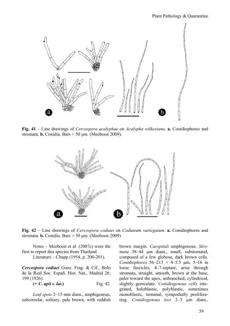
Fig. 40 – L<strong>in</strong>e draw<strong>in</strong>gs of <strong>Cercospora</strong> cocc<strong>in</strong>iae on Cocc<strong>in</strong>ia gr<strong>and</strong>is. a. Conidia. b. Conidiophores <strong>and</strong> stromata. Bars = 50 μm. (Meeboon 2009). ferat<strong>in</strong>g. Conidiogenous loci 1.5–2.5 μm diam., conspicuous, thickened <strong>and</strong> darkened. Conidia 41–102 × 2.5–5 μm, solitary, obclavatecyl<strong>in</strong>dric, straight to mildly curved, hyal<strong>in</strong>e, 5– 10-septate, very variable <strong>in</strong> length, smooth obconically truncate at the base, taper<strong>in</strong>g toward a subacute apex, hila 1.5–2.5 μm diam., thickened <strong>and</strong> darkened. Specimen exam<strong>in</strong>ed – THAILAND, Chiang Mai Prov<strong>in</strong>ce, Chiang Mai University, Faculty of Agriculture, on leaves of Cocc<strong>in</strong>ia gr<strong>and</strong>is (L.) Voigt (Cucurbitaceae), 29 February 2008, Jamjan Meeboon (BBH 23564). Hosts – Cocc<strong>in</strong>ia <strong>in</strong>dica, Momordica charantia (Cucurbitaceae) (Crous & Braun 2003), Cocc<strong>in</strong>ia gr<strong>and</strong>is (Meeboon 2009). Distribution – Brunei, India, Pakistan, <strong>Thail<strong>and</strong></strong> (Crous & Braun 2003, Meeboon 2009). Notes – This specimens is typical of C. cocc<strong>in</strong>iae is hav<strong>in</strong>g obclavate conidia. <strong>Cercospora</strong> cocc<strong>in</strong>iae was first reported from <strong>Thail<strong>and</strong></strong> by Meeboon (2009). 58 Euphorbiaceae <strong>Cercospora</strong> acalyphae Peck, Rep. (Annual) New York State Mus. Nat. Hist. 34: 48 (1881). = <strong>Cercospora</strong> acalypharum Tharp, Mycologia 9: 106 (1917). ≡ Cercospor<strong>in</strong>a acalypharum (Tharp) Sacc., Syll. Fung. 25: 902 (1931). Fig. 41 Leaf spots 15–30 mm diam., amphigenous, circular or subcircular, symptoms at first pale greenish to ochraceous, later brown to dark brown, f<strong>in</strong>ally <strong>with</strong> grayish brown at the centre, surrounded by a dark marg<strong>in</strong>. Caespituli amphigenous. Stromata 25–47 μm diam., <strong>in</strong>traepidermal, well-developed, composed of globose to subglobose, brown to blackish brown cells. Conidiophores 48.5–83.5 × 4–6 μm, 2–8 <strong>in</strong> loose fascicules, 1–3-septate, aris<strong>in</strong>g from stomata, simple, straight, erect to decumbent, smooth, pale yellow to pale brown, sometimes branched, subcyl<strong>in</strong>drical, slightly geniculate. Conidiogenous cells <strong>in</strong>tegrated, term<strong>in</strong>al, holoblastic, mostly monoblastic, sometimes polyblastic, sympodially proliferat<strong>in</strong>g. Conidiogenous loci 2.5–3.5 μm diam., conspicuous, thickened <strong>and</strong> darkened. Conidia 44–256 × 1.5–3 μm, solitary, narrowly obclavate to subacicular, straight, hyal<strong>in</strong>e, 4–18septate, smooth, base obconically truncate, <strong>with</strong> subacute apex, hila 2–3 μm diam., thickened <strong>and</strong> darkened. Specimens exam<strong>in</strong>ed – THAILAND, Phetchabun Prov<strong>in</strong>ce, Nam Nao National Park, on leaves of Acalypha wilkesiana Mull. Arg. (Euphorbiaceae), 24 November 2004, Chiharu Nakashima <strong>and</strong> Jamjan Meeboon (CMU 27898); Chiang Mai Prov<strong>in</strong>ce, A. Mae Jo, Sansai, Farm<strong>in</strong>g area, on leaves of A. wilkesiana, 31 July 2008, Jamjan Meeboon (BBH 23694). Hosts – Acalypha alopecurioides, A. australis, A. ciliata, A. gracilens, A. gr<strong>and</strong>is, A. hispida, A. <strong>in</strong>dica, A. marg<strong>in</strong>ata, A. marvorata, A. ostryifolia, A. rhomboidea, A. virg<strong>in</strong>ica, A. wilkesiana, Acalypha sp. (Euphorbiaceae) (Crous & Braun 2003, Meeboon et al. 2007c). Distribution – Brazil, Ch<strong>in</strong>a, Cuba, India, Jamaica, Japan, Solomon Isl<strong>and</strong>s, <strong>Thail<strong>and</strong></strong>, USA, Venezuela (Crous & Braun 2003, Meeboon et al. 2007c).
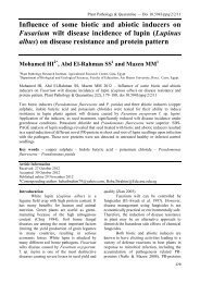
Plant Pathology & Quarant<strong>in</strong>e Fig. 41 – L<strong>in</strong>e draw<strong>in</strong>gs of <strong>Cercospora</strong> acalyphae on Acalypha wilkesiana. a. Conidiophores <strong>and</strong> stromata. b. Conidia. Bars = 50 μm. (Meeboon 2009). Fig. 42 – L<strong>in</strong>e draw<strong>in</strong>gs of <strong>Cercospora</strong> codiaei on Codiaeum variegatum. a. Conidiophores <strong>and</strong> stromata. b. Conidia. Bars = 50 μm. (Meeboon 2009) Notes – Meeboon et al. (2007c) were the first to report this species from <strong>Thail<strong>and</strong></strong>. Literature – Chupp (1954, p. 200-201). <strong>Cercospora</strong> codiaei Gonz. Frag. & Cif., Boln de la Real Soc. Españ. Hist. Nat., Madrid 26: 199 (1926). (= C. apii s. lat.) Fig. 42 Leaf spots 2–15 mm diam., amphigenous, subcircular, solitary, pale brown, <strong>with</strong> reddish brown marg<strong>in</strong>. Caespituli amphigenous. Stromata 38–44 μm diam., small, substomatal, composed of a few globose, dark brown cells. Conidiophores 56–213 × 4–5.5 μm, 5–16 <strong>in</strong> loose fascicles, 4–7-septate, arise through stromata, straight, smooth, brown at the base, paler toward the apex, unbranched, cyl<strong>in</strong>drical, slightly geniculate. Conidiogenous cells <strong>in</strong>tegrated, holoblastic, polyblastic, sometimes monoblastic, term<strong>in</strong>al, sympodially proliferat<strong>in</strong>g. Conidiogenous loci 2–3 μm diam., 59
- Page 1 and 2: Plant Pathology & Quarantine Genus
- Page 3 and 4: etter workable system. Braun (1993)
- Page 5 and 6: Plant Pathology & Quarantine Fig. 1
- Page 7 and 8: Plant Pathology & Quarantine Fig. 4
- Page 9 and 10: Plant Pathology & Quarantine Fig. 5
- Page 11 and 12: Pseudocercospora based on ITS regio
- Page 13 and 14: Plant Pathology & Quarantine Fig. 8
- Page 15 and 16: Plant Pathology & Quarantine Fig. 9
- Page 17 and 18: Key to the treated species in Thail
- Page 19 and 20: Hydrangeaceae A single species, on
- Page 21 and 22: Distribution - China, Thailand (Cro
- Page 23 and 24: Hosts - Adiantum philippense, Doryo
- Page 25 and 26: Stromata 19-29 µm diam., small, co
- Page 27 and 28: smooth, pale brown to brown, unbran
- Page 29 and 30: Plant Pathology & Quarantine Fig. 2
- Page 31 and 32: Plant Pathology & Quarantine Fig. 2
- Page 33 and 34: Fig. 25 - Line drawings of Cercospo
- Page 35 and 36: Plant Pathology & Quarantine Fig. 2
- Page 37 and 38: Plant Pathology & Quarantine Fig. 2
- Page 39 and 40: diam., substomatal to intraepiderma
- Page 41 and 42: Conidia 17-93 × 3-5 μm, solitary,
- Page 43 and 44: Carica papaya L. (Caricaceae), 12 S
- Page 45 and 46: Fig. 37 - Line drawings of Cercospo
- Page 47: Fig. 39 - Line drawings of Cercospo
- Page 51 and 52: Hosts - Phyllanthus niruri, Sauropu
- Page 53 and 54: Fig. 45 - Line drawings of Cercospo
- Page 55 and 56: Fungi imperfecti parasitici 1. Hyph
- Page 57 and 58: Plant Pathology & Quarantine Fig. 4
- Page 59 and 60: Plant Pathology & Quarantine Fig. 5
- Page 61 and 62: Fig. 53 - Line drawings of Cercospo
- Page 63 and 64: Distribution - Thailand (type local
- Page 65 and 66: ase, paler toward the apex, unbranc
- Page 67 and 68: Plant Pathology & Quarantine Fig. 6
- Page 69 and 70: Leaf spots 2-15 mm diam., amphigeno
- Page 71 and 72: obconically truncate, with subacute
- Page 73 and 74: Plant Pathology & Quarantine Fig. 6
- Page 75 and 76: and tolerant Cercospora beticola. J
- Page 77: Pollack FG. 1987 - An annotated com