Developmental Biology BY1101 Lectures 4 and 5 Cleavage-
Developmental Biology BY1101 Lectures 4 and 5 Cleavage-
Developmental Biology BY1101 Lectures 4 and 5 Cleavage-

You also want an ePaper? Increase the reach of your titles
YUMPU automatically turns print PDFs into web optimized ePapers that Google loves.
<strong>Developmental</strong> <strong>Biology</strong> <strong>BY1101</strong><br />
P. Murphy<br />
<strong>Lectures</strong> 4 <strong>and</strong> 5<br />
The first steps to forming a new organism<br />
Descriptive embryology 2<br />
<strong>Cleavage</strong>, Gastrulation, Neurulation <strong>and</strong> Organogenesis<br />
Early animal development can be divided into the following key events<br />
Fertilisation – cleavage – gastrulation – neurulation – organogenesis<br />
Lecture 3 Lecture 4 <strong>and</strong> Lecture 5<br />
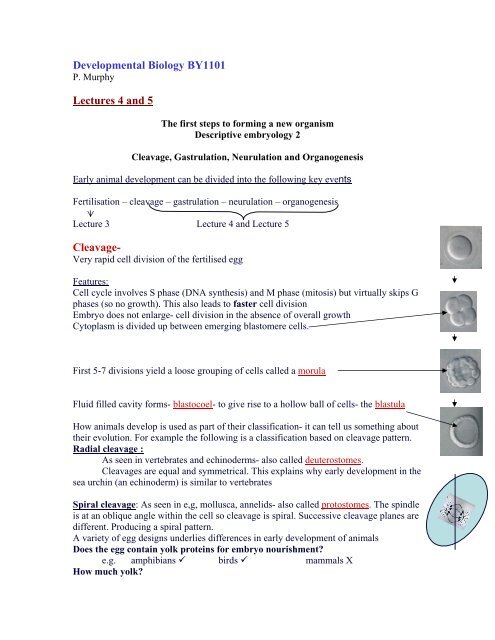
<strong>Cleavage</strong>-<br />
Very rapid cell division of the fertilised egg<br />
Features:<br />
Cell cycle involves S phase (DNA synthesis) <strong>and</strong> M phase (mitosis) but virtually skips G<br />
phases (so no growth). This also leads to faster cell division<br />
Embryo does not enlarge- cell division in the absence of overall growth<br />
Cytoplasm is divided up between emerging blastomere cells.<br />
First 5-7 divisions yield a loose grouping of cells called a morula<br />
Fluid filled cavity forms- blastocoel- to give rise to a hollow ball of cells- the blastula<br />
How animals develop is used as part of their classification- it can tell us something about<br />
their evolution. For example the following is a classification based on cleavage pattern.<br />
Radial cleavage :<br />
As seen in vertebrates <strong>and</strong> echinoderms- also called deuterostomes.<br />
<strong>Cleavage</strong>s are equal <strong>and</strong> symmetrical. This explains why early development in the<br />
sea urchin (an echinoderm) is similar to vertebrates<br />
Spiral cleavage: As seen in e,g, mollusca, annelids- also called protostomes. The spindle<br />
is at an oblique angle within the cell so cleavage is spiral. Successive cleavage planes are<br />
different. Producing a spiral pattern.<br />
A variety of egg designs underlies differences in early development of animals<br />
Does the egg contain yolk proteins for embryo nourishment?<br />
e.g. amphibians birds mammals X<br />
How much yolk?
e.g. most plentiful in birds, reptiles, many fishes <strong>and</strong> insects.<br />
less in sea urchins<br />
How is the yolk distributed? Basis of differences between frog <strong>and</strong> chick.<br />
<strong>Cleavage</strong> in the frog<br />
The egg has polarity<br />
Upper pigmented side- animal pole; lower yolky side- vegetal pole.<br />
The movement of molecules in the egg -triggered by fertilisation<br />
→ appearance of “gray crescent”<br />
First two cleavages are vertical<br />
<strong>Cleavage</strong> 3 is horizontal<br />
Yolk content displaces plane of division toward animal pole.<br />
⇒More cells <strong>and</strong> blastocoel in animal half<br />
Fig 47.7<br />
This stage is the blastula<br />
<strong>Cleavage</strong> pattern is similar in all animals classified as deuterostomes-<br />
But in eggs with less yolk e.g. sea urchin, the blastomeres are of more even size <strong>and</strong> the<br />
blastocoel is more centrally located<br />
<strong>Cleavage</strong> in the chick<br />
•The entire “yolk” of the chicken egg is the egg cell- packed full of yolk proteins.<br />
•The “white” - a protein rich solution bathing the cell- also used for nutrition<br />
•All the cytoplasm is contained in a tiny disk at the animal pole of the yolk.<br />
<strong>Cleavage</strong> planes cannot penetrate the dense yolk so only the animal pole<br />
cytoplasm is cleaved. This incomplete cleavage is know as meroblastic cleavage<br />
(complete cleavage is called holoblastic)<br />
<strong>Cleavage</strong> produces a cap of cells sitting on top of yolk- called blastoderm<br />
So the blastoderm in the chick is the equivalent of the blastula in<br />
the frog- the shape is related to egg architecture (yolk content <strong>and</strong> distribution).<br />
The cavity that forms (blastocoel) divides two layers of cells: the<br />
hypoblast <strong>and</strong> the epiblast. The embryo will emerge entirely from<br />
the epiblast.<br />
Therefore the embryo will emerge from a simple flat layer of cells
<strong>Cleavage</strong> in different species can produce a ball or disk of cells. How does that turn into<br />
an organised embryo?<br />
The next step is gastrulation- Literally means “making a gut”<br />
Gastrulation<br />
Marked by extensive cell movement where the cells of the blastula become organised<br />
into three layers. This 3 layered embryo has a primitive gut.<br />
The details of the movements are different in different organisms (again related to the<br />
architecture of the egg) but the outcome is the same- a 3 layered embryo<br />
The 3-layered embryo is called a gastrula<br />
Morula blastula gastrula<br />
A ball of cells hollow ball 3-layered<br />
hollow ball<br />
Gastrulation best studied in the frog.<br />
How could the early embryologists figure out the details of cell movement in a tiny<br />
embryo?<br />
1. Frog embryo is not so tiny<br />
2. The obviously visible poles aid visualization of movements<br />
3. They used methods to mark cells <strong>and</strong> follow their movements <strong>and</strong> their fate- cell<br />
lineage marking / cell fate mapping<br />
Cell lineage marking / Cell fate mapping<br />
Mark a cell early in development <strong>and</strong> observe where it <strong>and</strong> its descendent cells end up.<br />
This is how cell fate maps are made <strong>and</strong> how we can<br />
follow the movements <strong>and</strong> fates of all the cells<br />
Gastrulation occurs in all eumetazoa (animals with an organised body plan)<br />
Animals that do not undergo gastrulation are the most “simple” animals with no organs<br />
e.g. jelly fish.<br />
“triploblastic” animals – gastrulation produces 3 germ layers.<br />
Some animals are diploblastic (e.g. hydra- ;there are only two layers)<br />
The 3 layers formed are called the embryonic germ layers<br />
Ectoderm “ecto”: outer<br />
Mesoderm “meso”: middle<br />
Endoderm “endo”: inside (Note: the colour scheme used here is the classical<br />
scheme for mapping ectoderm (blue), mesoderm (red) <strong>and</strong> endoderm (yellow))<br />
Eventually these 3 germ layers develop into all the tissues <strong>and</strong> organs of the adult animal
The sea urchin- gastrulation on a simple scale<br />
The blastula is a single layered hollow ball<br />
1. Gastrulation begins at the vegetal pole- individual cells detach <strong>and</strong><br />
enter the blastocoel- first mesenchyme cells (top arrow)<br />
2. The vegetal plate destined to form endoderm- flattens <strong>and</strong> buckles<br />
inward- invagination (arrow)<br />
3. Invagination deepens to form a tube called the Archenteron-<br />
this is the primitive gut (arrow).<br />
4. Extensions from the mesenchyme cells called filopodia- drag the<br />
archenteron toward the blastocoel wall (arrow)<br />
5. Archenteron fuses with blastocoel wall - forms digestive tube with<br />
mouth <strong>and</strong> anus- 3 layers - mesenchyme forms future skeleton<br />
Frog gastrulation<br />
Gastrulation is much more complex in the frog because there are more<br />
cells <strong>and</strong> there are large yolk laden cells in the vegetal pole, but the<br />
process also produces a 3-layered gastrula with an archenteron as a<br />
primitive gut.<br />
In Fig 47.10 panel 1 you see the fate map of where the future<br />
ectoderm, mesoderm <strong>and</strong> endoderm are located at the start of gastrulation.<br />
Gastrulation begins when a line of cells begin to change shape <strong>and</strong><br />
invaginate. This occurs at the earlier site of the gray crescent <strong>and</strong> is<br />
called the dorsal lip of the blastopore.<br />
Panel 2: Future mesoderm cells <strong>and</strong> endoderm cells at the site of the dorsal<br />
lip then begin to roll over the lip <strong>and</strong> involute into the interior of the embryo.
Inside, these cells move away from the blastopore <strong>and</strong> become organised into layers of<br />
endoderm <strong>and</strong> mesoderm.<br />
Panel 3: The original cavity- the blastocoel- is collapsed by the movement<br />
<strong>and</strong> a new cavity is created- this is the archenteron <strong>and</strong> will form the future<br />
gut. The yolk rich cells are internalised in the region of the future gut.<br />
Meanwhile the remaining cells on the surface that will form the ectoderm<br />
(blue) move down around the surface to completely cover the embryo.<br />
As in the sea urchin the blastopore is the future site of the anus <strong>and</strong> the<br />
mouth will form at the opposite side.<br />
As a result of all the cell movement the embryo changes shape- elongates as the cells<br />
meet <strong>and</strong> converge inside the embryo<br />
Convergent extension<br />
-like lanes of traffic meeting <strong>and</strong> converging - leading to elongation (extension)<br />
Campbell <strong>and</strong> Reece Fig 47.16<br />
The important features of gastrulation:<br />
Elaborate cell movement.<br />
Differentiation of cells into 3 broad types: ectodermal, mesodermal <strong>and</strong> endodermal,<br />
placed in the right position with respect to each other.<br />
Allows communication between neighbouring cells that are now set apart as different to<br />
each other.<br />
Mechanisms involved in bringing about gastrulation- what is needed?<br />
• changes in cell motility<br />
• changes in cell adhesion (therefore molecules on the outside of the cells).<br />
Gastrulation may vary in detail from organism to organism but the result<br />
is similar:<br />
the production of a three layered gastrula with organised groups of<br />
cells that interact <strong>and</strong> co-operate to form the embryo.<br />
The positioning of the cell layers in the gastrula allows cells to interact in new ways.<br />
Gastrulation brought all the cells into the correct position within the embryo to facilitate<br />
local development of the organs- organogenesis.
While gastrulation involves massive cell movements, organogenesis involves more local<br />
interactions between cells <strong>and</strong> morphogenetic changes in tissue <strong>and</strong> cell shape- <strong>and</strong> in<br />
cell arrangement.<br />
Note that cell differentiation is happening continuously throughout these processes <strong>and</strong><br />
cells are getting closer to their final characteristics <strong>and</strong> functions.<br />
Many of the changes in organogenesis are brought about by Induction:- the influence of<br />
one group of cells on another changing the way in which the responding cells develop<br />
(more in lecture 8).<br />
The process of Neurulation is a very special type of organogenesis<br />
• It sets aside the cells for <strong>and</strong> forms the rudiments of the entire nervous system.<br />
• It is the first event in organogenesis.<br />
The first event in neurulation starts as gastrulation is still underway <strong>and</strong> the future<br />
mesoderm cells are moving inside the embryo. A particular group of these migrating<br />
mesoderm cells assemble into a rod-like structure along the dorsal midline of the future<br />
embryo.<br />
These special mesoderm cells form the notochord <strong>and</strong> have very special characteristics-<br />
they have very potent ability to induce neighbouring cells.<br />
Signals sent from the notochord to the ectoderm lying above induce the ectoderm to<br />
thicken <strong>and</strong> form the neural plate (green on the figure below) the rudiment of the<br />
nervous system<br />
See figure 47.12 <strong>and</strong> images below- note the notochord <strong>and</strong> the neural plate<br />
Cross section<br />
Dorsal view of frog embryo<br />
undergoing neurulation<br />
The outer edges of the neural plate then begin to fold upward- forming the neural folds.<br />
Eventually the two sides of the neural folds fuse to enclose the neural tube (see figure<br />
below).<br />
As the neural tube fuses the adjacent surface ectoderm (blue) also fuses to enclose the<br />
neural tube, which forms the rudiment of the brain <strong>and</strong> the spinal chord. The neural tube<br />
runs the length of the embryo from anterior where it forms the vesicles of the brain.
By the end of gastrulation <strong>and</strong> neurulation, the embryo has elongated with a head end <strong>and</strong><br />
a tail end <strong>and</strong> has an internalised neural tube running along the dorsal midline. In the<br />
head region this will form the brain chambers <strong>and</strong> elsewhere the spinal chord.<br />
head tail<br />
Cross-section<br />
As neurulation proceeds, other organs <strong>and</strong> structures are also starting to form.<br />
A special set of cells form at the dorsal edge of the neural tube- the neural crest cells<br />
(see below).
These migrate away from the neural tube to many destinations <strong>and</strong> form peripheral<br />
nerves, teeth, skull bones <strong>and</strong> other cell types.<br />
Note the coelom in the top figure – this is a fluid-filled cavity which forms by splitting<br />
the mesoderm- it is by definition surrounded by mesoderm on all sides<br />
Many of the organs as they form will push into this space- although contained within the<br />
peritoneum.<br />
The somites are another set of structures forming at this time.<br />
The somites are mesoderm cells that form into blocks on either side of the neural tube<br />
(see figure below). These are transitory structures that later dissociate to form different<br />
cell types.<br />
1. The muscle blocks of the body <strong>and</strong> muscles attached to the skeleton<br />
2. The vertebrae of the back-bone. The cells from within the somite that will form<br />
the vertebrae (blue on lower diagram) leave the somite <strong>and</strong> migrate around the<br />
notochord <strong>and</strong> neural tube. The notochord later degenerates; the only remnants<br />
forming the “disks” between the vertebrae.<br />
Blue: cells migrate around<br />
the notochord <strong>and</strong> neural<br />
tube <strong>and</strong> form the vertebrae<br />
Red: Muscle cells migrate<br />
around trunk <strong>and</strong> into the<br />
limb
The somites show that vertebrates are partially segmented.<br />
In organogenesis cells of one germ layer often induce neighbouring cells of another germ<br />
layer to change character <strong>and</strong> begin to form a tissue or part of an organ-<br />
-this is precisely why bringing the germ layers into the right position during<br />
gastrulation is so vital for development of the embryo.<br />
-More about induction <strong>and</strong> its molecular basis in lecture 8<br />
Gastrulation in the chick<br />
The process looks very different because the early embryo is a flat disk of cells with<br />
masses of yolk underneath<br />
The entire embryo will form from one cell layer- the epiblast. Gastrulation involves cells<br />
from the epiblast moving underneath through a line at the midline of the disk called the<br />
primitive streak. Some of these involuting cells form endoderm <strong>and</strong> some form<br />
mesoderm. The primitive streak marks the future anterior-posterior axis of the embryo.<br />
The primitive streak in the chick is equivalent to the blastopore in the frog (site through<br />
which the cells involute).<br />
In the chick, organogenesis proceeds much as it does in the frog but appears different<br />
because the chick embryo is flat <strong>and</strong> the frog round.<br />
Organogenesis in the chick: To achieve the same “tube within a tube” (gut within the<br />
body wall) structure of other embryos, the edges of the blastodisk (flat chick embryo)<br />
fold ventrally to enclose the gut - but the gut is still joined in the middle of the body to<br />
the yolk in the yolk sac. See Figure 47.13(a) reproduced below<br />
Note the neural tube, notochord, somites <strong>and</strong> endoderm of the future gut.<br />
Future gut
By 2-3 days of development the rudiments of all the major organs is seen in the<br />
chick embryo. See Figure 47.13(b) below. A movie showing the beating heat of<br />
the chick embryo at this stage was viewed.<br />
A movie of organogenesis in zebrafish was shown<br />
Fertilization<br />
<strong>Cleavage</strong><br />
Gastrulation<br />
Neurulation<br />
The figure on the right shows illustrations Organogenesis<br />
of embryos during each of the<br />
developmental processes that we have discussed<br />
Note it is a continuous series of processes<br />
A movie of the continuous processes listed above in zebrafish was viewed again<br />
The story so far- has led us from quite different egg designs to very similar<br />
embryos among vertebrates- as shown in the figure below for frog (Xenopus), chick,<br />
mouse <strong>and</strong> zebrafish
This was first noted by Karl Ernst von Baer in the early 19th century- he called the stage<br />
when all vertebrate embryos look alike (2 nd row above) the phylotypic stage- typical of<br />
the phylum. At this stage the general characteristics of the phylum are established-<br />
notochord, neural tube, somites <strong>and</strong> the very beginnings of the limbs.<br />
Von Baer devised some laws to express this observation: he stated in these laws that<br />
general characteristics of a group appear first while those that distinguish a species<br />
appear later<br />
But why is this? He couldn’t really explain it – there is something he didn’t know<br />
but we know today….<br />
….organisms share similar developmental mechanisms.<br />
The mechanisms driving the changes you have just seen will be explored in future<br />
lectures<br />
Lecture 7: cell differentiation<br />
Lecture 8: cell communication<br />
<strong>Lectures</strong> 9-10: developmental genetics<br />
Key concepts in lectures 4 <strong>and</strong> 5<br />
1. <strong>Cleavage</strong> refers to the very rapid cell division of the fertilised egg. It is marked by little<br />
or no protein synthesis <strong>and</strong> no growth of the embryo.<br />
2. The first divisions form a loose group of cells called a morula.<br />
3. After further divisions a cavity forms (blastocoel) – the embryo is then called a blastula<br />
4. <strong>Cleavage</strong> is holoblastic if it divides the entire egg into blastomeres (cells) but<br />
meroblastic if only the cytoplasm is divided <strong>and</strong> the yolk proteins are excluded (e.g.<br />
chick).<br />
5. The appearance of cleavage <strong>and</strong> the resulting embryo depends on the amount <strong>and</strong><br />
distribution of yolk. <strong>Cleavage</strong> produces a ball or disk of cells. Gastrulation then turns that<br />
ball or disk into an organised embryo.<br />
6. Gastrulation is a fundamental event in all complex animals (eumetazoa) with organs. It<br />
leads to the formation of a 3 germ layered embryo (for triploblastic animals)- the three<br />
germ layers will give rise to all the future organs <strong>and</strong> structures.<br />
7. The three germ layers, the ectoderm, mesoderm <strong>and</strong> endoderm contain 3 broadly<br />
different cell types.
8. Gastrulation is characterised by elaborate cell movement so that the three germ layers<br />
end up in the correct position with respect to each other.<br />
9. This sets groups of cells apart as different to each other <strong>and</strong> allows communication<br />
between neighbouring cells that will be so important in future events- organogenesis.<br />
10. Changes in cell motility <strong>and</strong> changes in cell adhesion (therefore molecules on the<br />
outside of the cells) are important in bringing about the cell movements of gastrulation.<br />
11. Gastrulation brought all the cells into the correct position within the embryo to<br />
facilitate local development of the organs- organogenesis.<br />
12. While gastrulation involved massive cell movements, organogenesis involves more<br />
local interactions between cells <strong>and</strong> morphogenetic changes in tissue <strong>and</strong> cell shape, <strong>and</strong><br />
in cell arrangement.<br />
13. Many of the changes in organogenesis are brought about by Induction- cells of one<br />
germ layer often induce neighbouring cells of another germ layer to change character <strong>and</strong><br />
begin to form a tissue or part of an organ- underlines the importance of bringing the germ<br />
layers into the right position during gastrulation.<br />
14. The process of Neurulation- a special event in organogenesis that sets aside the cells<br />
for <strong>and</strong> forms the rudiments of the entire nervous system.<br />
(Make sure you can describe the events involved in neurulation)<br />
15. By the end of gastrulation <strong>and</strong> neurulation, the embryo has elongated with a head end<br />
<strong>and</strong> a tail end <strong>and</strong> has an internalised neural tube running along the dorsal midline. In the<br />
head region this will form the brain chambers <strong>and</strong> elsewhere the spinal chord.<br />
16. The somites are mesoderm cells that form into blocks on either side of the neural<br />
tube- transitory structures that later dissociate to form different cell types including the<br />
muscle blocks <strong>and</strong> the vertebrae<br />
17. In the chick, organogenesis proceeds much as it does in the frog but appears different<br />
because the chick embryo is flat <strong>and</strong> the frog round.<br />
18. The processes that form the embryo (cleavage, gastrulation <strong>and</strong> organogenesis) occur<br />
as a continuous series of interlinked events- they are not discrete.<br />
19. The early observation by von Baer that all vertebrate embryos look similar at the end<br />
of gastrulation <strong>and</strong> early organogenesis reflects the fact that molecular <strong>and</strong> cellular events<br />
that underlie early development are very similar across all animals.<br />
Lecture 4 <strong>and</strong> Lecture 5: Learning outcomes: you should be able to….<br />
A) State what cleavage is <strong>and</strong>, using appropriate terminology, describe cleavage in the<br />
frog <strong>and</strong> differences in the chick.
B) State what gastrulation is <strong>and</strong> describe its fundamental importance in all complex<br />
animals. Describe gastrulation in frog but be able to discuss the differences between frog<br />
<strong>and</strong> chick <strong>and</strong> how the differences in detail are related to structure of the egg- but that the<br />
outcome is the same.<br />
C) Distil the important features of gastrulation from the descriptions <strong>and</strong> relate this to its<br />
important outcomes.<br />
D) State what organogenesis is <strong>and</strong> contrast it with the earlier events of gastrulation.<br />
E) Describe neurulation as a very early event in organogenesis <strong>and</strong> the importance of this<br />
early event as part of establishing the body plan.<br />
Key terms to be familiar with: <strong>Cleavage</strong>, blastomere cells, morula, blastocoel, blastula,<br />
gray crescent, holoblastic cleavage, meroblastic cleavage, blastoderm, epiblast,<br />
hypoblast, gastrulation, gastrula, fate map, germ layers, ectoderm, mesoderm, endoderm,<br />
invagination, archenteron, blastopore, involution, convergent extension, primitive streak,<br />
Henson’s node,