

INTRODUCTION Granulomatous inflammation is a distinctive ...
INTRODUCTION Granulomatous inflammation is a distinctive ...
INTRODUCTION Granulomatous inflammation is a distinctive ...

You also want an ePaper? Increase the reach of your titles
YUMPU automatically turns print PDFs into web optimized ePapers that Google loves.
Occasionally, granulomas coalesce to form mass like lesions, particularly in the region of the<br />
chiasma, floor of the third ventricle, and pituitary stalk. Clinical symptoms correlate with the<br />
location of the lesion on MR images. Furthermore, it was found that these lesions recur frequently<br />
(Ando et al., 1985). D<strong>is</strong>ease entities that can involve the basal leptomeninges and mimic<br />
leptomeningeal sarcoidos<strong>is</strong> on imaging include granulomatous d<strong>is</strong>ease (such as TB), Wegener<br />
granulomatos<strong>is</strong> and fungal meningit<strong>is</strong>, pyogenic meningit<strong>is</strong>, leptomeningeal lymphoma,<br />
demyelination, meningoangiomatos<strong>is</strong>, acute lymphocytic leukemia, and leptomeningeal<br />
carcinomatos<strong>is</strong> (Brooks et al., 1982).<br />
Dural/epidural mass lesions have an imaging appearance similar to that of meningioma/epidural<br />
lymphoma and are not associated with intraparenchymal extension. Dural/epidural sarcoidos<strong>is</strong><br />
probably represents blood borne deposits of the d<strong>is</strong>ease in the epidural spaces. The annual<br />
incidence of these lesions exceeds the annual incidence of meningiomas (2.3 per 100,000) by 130<br />
times. These lesions tend to be <strong>is</strong>ointense with gray matter on T1-weighted MR images and<br />
hypointense on T2-<br />
weighted images, and they enhance uniformly (Figure 19). Th<strong>is</strong> hypointensity on T2-weighted<br />
images has been reported previously (Chaflenor et al., 1984) and <strong>is</strong> thought to be related to<br />
fibrocollagenous buildup. Unfortunately, th<strong>is</strong> <strong>is</strong> not a unique finding. Eighteen percent of<br />
meningiomas (typically fibroblastic or transitional types) demonstrate low signal on T2-weighted<br />
images.<br />
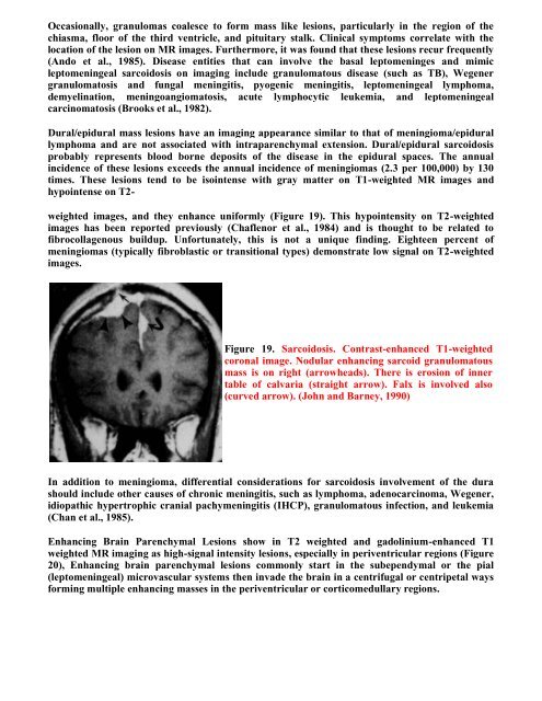
Figure 19. Sarcoidos<strong>is</strong>. Contrast-enhanced T1-weighted<br />
coronal image. Nodular enhancing sarcoid granulomatous<br />
mass <strong>is</strong> on right (arrowheads). There <strong>is</strong> erosion of inner<br />
table of calvaria (straight arrow). Falx <strong>is</strong> involved also<br />
(curved arrow). (John and Barney, 1990)<br />
In addition to meningioma, differential considerations for sarcoidos<strong>is</strong> involvement of the dura<br />
should include other causes of chronic meningit<strong>is</strong>, such as lymphoma, adenocarcinoma, Wegener,<br />
idiopathic hypertrophic cranial pachymeningit<strong>is</strong> (IHCP), granulomatous infection, and leukemia<br />
(Chan et al., 1985).<br />
Enhancing Brain Parenchymal Lesions show in T2 weighted and gadolinium-enhanced T1<br />
weighted MR imaging as high-signal intensity lesions, especially in periventricular regions (Figure<br />
20), Enhancing brain parenchymal lesions commonly start in the subependymal or the pial<br />
(leptomeningeal) microvascular systems then invade the brain in a centrifugal or centripetal ways<br />
forming multiple enhancing masses in the periventricular or corticomedullary regions.