INTRODUCTION Granulomatous inflammation is a distinctive ...
INTRODUCTION Granulomatous inflammation is a distinctive ...
INTRODUCTION Granulomatous inflammation is a distinctive ...
You also want an ePaper? Increase the reach of your titles
YUMPU automatically turns print PDFs into web optimized ePapers that Google loves.
deficit. CSF <strong>is</strong> largely normal or may show a slight increase in proteins and cells. Pathologically,<br />
there <strong>is</strong> diffuse oedema of cerebral white matter with loss of neurons in grey matter. A picture<br />
resembling haemorrhagic leukoencephalopathy or a post-infectious demyelinating<br />
encephalomyelit<strong>is</strong> may be observed (Udani and Dastur, 1970; Dastur et al., 1995).<br />
Pott’s spine and Pott’s paraplegia<br />
It <strong>is</strong> estimated that involvement of the spine occurs in less than 1% of patients with TB. It <strong>is</strong> a<br />
leading cause of paraplegia in developing nations. Infection in the vertebral bodies usually starts<br />
in cancellous bone adjacent to an intervertebral d<strong>is</strong>c or anteriorly under the periosteum of the<br />
vertebral body; the neural arch <strong>is</strong> rarely affected. Vertebral destruction leads to collapse of the<br />
body of the vertebra along with anterior wedging. Spinal cord compression in Pott’s spine <strong>is</strong><br />
mainly caused by pressure from a paraspinal abscess which <strong>is</strong> retropharyngeal in the cervical<br />
region, and spindle shaped in thoracic and thoracolumbar regions. Neurological deficits may also<br />
result from dural invasion by granulation t<strong>is</strong>sue and compression from the debr<strong>is</strong> of sequestrated<br />
bone, a destroyed intervertebral d<strong>is</strong>c, or a d<strong>is</strong>located vertebra. Rarely, vascular insufficiency in<br />
the territory of the anterior spinal artery has also been suggested. Neurological involvement can<br />
occur at any stage of Pott’s spine and even years later, when there has been apparent healing,<br />
because of stretching of the cord in the deformed spinal canal. The thoracic spine <strong>is</strong> involved in<br />
about 65% of cases, and the lumbar, cervical and thoracolumbar spine in about 20%, 10% and<br />
5%, respectively. The atlanto-axial region may also be involved in less than 1% of cases. Males are<br />
affected more often than females in most series, and the d<strong>is</strong>ease generally affects young persons<br />
(Vidyasagar and Murthy, 1994; Nussbaum et al.,1995; Razai et al., 1995;).<br />
Typically, there <strong>is</strong> a h<strong>is</strong>tory of local pain, tenderness over the affected spine or even overlying<br />
bony deformity. Paravertebral abscess may be palpated on the back of a number of patients.<br />
These patients usually have acute or subacute, progressive, spastic type of sensorimotor<br />
parapares<strong>is</strong>. The incidence of parapares<strong>is</strong> in patients with Pott’s spine varies from 27% to 47%.<br />
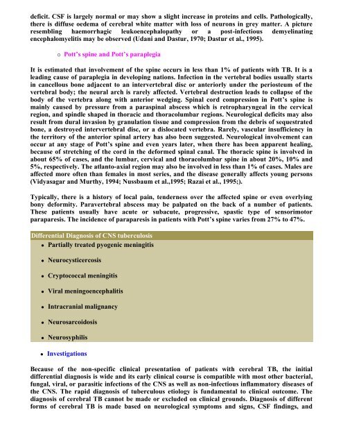
Differential Diagnos<strong>is</strong> of CNS tuberculos<strong>is</strong><br />
Partially treated pyogenic meningit<strong>is</strong><br />
Neurocysticercos<strong>is</strong><br />
Cryptococcal meningit<strong>is</strong><br />
Viral meningoencephalit<strong>is</strong><br />
Intracranial malignancy<br />
Neurosarcoidos<strong>is</strong><br />
Neurosyphil<strong>is</strong><br />
Investigations<br />
Because of the non-specific clinical presentation of patients with cerebral TB, the initial<br />
differential diagnos<strong>is</strong> <strong>is</strong> wide and its early clinical course <strong>is</strong> compatible with most other bacterial,<br />
fungal, viral, or parasitic infections of the CNS as well as non-infectious inflammatory d<strong>is</strong>eases of<br />
the CNS. The rapid diagnos<strong>is</strong> of tuberculous etiology <strong>is</strong> fundamental to clinical outcome. The<br />
diagnos<strong>is</strong> of cerebral TB cannot be made or excluded on clinical grounds. Diagnos<strong>is</strong> of different<br />
forms of cerebral TB <strong>is</strong> made based on neurological symptoms and signs, CSF findings, and


