Chromatofocusing - Umbc
Chromatofocusing - Umbc
Chromatofocusing - Umbc

Create successful ePaper yourself
Turn your PDF publications into a flip-book with our unique Google optimized e-Paper software.
<strong>Chromatofocusing</strong><br />
Position and Width of Protein Bands in<br />
the Column Effluent<br />
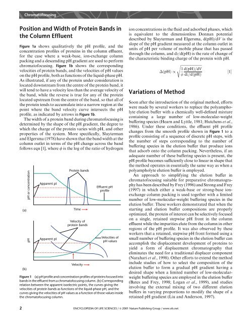
Figure 1a shows qualitatively the pH profile, and the<br />
concentration profiles of proteins in the column effluent,<br />
for the case where a weak-base, ion-exchange column<br />
packing and a descending pH gradient are used to perform<br />
chromatofocusing. Figure 1b shows the corresponding<br />
velocities of protein bands, and the velocities of pH values<br />
on the pH profile, both as functions of the liquid-phase pH.<br />
As illustrated, if any of the protein under consideration is<br />
located downstream from the centre of the protein band, it<br />
will tend to have a velocity less than the average velocity of<br />
the band, while the reverse is true for any of the protein<br />
located upstream from the centre of the band, so that all of<br />
the protein tends to accumulate into a narrow region at the<br />
point where the band velocity curve intersects the pH<br />
profile, as indicated by arrows in Figure 1b.<br />
The width of a protein band during chromatofocusing is<br />
determined by the shape of the pH gradient, the degree to<br />
which the charge of the protein varies with pH, and other<br />
properties of the system. More specifically, Sluyterman<br />
and Elgersma (1978) have shown that the band width at the<br />
column outlet in terms of the pH change across the band<br />
follows eqn [1], where f is the log of the ratio of hydrogen<br />
(a)<br />
(b)<br />
pH or concentration<br />
pH<br />
Apparent pI<br />
Apparent pI<br />
Protein bands<br />
Time<br />
Velocity of<br />
protein bands<br />
Velocity<br />
Effluent pH<br />
profile<br />
Velocities of<br />
pH values<br />
Figure 1 (a) pH profile and concentration profiles of proteins focused into<br />
bands in the effluent from a chromatofocusing column. (b) Corresponding<br />
relation between the apparent isoelectric points, the curves giving the<br />
velocities of protein bands as functions of the liquid-phase pH, and the<br />
curves giving the velocities of pH values as a function of those values inside<br />
the chromatofocusing column.<br />
ion concentrations in the fluid and adsorbed phases, which<br />
is equivalent to the dimensionless Donnan potential<br />
described by Sluyterman and Elgersma, d(pH)/dV is the<br />
slope of the pH gradient measured at the column outlet in<br />
units of pH per volume of mobile phase that has passed<br />
through the column, and dz/d(pH) is the rate of change of<br />
the characteristic binding charge of the protein with pH.<br />
s<br />
1 d…pH†=dV<br />
…pH† / ‰1Š<br />
dz=d…pH†<br />
Variations of Method<br />
2 ENCYCLOPEDIA OF LIFE SCIENCES / & 2001 Nature Publishing Group / www.els.net<br />
Soon after the introduction of the original method, efforts<br />
were made by several workers to replace the polyampholyte<br />
elution buffer with a chemically well-defined mixture<br />
containing a large number of low-molecular-weight<br />
buffering species (Hearn and Lyttle, 1981;Hutchens et al.,<br />
1986). Under these conditions, the effluent pH profile<br />
changes from the smooth profile shown in Figure 1 to a<br />
profile consisting of a sequence of discrete pH steps, with<br />
the number of steps corresponding to the number of<br />
buffering species in the elution buffer that produce ions<br />
that adsorb onto the column packing. Nevertheless, if an<br />
adequate number of these buffering species is present, the<br />
pH profile becomes sufficiently close to linear in shape that<br />
the method operates in essentially the same way as when a<br />
polyampholyte elution buffer is employed.<br />
An approach to simplifying the elution buffer in<br />
chromatofocusing suitable for preparative chromatography<br />
has been described by Frey (1996) and Strong and Frey<br />
(1997) in which either a weak-base or strong-base ionexchange<br />
column packing is used together with a limited<br />
number of low-molecular-weight buffering species in the<br />
elution buffer. These workers demonstrated that when the<br />
starting and elution buffer compositions are properly<br />
optimized, the protein of interest can be selectively focused<br />
on a single, retained stepwise pH front in the column<br />
effluent while the impurities elute from the column in other<br />
regions of the pH profile. It was also observed by these<br />
workers that a retained, stepwise pH front formed using a<br />
small number of buffering species in the elution buffer can<br />
accomplish the displacement development of proteins to<br />
yield a form of displacement chromatography that<br />
eliminates the need for a traditional displacer component<br />
(Narahari et al., 1998). Other efforts to extend the method<br />
include studies of how to select the composition of the<br />
elution buffer to form a gradual pH gradient having a<br />
desired shape when a limited number of low-molecularweight<br />
buffering species are employed in the elution buffer<br />
(Bates and Frey, 1998;Logan et al., 1999), and studies<br />
involving the external mixing of two different elution<br />
buffers in varying proportions to modify the shape of a<br />
retained pH gradient (Liu and Anderson, 1997).