Chromatofocusing - Umbc
Chromatofocusing - Umbc
Chromatofocusing - Umbc

Create successful ePaper yourself
Turn your PDF publications into a flip-book with our unique Google optimized e-Paper software.
<strong>Chromatofocusing</strong><br />
Douglas D Frey, University of Maryland Baltimore County, Maryland, USA<br />
Chittoor R Narahari, University of Maryland Baltimore County, Maryland, USA<br />
Ronald C Bates, University of Maryland Baltimore County, Maryland, USA<br />
<strong>Chromatofocusing</strong> is a form of gradient elution chromatography performed using an ionexchange<br />
column packing and an internally generated pH gradient that travels through<br />
the column as a retained pH front.<br />
Introduction<br />
The technique of chromatofocusing is used to separate<br />
amphoteric substances, most commonly proteins, and was<br />
originally developed by Sluyterman and his colleagues<br />
(Sluyterman and Elgersma, 1978;Sluyterman and Wijdeness,<br />
1978). In contrast to the related technique of<br />
isoelectric focusing, chromatofocusing does not utilize an<br />
electric field. Instead, a pH gradient is made to propagate<br />
inside an ion-exchange chromatography column as a<br />
retained front owing to the adsorption behaviour of the<br />
buffering species in the elution buffer. The separation<br />
achieved is based on charge differences between proteins.<br />
Proteins elute from a chromatofocusing column at a pH,<br />
generally termed the apparent isoelectric point, that is<br />
often close to the actual isoelectric point. <strong>Chromatofocusing</strong><br />
is used both as an analytical and as a preparative<br />
method, and is capable of high resolution, with separations<br />
reported between protein isoforms differing by a single<br />
amino acid residue and by less than 0.05 pH units in<br />
apparent isoelectric points.<br />
Outline and Applications of Method<br />
The method as most commonly practised employs a weakbase,<br />
ion-exchange column packing and a polyampholyte<br />
Table 1 Illustrative applications of chromatofocusing<br />
Application Reference<br />
. Introduction<br />
Secondary article<br />
Article Contents<br />
. Outline and Applications of Method<br />
. Position and Width of Protein Bands in the Column<br />
Effluent<br />
. Variations of Method<br />
. Conclusions<br />
elution buffer containing a mixture of polymeric buffering<br />
species that buffers a broad pH range. To perform the<br />
method, the column is first equilibrated at an initial pH<br />
using a starting buffer typically containing a single, weakbase<br />
buffering species. After the sample containing the<br />
proteins to be separated is injected into the column, the<br />
polyampholyte elution buffer, which has been titrated to a<br />
pH lower than the initial pH, is introduced into the column<br />
as a step change at the column entrance. The interaction of<br />
the column packing with the elution buffer produces a<br />
gradual, decreasing pH gradient that travels through the<br />
column as a retained front. The effect of the pH gradient on<br />
the adsorption behaviour of the proteins in the sample<br />
causes the analyte proteins to separate in the column<br />
effluent. Although early studies of the method employed<br />
column packings derivatized with diethylaminoethyl<br />
groups to provide the ion-exchange functionality, it was<br />
soon recognized that linear pH gradients could be<br />
produced more easily with polylethylenimine derivatized<br />
column packings. Currently, several commercial suppliers<br />
produce a variety of weak-base ion-exchange column<br />
packings and polyampholyte buffers with properties<br />
optimized for use in chromatofocusing. Illustrative applications<br />
of the method using these common procedures and<br />
materials are summarized in Table 1.<br />
Separation of cortisol–bovine serum albumin conjugates Giraudi G and Baggiani C (1990) Analyst 115: 1531–1534<br />
Separation of two isoforms of the protein b 2-microglobulin<br />
that differ by a single amino acid residue<br />
Preparative-scale separation and purification of the peptides<br />
thymosin b4 and thymosin b9 from bovine tissue<br />
Purification and concentration of proteins produced by<br />
Haemophilus influenzae for use in proteome analysis<br />
Odani H, Oyama R, Titani K, Ogawa H and Saito A (1990)<br />
Biochemical and Biophysical Research Communications<br />
168(3): 1223–1229<br />
Roboti A, Livaniou E, Evangelatos GP et al. (1994) Journal of<br />
Chromatography A 662: 27–34<br />
Fountoulakis M, Langen H, Gray C and Takacs B (1998)<br />
Journal of Chromatography A 806: 279–291<br />
ENCYCLOPEDIA OF LIFE SCIENCES / & 2001 Nature Publishing Group / www.els.net<br />
1
<strong>Chromatofocusing</strong><br />
Position and Width of Protein Bands in<br />
the Column Effluent<br />
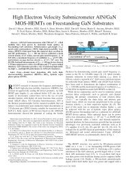
Figure 1a shows qualitatively the pH profile, and the<br />
concentration profiles of proteins in the column effluent,<br />
for the case where a weak-base, ion-exchange column<br />
packing and a descending pH gradient are used to perform<br />
chromatofocusing. Figure 1b shows the corresponding<br />
velocities of protein bands, and the velocities of pH values<br />
on the pH profile, both as functions of the liquid-phase pH.<br />
As illustrated, if any of the protein under consideration is<br />
located downstream from the centre of the protein band, it<br />
will tend to have a velocity less than the average velocity of<br />
the band, while the reverse is true for any of the protein<br />
located upstream from the centre of the band, so that all of<br />
the protein tends to accumulate into a narrow region at the<br />
point where the band velocity curve intersects the pH<br />
profile, as indicated by arrows in Figure 1b.<br />
The width of a protein band during chromatofocusing is<br />
determined by the shape of the pH gradient, the degree to<br />
which the charge of the protein varies with pH, and other<br />
properties of the system. More specifically, Sluyterman<br />
and Elgersma (1978) have shown that the band width at the<br />
column outlet in terms of the pH change across the band<br />
follows eqn [1], where f is the log of the ratio of hydrogen<br />
(a)<br />
(b)<br />
pH or concentration<br />
pH<br />
Apparent pI<br />
Apparent pI<br />
Protein bands<br />
Time<br />
Velocity of<br />
protein bands<br />
Velocity<br />
Effluent pH<br />
profile<br />
Velocities of<br />
pH values<br />
Figure 1 (a) pH profile and concentration profiles of proteins focused into<br />
bands in the effluent from a chromatofocusing column. (b) Corresponding<br />
relation between the apparent isoelectric points, the curves giving the<br />
velocities of protein bands as functions of the liquid-phase pH, and the<br />
curves giving the velocities of pH values as a function of those values inside<br />
the chromatofocusing column.<br />
ion concentrations in the fluid and adsorbed phases, which<br />
is equivalent to the dimensionless Donnan potential<br />
described by Sluyterman and Elgersma, d(pH)/dV is the<br />
slope of the pH gradient measured at the column outlet in<br />
units of pH per volume of mobile phase that has passed<br />
through the column, and dz/d(pH) is the rate of change of<br />
the characteristic binding charge of the protein with pH.<br />
s<br />
1 d…pH†=dV<br />
…pH† / ‰1Š<br />
dz=d…pH†<br />
Variations of Method<br />
2 ENCYCLOPEDIA OF LIFE SCIENCES / & 2001 Nature Publishing Group / www.els.net<br />
Soon after the introduction of the original method, efforts<br />
were made by several workers to replace the polyampholyte<br />
elution buffer with a chemically well-defined mixture<br />
containing a large number of low-molecular-weight<br />
buffering species (Hearn and Lyttle, 1981;Hutchens et al.,<br />
1986). Under these conditions, the effluent pH profile<br />
changes from the smooth profile shown in Figure 1 to a<br />
profile consisting of a sequence of discrete pH steps, with<br />
the number of steps corresponding to the number of<br />
buffering species in the elution buffer that produce ions<br />
that adsorb onto the column packing. Nevertheless, if an<br />
adequate number of these buffering species is present, the<br />
pH profile becomes sufficiently close to linear in shape that<br />
the method operates in essentially the same way as when a<br />
polyampholyte elution buffer is employed.<br />
An approach to simplifying the elution buffer in<br />
chromatofocusing suitable for preparative chromatography<br />
has been described by Frey (1996) and Strong and Frey<br />
(1997) in which either a weak-base or strong-base ionexchange<br />
column packing is used together with a limited<br />
number of low-molecular-weight buffering species in the<br />
elution buffer. These workers demonstrated that when the<br />
starting and elution buffer compositions are properly<br />
optimized, the protein of interest can be selectively focused<br />
on a single, retained stepwise pH front in the column<br />
effluent while the impurities elute from the column in other<br />
regions of the pH profile. It was also observed by these<br />
workers that a retained, stepwise pH front formed using a<br />
small number of buffering species in the elution buffer can<br />
accomplish the displacement development of proteins to<br />
yield a form of displacement chromatography that<br />
eliminates the need for a traditional displacer component<br />
(Narahari et al., 1998). Other efforts to extend the method<br />
include studies of how to select the composition of the<br />
elution buffer to form a gradual pH gradient having a<br />
desired shape when a limited number of low-molecularweight<br />
buffering species are employed in the elution buffer<br />
(Bates and Frey, 1998;Logan et al., 1999), and studies<br />
involving the external mixing of two different elution<br />
buffers in varying proportions to modify the shape of a<br />
retained pH gradient (Liu and Anderson, 1997).
Conclusions<br />
Since its introduction in the late 1970s, chromatofocusing<br />
has found widespread use as a high-resolution chromatographic<br />
procedure for separating proteins according to<br />
their apparent isoelectric points. Recent development<br />
efforts aimed at reducing its reliance on polyampholyte<br />
buffers and specialized column packings are likely to<br />
expand the future range of applications for the method,<br />
especially as a preparative separation technique.<br />
References<br />
Bates R and Frey DD (1998) Quasi-linear pH gradients for chromatofocusing<br />
using simple buffer mixtures: local equilibrium theory and<br />
experimental verification. Journal of Chromatography A 814: 43–54.<br />
Frey DD (1996) Local-equilibrium behavior of retained pH and ionic<br />
strength gradients in preparative chromatography. Biotechnology<br />
Progress 12: 65–72.<br />
Hearn MTW and Lyttle D (1981) Buffer-focusing chromatography<br />
using multicomponent electrolyte elution systems. Journal of Chromatography<br />
218: 483–495.<br />
Hutchens TW, Li CM and Besch PK (1986) Performance evaluation of a<br />
focusing buffer developed for chromatofocusing on high-performance<br />
anion exchange columns. Journal of Chromatography 359: 169–179<br />
Liu Y and Anderson DJ (1997) <strong>Chromatofocusing</strong> high-performance<br />
liquid chromatography: 1. Practical aspects. Journal of Chromatography<br />
A 762: 207–217.<br />
Logan KA, Lagerlund I and Chamow SM (1999) A simple, twocomponent<br />
buffer enhances use of chromatofocusing for processing of<br />
therapeutic proteins. Biotechnology and Bioengineering 62(2): 208–<br />
215.<br />
Narahari CR, Strong JC and Frey DD (1998) Displacement chromatography<br />
of proteins using a self-sharpening pH front formed by<br />
adsorbed buffering species as the displacer. Journal of Chromatography<br />
A 825(2): 115–126.<br />
Sluyterman LA AE and Elgersma O (1978) <strong>Chromatofocusing</strong>: isoelectric<br />
focusing on ion-exchange columns: 1. General principles.<br />
Journal of Chromatography 150: 17–30.<br />
Sluyterman, LA AE and Wijdeness J (1978) <strong>Chromatofocusing</strong>:<br />
isoelectric focusing on ion-exchange columns: 2. Experimental<br />
verification. Journal of Chromatography 150: 31–44.<br />
Strong JC and Frey DD (1997) Experimental and numerical studies of<br />
the chromatofocusing of dilute proteins using retained pH gradients<br />
formed on a strong-base anion-exchange column. Journal of<br />
Chromatography A 769: 129–143.<br />
Further Reading<br />
ENCYCLOPEDIA OF LIFE SCIENCES / & 2001 Nature Publishing Group / www.els.net<br />
<strong>Chromatofocusing</strong><br />
Amersham Pharmacia Biotechnology (1987) <strong>Chromatofocusing</strong> with<br />
Polybuffer and PBE. Uppsala: Amersham.<br />
Giri L (1990) <strong>Chromatofocusing</strong>. Methods in Enzymology 182: 380–392.<br />
Hutchens TW (1989) <strong>Chromatofocusing</strong>. In: Janson J and Ryden L (eds)<br />
Protein Purification, chap. 5, pp. 149–174. New York: VCH.<br />
Li CM and Hutchens TW (1992) <strong>Chromatofocusing</strong>. Methods in<br />
Molecular Biology, 11: 237–248.<br />
3