A Klinefelter bull with a 1;29 translocation born to a fertile 61,XXX cow
A Klinefelter bull with a 1;29 translocation born to a fertile 61,XXX cow
A Klinefelter bull with a 1;29 translocation born to a fertile 61,XXX cow

Create successful ePaper yourself
Turn your PDF publications into a flip-book with our unique Google optimized e-Paper software.
BRIEF COMMUNICATIONS<br />
COMMUNICATIONS BREVES<br />
A <strong>Klinefelter</strong> <strong>bull</strong> <strong>with</strong> a 1;<strong>29</strong> <strong>translocation</strong> <strong>born</strong><br />
<strong>to</strong> a <strong>fertile</strong> <strong>61</strong>,<strong>XXX</strong> <strong>cow</strong><br />
<strong>Klinefelter</strong>'s syndrome is caused by a chromosomal<br />
anomaly in which individuals typically possess two<br />
X chromosomes and a Y chromosome. This syndrome<br />
has previously been reported in cattle (1-3). The chromosomal<br />
anomaly is generally thought <strong>to</strong> be the result<br />
of nondisjunction during meiosis in one of the parents.<br />
Generally, most mammals <strong>with</strong> <strong>Klinefelter</strong>'s syndrome<br />
are expected <strong>to</strong> be phenotypically male and in<strong>fertile</strong>.<br />
Libido and service behavior may be completely normal<br />
(3); however, ejaculates are azoospermic.<br />
The <strong>bull</strong> described in this report was discovered<br />
during a study involving the karyotyping of a complete<br />
herd of purebred Charolais (4). He was <strong>born</strong> on<br />
April 6, 1990 <strong>to</strong> a three-year-old dam, as the result of her<br />
first breeding at two years of age by natural service. In<br />
the fall of 1990, he was taken <strong>to</strong> the university's feedlot<br />
for finishing. He weighed 786 kg (1730 lb) and was<br />
142 cm tall at the shoulder at 25 months of age. His scrotal<br />
circumference was 19 cm at 13 months of age and<br />
18 cm at 25 months of age, compared <strong>to</strong> an average of<br />
33.1 cm and 36.3 cm for one and two-year old Charolais<br />
<strong>bull</strong>s, respectively (5). The length of the extended penis<br />
at electroejaculation measured 24 cm, and the penis<br />
appeared <strong>to</strong> be of normal size and conformation. The urethra,<br />
prostate gland, and vesicular glands were felt <strong>to</strong> be<br />
in the normal size range, as determined by transrectal palpation.<br />
A seminal fluid sample collected by electroejaculation<br />
contained no sperma<strong>to</strong>zoa.<br />
Blood was aseptically drawn by venipuncture, and a<br />
culture of lymphocytes was established on the day of collection<br />
<strong>with</strong> 0.5 mL of blood in 10 mL of Ham's FIO<br />
medium, containing 15% fetal calf serum and 0.2 mL of<br />
phy<strong>to</strong>hemaglutinin (Wellmark Diagnostics, Guelph,<br />
Ontario). A small skin sample was surgically taken<br />
under local anesthetic, and fibroblast cultures were<br />
established (6). Samples of myocardium, pericardium,<br />
liver, kidney, and skeletal muscle were collected at<br />
slaughter, and cell cultures were similarly established<br />
from these tissues. Cell harvest, slide preparation, and<br />
pho<strong>to</strong>microscopy for karyotyping was done in the usual<br />
manner (6). Trypsin G banding (7) was done on the<br />
Can Vet J 1994; 35: 182-184<br />
Department of Animal and Poultry Science (Schmutz, Moker)<br />
and Department of Herd Medicine and Theriogenology (Barth),<br />
University of Saskatchewan, Saska<strong>to</strong>on, Saskatchewan<br />
S7N OWO.<br />
182<br />
Sheila M. Schmutz, Albert D. Barth, Jane S. Moker<br />
x-X- ----<br />
0 ~~~, f1 P ft ^ ;";0"X<br />
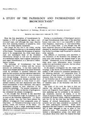
Figure 1. Trypsin G banded karyotype of a Charolais <strong>bull</strong>,<br />
60,XXY,t( 1 ;<strong>29</strong>).<br />
slide preparations from lymphocytes. A karyotype of<br />
60,XXY,t(1;<strong>29</strong>) (Figure 1) was found in preparations<br />
from all lymphocytes and all fibroblasts examined from<br />
skin, heart muscle, pericardium, liver, kidney, and<br />
skeletal muscle.<br />
Blood samples for serum tes<strong>to</strong>sterone analysis were<br />
taken from an indwelling catheter in the external jugular<br />
vein every 30 min over a six-hour period. This was<br />
followed immediately by a gonadotropin releasing hormone<br />
(GnRH) stimulation test, when 250 jig of GnRH<br />
(Factrel, Ayerst Labora<strong>to</strong>ries, Montreal, Quebec) were<br />
injected intravenously and blood samples were taken<br />
every 30 min for a two-hour period. Serum was harvested<br />
from the blood samples and frozen for tes<strong>to</strong>sterone<br />
assay at a later date. Tes<strong>to</strong>sterone was measured using<br />
a modification of a radioimmunoassay (8); assay standards<br />
were prepared in charcoal-stripped serum (0.02 ng/mL<br />
<strong>to</strong> 10 ng/mL) and extracted <strong>with</strong> ether before assay,<br />
rather than being put directly in<strong>to</strong> assay tubes. Fifty<br />
microliter aliquots (instead of 200 ,uL) of standards<br />
and samples were extracted in 2 mL diethyl ether. The<br />
sensitivity of the assay was 0.02 ng/mL or 0.07 nmol/L.<br />
When 1.0 ng or 2.5 ng of tes<strong>to</strong>sterone were added <strong>to</strong><br />
I mL of serum, the values obtained after correction<br />
for endogenous tes<strong>to</strong>sterone were 3.75 ± 0.52 nmol/L and<br />
8.74 ± 0.97 nmol/L (x ± SEM), respectively. All samples<br />
were analyzed in a single assay and the intra-assay<br />
coefficients of variation were 5.6% (n=4) and 7.7%<br />
(n=4) for sera <strong>with</strong> tes<strong>to</strong>sterone concentrations of 8.71<br />
nmol/L and 4.44 nmol/L, respectively. Tes<strong>to</strong>sterone<br />
Can Vet J Volume 35, March 1994
Figure 2. Section of testicular tissue from an 60,XXY,t(1 ;<strong>29</strong>)<br />
Charolais <strong>bull</strong> stained <strong>with</strong> hema<strong>to</strong>xylin-periodic acid-Schiff.<br />
Seminiferous tubules are partially collapsed, contain few<br />
Ser<strong>to</strong>li cells, and are devoid of germinal epithelium. The<br />
interstitial tissue contains normal appearing Leydig cells,<br />
which are proportionately inordinately abundant compared <strong>to</strong><br />
seminiferous tubules. Bar scale depicts 100 l,m.<br />
concentrations measured over the six-hour period ranged<br />
from 4.82 nmol/L <strong>to</strong> 12.01 nmol/L. There was a good<br />
response <strong>to</strong> the GnRH stimulation test, <strong>with</strong> tes<strong>to</strong>sterone<br />
concentrations increasing from 12.01 nmol/L at<br />
zero time <strong>to</strong> 17.88 nmol/L at two hours after injection of<br />
the GnRH.<br />
The day after the blood sampling for the tes<strong>to</strong>sterone<br />
analysis and the GnRH stimulation test was completed,<br />
the <strong>bull</strong> was castrated. Tissue segments were taken in the<br />
field from the dorsal, central, and ventral regions of<br />
each testis and from the caput, corpus, and cauda epididymis.<br />
They were immediately fixed in Bouin's and<br />
Helly's solutions. The fixed tissue segments were paraffin<br />
embedded, sectioned, and stained for his<strong>to</strong>logical study<br />
by light microscopy according <strong>to</strong> routine methods.<br />
The testicles obtained at castration and dissected<br />
free of the vaginal tunics and epididymides had a combined<br />
weight 80.3 g. The combined weight of the epididymides<br />
was 30.3 g. The left testis was 3.5 X 3.2 X<br />
6.0 cm and the right 3.3 X 3.1 X 6.2 cm in size.<br />
His<strong>to</strong>logically, the testicular tissue was mainly composed<br />
of normal appearing Leydig cells <strong>with</strong> a minor<br />
component of poorly developed, partially collapsed,<br />
seminiferous tubules. The seminiferous tubules had a<br />
thick wrinkled basement membrane and contained only<br />
a few Ser<strong>to</strong>li cells. No germinal cells could be found in<br />
any of the seminiferous tubules (Figure 2). His<strong>to</strong>logically,<br />
cauda epididymal tissue had a normal appearance; however,<br />
the lumen was completely devoid of sperma<strong>to</strong>zoa.<br />
Can Vet J Volume 35, February 1994<br />
In comparison <strong>to</strong> his male herd mates and <strong>to</strong> male<br />
members of his breed in general, the <strong>bull</strong> appeared<br />
relatively short in height and length. However, no objective<br />
data were obtained on the <strong>bull</strong>'s length, and a shoulder<br />
height of 142 cm is at the low end of normal range.<br />
The <strong>bull</strong>'s masculine facial appearance, general muscling,<br />
and heavy muscling of the dorsal neck region was in contrast<br />
<strong>to</strong> the feminine appearance of a <strong>61</strong>,XXY <strong>bull</strong> seen<br />
by Dunn et al (2).<br />
The small scrotal circumference measurement is<br />
likely attributable <strong>to</strong> a decrease in seminiferous tubule<br />
length and diameter, as reported for a <strong>61</strong>,XXY <strong>bull</strong> by<br />
Logue et al (3). The weights of the testicles and epididymides<br />
were comparable <strong>to</strong> those reported by Dunn<br />
et al (2). The extremely small testicle size was most<br />
likely due <strong>to</strong> a partial lack of development of seminiferous<br />
tubules and a <strong>to</strong>tal lack of development of germinal<br />
epithelium <strong>with</strong>in the seminiferous tubules. The complete<br />
absence of sperma<strong>to</strong>zoa in a seminal fluid sample confirms<br />
the <strong>to</strong>tal lack of germinal tissue in this <strong>bull</strong>'s testicles<br />
and is in agreement <strong>with</strong> Logue et al (3) and<br />
Dunn et al (2) who reported azoospermia in a <strong>61</strong>,XXY<br />
Friesian and a <strong>61</strong>,XXY Hereford, respectively. The relative<br />
abundance of normal appearing Leydig cells in<br />
comparison <strong>to</strong> that seen in normal testis tissue is similar<br />
<strong>to</strong> the situation in the <strong>bull</strong> reported by Logue et al (3).<br />
The normal size of the <strong>bull</strong>'s penis and accessory<br />
sex glands indicates that there were sufficient levels of<br />
circulating tes<strong>to</strong>sterone for the development of these male<br />
characteristics. The serum tes<strong>to</strong>sterone concentrations<br />
over the six-hour bleeding period (range 4.82 nmol/L <strong>to</strong><br />
12.01 nmol/L) were similar <strong>to</strong> those of 12 normal <strong>bull</strong>s<br />
(range 1.87 nmol/L <strong>to</strong> 21.90 nmol/L) bled over an eighthour<br />
period. The results of the GnRH stimulation test<br />
were also similar <strong>to</strong> those of 12 normal <strong>bull</strong>s tested in a<br />
similar manner (unpublished observations) and suggest<br />
a normal responsiveness of Leydig cell tissue in the<br />
secretion of tes<strong>to</strong>sterone. Although quantitative estimates<br />
of Leydig cell tissue were not done in this case,<br />
Logue et al (3) reported that Leydig cell volume in a<br />
<strong>61</strong>,XXY <strong>bull</strong> was in the normal range.<br />
The karyotype of the <strong>bull</strong>'s dam was <strong>61</strong>,<strong>XXX</strong> and<br />
that of his sire 59,XY,t(1;<strong>29</strong>). Therefore, we presume that<br />
he inherited the extra X chromosome maternally and the<br />
<strong>translocation</strong> paternally. Trisomy X cattle have previously<br />
been reported (9-13) as exhibiting limited fertility or,<br />
more commonly, sterility. This dam was obviously <strong>fertile</strong><br />
since she produced this calf. She remained on her<br />
home ranch until recently and subsequently produced a<br />
<strong>bull</strong> calf in both of 1991 and 1992. In January 1993, she<br />
was observed <strong>to</strong> be in heat after several months <strong>with</strong>out<br />
heats, when, presumably, she was pregnant. By contrast,<br />
Pinheiro et al (14) described a heifer <strong>with</strong> a 60,<strong>XXX</strong>,t(1;<strong>29</strong>)<br />
karyotype that appeared <strong>to</strong> be in<strong>fertile</strong>, since she was<br />
never observed in heat.<br />
<strong>Klinefelter</strong>'s syndrome appears <strong>to</strong> be a very rare occurrence.<br />
The few cases (1-3) discussed here were all<br />
cited as rare occurrences, found by chance, and in a chromosome<br />
study of young <strong>bull</strong>s, including 51 <strong>with</strong> smaller<br />
than average testicles, we found no cases (15). However,<br />
we recently found a case of <strong>61</strong>,XXY in a Simmental calf<br />
during screening of a herd for the frequency of 14;20<br />
<strong>translocation</strong> (unpublished data). The previously reported<br />
cases (1-3) and the Simmental appear <strong>to</strong> be the result of<br />
183
nondisjunction during game<strong>to</strong>genesis, whereas the<br />
Charolais described in this paper inherited the extra<br />
X chromosome from his dam. His clinical phenotype<br />
appears <strong>to</strong> be no different from those described<br />
previously, however, even though he also carries a 1;<strong>29</strong><br />
<strong>translocation</strong>.<br />
Acknowledgments<br />
We thank Susan Cook for conducting the tes<strong>to</strong>sterone<br />
assays and Bill Kowalenko for supervising weighing and<br />
care of the <strong>bull</strong> at the Saska<strong>to</strong>on R.O.P. Test Station. We<br />
also thank the veterinarians and staff at Intercontinental<br />
Packers for assistance in collecting samples at slaughter.<br />
cvI<br />
References<br />
1. Finger KH, Herzog A, Hohn H, Rieck GW. Fortschritte auf dem<br />
Gebiet der Zy<strong>to</strong>genetik (Chromosomenpathologie) in der<br />
Veterinarmedizin. Giessner Beitr Erbpath Zuchthyg 1991;<br />
2/3: 13-30.<br />
2. Dunn HO, Lein DH, McEntee K. Testicular hypoplasia in a<br />
Hereford <strong>bull</strong> <strong>with</strong> <strong>61</strong>,XXY karyotype: the bovine counterpart of<br />
human <strong>Klinefelter</strong>'s syndrome. Cornell Vet 1980; 70: 137-146.<br />
3. Logue DN, Harvey MJA, Munro CD, Lennox B. Hormonal and his<strong>to</strong>logical<br />
studies in a 6lXXY <strong>bull</strong>. Vet Rec 1979; 104: 500-503.<br />
4. Schmutz SM, Moker JS. Impact of a 1;<strong>29</strong> Robertsonian <strong>translocation</strong><br />
on a herd of purebred beef cattle. Can J Anim Sci 1989;<br />
69: 891-896.<br />
5. Coulter GH, Maple<strong>to</strong>ft RJ, Kozub GC, Cates WF. Scrotal circumference<br />
of two-year-old <strong>bull</strong>s of several beef breeds. Theriogenology<br />
1987; 27: 485-491.<br />
6. Coates JW, Schmutz SM, Rousseaux CG. A survey of malformed<br />
aborted bovine fetuses, stillbirths and nonviable neonates for<br />
abnormal karyotypes. Can J Vet Res 1988; 52: 258-263.<br />
7. Seabright M. A rapid banding technique for human chromosomes.<br />
Lancet 1971; 2: 971-972.<br />
8. Cook SJ, Rawlings NC, Kennedy RI. Quantitation of six androgens<br />
by combined high performance liquid chroma<strong>to</strong>graphy and<br />
radioimmunoassay. Steroids 1982; 40: 369-380.<br />
9. Rieck GW, Hohn H, Herzog A. X-trisomie beim Rind mit<br />
Anzeichen familiarer Disposition fur Meioses<strong>to</strong>rungen.<br />
Cy<strong>to</strong>genetics 1970; 9: 401-409.<br />
10. Norberg HS, Refsdal AO, Garm ON, Nes N. A case report on<br />
X-trisomy in cattle. Hereditas 1976; 82: 69-72.<br />
11. Buoen LC, Seguin BE, Weber AF, Shoffner RN. X-trisomy<br />
karyotype and associated infertility in a Holstein heifer. J Am Vet<br />
Med Assoc 1981; 179: 808-811.<br />
12. Swartz HA, Vogt DW. Chromosome abnormalities as a cause<br />
of reproductive inefficiency in heifers. J Hered 1983; 74: 320-324.<br />
13. King WA, Linares T. A cy<strong>to</strong>genetic study of repeat-breeder<br />
heifers and their embryos. Can Vet J 1983; 24: 112-115.<br />
14. Pinheiro LEL, Almeida IL Jr, Garcia JM, Basrur PK. Trisomy X<br />
and 1/<strong>29</strong> <strong>translocation</strong> in in<strong>fertile</strong> heifers. Theriogenology 1987;<br />
28: 891-898.<br />
15. Schmutz SM, Flood P, Moker J, Barth A, Maple<strong>to</strong>ft R, Cates W.<br />
Incidence of chromosomal anomalies among western Canadian beef<br />
cattle. Can J Anim Sci 1990; 70: 779-783.<br />
_ ~TO GAIN APPROVAL<br />
Excenel' the new generation cephalosporin is now indicated for swine.<br />
With this claim, Excenel offers you the flexibflity and confidence of proven<br />
therapy for bovine, equine and swine respira<strong>to</strong>ry disease<br />
Excenel gains approval <strong>with</strong> your clients by offering effective therapy<br />
<strong>with</strong> min injection site discomfort In addition, Excenel offers<br />
no meat <strong>with</strong>drawal and no mik discard in cattle and only one day<br />
meat <strong>with</strong>drawal in swine.<br />
Excenel The leding animal health antibiotic in North Americal<br />
Consider it your first line of treatment.<br />
E~~~lxcenelt<br />
4 _ STERILE POWDER (cefiolur sodium)<br />
The UpjohnCompay - Anial Hlk Divso of Upjohnlner-AmerfanCorponion. jNgm<br />
Onrenile, Ont. L9W 3T3 EXNL-2E93 IPAAB HEAIIH<br />
(141992. aks data on file. A<br />
184 Can Vet J Volume 35, February 1994