ticular connective tissue fibers from the collagenous fibers, reticulum ...
ticular connective tissue fibers from the collagenous fibers, reticulum ...
ticular connective tissue fibers from the collagenous fibers, reticulum ...
You also want an ePaper? Increase the reach of your titles
YUMPU automatically turns print PDFs into web optimized ePapers that Google loves.
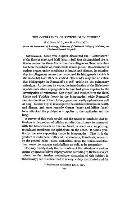
THE OCCURRENCE OF RETICULUM IN TUMORS *<br />
N. C. FOOT, M.D., AND H. A. DAY, M.D.<br />
(From <strong>the</strong> Department of Pathology, University of Cincinnati College of Medicine, and<br />
Cincinnati General Hospit)<br />
Introduction. Since von Kupffer discovered <strong>the</strong> "Gitterfasern"<br />
of <strong>the</strong> liver in I876, and Mall (189I, I896) first distinguished <strong>the</strong> re<strong>ticular</strong><br />
<strong>connective</strong> <strong>tissue</strong> <strong>fibers</strong> <strong>from</strong> <strong>the</strong> <strong>collagenous</strong> <strong>fibers</strong>, <strong>reticulum</strong><br />
has been <strong>the</strong> subject of considerable investigation; its occurrence in<br />
various organs under conditions of health and disease, its relationship<br />
to <strong>collagenous</strong> <strong>connective</strong> <strong>tissue</strong>, and its histogenesis (which is<br />
still in doubt) have all been studied. The reader may find an extensive<br />
bibliography in Russakoff's (i908) article on <strong>the</strong> pulmonary<br />
<strong>reticulum</strong>. At <strong>the</strong> time he wrote, <strong>the</strong> introduction of <strong>the</strong> Bielschowsky-Maresch<br />
silver impregnation technic had given impetus to <strong>the</strong><br />
investigation of <strong>reticulum</strong>; Kon ('i9o8) had studied it in <strong>the</strong> liver,<br />
Rossle and Yoshida (I909) in <strong>the</strong> lymphnodes, while Russakoff<br />
examined sections of liver, kidney, pancreas, and lymphnodes as well<br />
as lung. Neuber (I9I2) investigated <strong>the</strong> cardiac <strong>reticulum</strong> in health<br />
and disease, and more recently Corner (I920) and Miller (1923)<br />
have attacked <strong>the</strong> problem as it applies to <strong>the</strong> capillaries and <strong>the</strong><br />
lung.<br />
A survey of this work would lead <strong>the</strong> reader to conclude that <strong>reticulum</strong><br />
is <strong>the</strong> product of cellular activity, that it may be connected<br />
with <strong>the</strong> blood-vessels on <strong>the</strong> one hand, or serve as a supporting,<br />
reticulated membrane for epi<strong>the</strong>lium on <strong>the</strong> o<strong>the</strong>r. It forms practically<br />
<strong>the</strong> sole supporting <strong>tissue</strong> in lymphnodes. That it is <strong>the</strong><br />
product of endo<strong>the</strong>lial cells and, occasionally, fibroblasts, seems to<br />
be <strong>the</strong> general belief; some authorities claim <strong>the</strong> reticuloendo<strong>the</strong>lium,<br />
some <strong>the</strong> vascular endo<strong>the</strong>lium as well, as its progenitor.<br />
One may readily study <strong>the</strong> distribution of <strong>the</strong> <strong>reticulum</strong> in various<br />
organs by means of silver impregnations according to Bielschowsky's<br />
technic, so that fur<strong>the</strong>r preliminary discussion of this subject is<br />
unnecessary; let it suffice that it is very widely distributed and its<br />
* Received for publication June i, 1925.<br />
431
432<br />
FOOT AND DAY<br />
importance is often overlooked. For example, its widespread occurrence<br />
in epi<strong>the</strong>lial organs, where it forms a dense basement membrane,<br />
is insufficiently recognized. That it may with time, and under<br />
ra<strong>the</strong>r uncertain conditions, become transformed into <strong>collagenous</strong><br />
fibrous <strong>tissue</strong> is becoming more universally believed. Russakoff,<br />
R6ssle and Yoshida, and Miller have all stressed this point. That<br />
it is <strong>the</strong> product of cellular activity on <strong>the</strong> part of endo<strong>the</strong>lial derivatives<br />
is, however, becoming a debatable question. Miller has<br />
pointed out <strong>the</strong> close relationship between newly-formed reticulin<br />
<strong>fibers</strong> and neighboring, preexisting <strong>reticulum</strong>; Baitsell (I9I5, I9I6)<br />
has advanced anew <strong>the</strong> <strong>the</strong>ory of purely extracellular origin; and<br />
one of us (Foot, I925) has recently stated his belief that we must<br />
look elsewhere than to cells for <strong>the</strong> origin of reticulin fibrils, basing<br />
this view on observations on <strong>the</strong> development of <strong>reticulum</strong> in experimental<br />
tubercles.<br />
As nothing appears to have been done in connection with <strong>the</strong> distribution<br />
and occurrence of <strong>reticulum</strong> in tumors, a hundred or more<br />
have been collected in <strong>the</strong> past three years' accumulation of laboratory<br />
material of this department, and <strong>the</strong>se have been studied with<br />
special reference to <strong>the</strong>ir <strong>reticulum</strong> content.<br />
TECHNIC<br />
In most cases Zenker-fixed material is available, but where it is<br />
not, formalin-fixed <strong>tissue</strong> has been used. Several serial sections were<br />
cut <strong>from</strong> each paraflin block, three being stained with <strong>the</strong> routine<br />
Harris' hematoxylin-eosin stain and two by a modification of <strong>the</strong><br />
Bielschowsky-Maresch technic devised by one of us last year. (Foot,<br />
I924.)<br />
Briefly summarized, <strong>the</strong> steps are as follows. Sections about five<br />
microns in thickness are cut in paraffin. Zenker fixation is superior<br />
to formalin, but <strong>the</strong> technic is <strong>the</strong> same in ei<strong>the</strong>r case. After removing<br />
<strong>the</strong> paraffin, treat five minutes with a weak alcoholic solution<br />
of iodine, removing this by a brief immersion in 5 per cent "hypo."<br />
Wash and treat for five minutes with 0.25 per cent potassium permanganate<br />
in water. Wash and place <strong>the</strong> sections in 5 per cent<br />
aqueous oxalic acid. Wash in distilled water and impregnate for<br />
forty-eight hours with a 2 per cent solution of silver nitrate in distilled<br />
water. Wash in distilled water and treat with Bielschowsky's<br />
silver-ammonium oxide solution for one half-hour. Rinse in distilled
RETICULUM IN TUMORS<br />
water and leave in 5 per cent formalin (2 per cent formaldehyde) for<br />
ano<strong>the</strong>r half-hour and <strong>the</strong>n, after washing in tap-water, tone <strong>the</strong><br />
sections for one hour in a one per cent aqueous solution of gold chloride,<br />
after which a two-minute immersion in 5 per cent "hypo" will<br />
remove any superfluity of <strong>the</strong> silver. The sections may be mounted<br />
at this point, but it is better to counterstain for <strong>the</strong> usual time in<br />
Harris' hematoxylin, wash, and stain for forty-five seconds in Van<br />
Gieson's picric-acid-acid-fuchsin. Run <strong>the</strong> sections immediately<br />
through ascending percentages of alcohol into xylol and mount in<br />
balsam, avoiding water as it decolorizes <strong>the</strong> fuchsin. After <strong>the</strong> silver<br />
impregnation <strong>the</strong> sections should be practically colorless, turning<br />
brownish-black in <strong>the</strong> formalin and fading to gray in <strong>the</strong> gold bath.<br />
The "hypo" turns <strong>the</strong>m old-rose and gray. The finished, counterstained<br />
sections show <strong>the</strong> <strong>reticulum</strong> in sharp black lines, <strong>the</strong> collagen<br />
in vermilion to crimson. The nuclei are brownish, <strong>the</strong> cytoplasm<br />
and muscle substance yellow. The silver impregnation is performed<br />
in subdued daylight, not in <strong>the</strong> dark.<br />
RETICULUm FIBERS IN TUmORS<br />
433<br />
Epi<strong>the</strong>lial Tumors<br />
Surface epi<strong>the</strong>lium. The epidermoid carcinoma is of paramount<br />
interest here, <strong>the</strong> benign epi<strong>the</strong>lial tumors of this type conforming<br />
closely to normal histology. In <strong>the</strong>se carcinomata <strong>the</strong>re is a varying<br />
amount of <strong>reticulum</strong>; plentiful in <strong>the</strong> corium, it forms a network of<br />
delicate fibrils about <strong>the</strong> advancing columns of tumor cells which<br />
dip down <strong>from</strong> <strong>the</strong> epidermis. Generally speaking, if <strong>the</strong> tumor be<br />
of rapid growth <strong>reticulum</strong> is plentiful, if slowly growing more collagen<br />
than <strong>reticulum</strong> develops. Usu,ally <strong>the</strong> latter forms a membrane-like<br />
plexus at <strong>the</strong> base of <strong>the</strong> epi<strong>the</strong>lial plugs, occasionally it<br />
penetrates <strong>the</strong>m superficially. A metastasis <strong>from</strong> such a tumor is<br />
shown in Fig. i. In <strong>the</strong> basal cell, or hair-matrix carcinoma <strong>the</strong> <strong>reticulum</strong><br />
forms a delicate basket-work around <strong>the</strong> cell masses, with<br />
radiating fibrils that are continuous with those of <strong>the</strong> corium.<br />
Glandular epi<strong>the</strong>lium. The common adenoma usually shows more<br />
collagen than <strong>reticulum</strong> in its stroma, <strong>the</strong> various types of fibroadenoma<br />
following this rule, but a narrow zone of <strong>reticulum</strong> is commonly<br />
found about <strong>the</strong> base of <strong>the</strong> acini, outlining <strong>the</strong>m in black.<br />
In a rapidly growing papillary adenoma <strong>the</strong> stroma is composed al-
434<br />
FOOT AND DAY<br />
most entirely of reticulin fibrils, to <strong>the</strong> comparative exclusion of<br />
collagen. If <strong>the</strong> tumor acini are closely compressed one observes<br />
reticulin fibrils between <strong>the</strong>m; lying almost, if not quite isolated and<br />
free <strong>from</strong> any great admixture of mesenchymal cells. Capillaries<br />
carry a sheath of <strong>reticulum</strong>, but frequently <strong>the</strong>re are but few vessels<br />
in this stroma and <strong>the</strong> <strong>reticulum</strong> appears to have been produced independently<br />
of cellular agency.<br />
The various types of adenocarcinoma and medullary carcinoma<br />
all follow <strong>the</strong> description just given; <strong>the</strong> cell-masses in <strong>the</strong> medullary<br />
type are usually quite free <strong>from</strong> penetration by <strong>the</strong> <strong>reticulum</strong>, but<br />
very rapidly growing tumors, with a tendency to cell dissociation,<br />
may show a good deal. Fig. 2 shows a typical adenocarcinoma. If<br />
<strong>the</strong> tumors have metastasized to lymphoid <strong>tissue</strong> <strong>the</strong> lymphoid <strong>reticulum</strong><br />
will, naturally, be found intimately intermingled with <strong>the</strong>ir<br />
cells. In <strong>the</strong> colloid type, those cells which remain at <strong>the</strong> center of<br />
<strong>the</strong> mucoid masses may have small, woolly plexuses of what seems<br />
to be <strong>reticulum</strong> about <strong>the</strong>m, but <strong>the</strong> production of fibrils resembling<br />
reticulin is quite common in necrotic areas in epi<strong>the</strong>lial tumors and<br />
it is doubtful if such fibrils constitute true <strong>reticulum</strong>. Scirrhous<br />
carcinoma, as one might expect <strong>from</strong> its slow, sclerotic growth,<br />
shows large amounts of collagen in its stroma and a very vanrable<br />
quantity of <strong>reticulum</strong>, which is usually scanty. The sarcomatoid<br />
type of pancreatic carcinoma, shown in Fig. 3, presents a moderate<br />
amount of <strong>reticulum</strong> in its stroma and <strong>the</strong>re is some penetration of<br />
this <strong>tissue</strong> into <strong>the</strong> more or less dissociated groups of tumor cells,<br />
which is also true of <strong>the</strong> similar type of carcinoma of <strong>the</strong> thyroid.<br />
The difference in <strong>the</strong> distribution of <strong>reticulum</strong> in <strong>the</strong>se tumors and<br />
that in lymphosarcomata is, however, so striking as to render a<br />
diagnosis comparatively simple in cases that would be quite obscure<br />
when stained in <strong>the</strong> usual way.<br />
Hypernephroma, or adrenal carcinoma, shows a very small<br />
amount of <strong>reticulum</strong> which does not penetrate into <strong>the</strong> alveolar cell<br />
masses. Although this tumor is found in <strong>the</strong> kidney, an organ rich in<br />
<strong>reticulum</strong>, <strong>the</strong>re is practically none in <strong>the</strong> alveoli which push <strong>the</strong><br />
preexisting <strong>fibers</strong> aside.<br />
The tumor section photographed for Fig. 4 was kindly sent to us<br />
<strong>from</strong> <strong>the</strong> Albany Hospital, by Dr. Victor Jacobson, and is worthy of<br />
special mention. It is <strong>from</strong> a medullary carcinoma of <strong>the</strong> uterus,<br />
which differs in no way <strong>from</strong> any o<strong>the</strong>r such carcinoma excepting
RETICULUM IN TUMORS<br />
435<br />
that its stroma shows an extraordinary anaplasia to what appears<br />
to be a true fibrosarcoma. Thus two malignant tumors are intimately<br />
combined in one section. The carcinoma is practically free<br />
<strong>from</strong> <strong>reticulum</strong> while its stroma shows an abundance of straight,<br />
coarse reticulin fibrils exactly analogous to those of <strong>the</strong> fibrosarcomata<br />
to be discussed presently.<br />
Endo<strong>the</strong>lial Tumors<br />
Vascular. Cavernous hemangiomata show a <strong>reticulum</strong> about <strong>the</strong>ir<br />
vessels and sinuses quite similar to that normally seen surrounding<br />
capillaries. A malignant peri<strong>the</strong>lioma appears to produce very little<br />
<strong>reticulum</strong>, as this is found only in <strong>the</strong> immediate proximity of <strong>the</strong><br />
vessels. As we could secure only one specimen, this negative finding<br />
should be confirmed.<br />
Reticuloendo<strong>the</strong>lial. Several types of malignant endo<strong>the</strong>lioma were<br />
examined, some showing comparatively little <strong>reticulum</strong>, some a<br />
great deal. Of <strong>the</strong> latter group, <strong>the</strong> primary lymphnode endo<strong>the</strong>lioma,<br />
so closely resembling medullary carcinoma, is a good example.<br />
Where <strong>the</strong> carcinoma shows little <strong>reticulum</strong> in its alveoli, <strong>the</strong> <strong>fibers</strong><br />
usually stopping short at <strong>the</strong>ir margin, or simply traversing <strong>the</strong>m in<br />
straight, unbranching lines, <strong>the</strong> endo<strong>the</strong>lioma exhibits an abundant,<br />
branching plexus of curving reticulin fibrils intimately associated<br />
with <strong>the</strong> cells of <strong>the</strong> tumor within its alveoli. The fibrils are distinctly<br />
plexiform and anastomotic in this case, which affords a good<br />
criterion for <strong>the</strong> diagnosis of doubtful alveolar tumors of <strong>the</strong> axillary<br />
lymphnodes in women, where primary endo<strong>the</strong>lioma is readily mistaken<br />
for <strong>the</strong> more common metastatic alveolar carcinoma of <strong>the</strong><br />
breast. We feel this to be <strong>the</strong> chief contribution of this paper.<br />
(Figs. 5 and 6.)<br />
Ano<strong>the</strong>r type of endo<strong>the</strong>lioma, supposedly originating in <strong>the</strong> reticuloendo<strong>the</strong>lium<br />
of lymphnodes, is composed of polygonal, anastomosing<br />
cells, and shows an abundant <strong>reticulum</strong> intimately related<br />
to <strong>the</strong>se cells and following <strong>the</strong>ir cytoplasmic processes very<br />
closely. A given fibril may, however, course over several cells without<br />
any interruption in its substance and <strong>the</strong> unbroken continuity<br />
of <strong>the</strong>se fibrils with those of <strong>the</strong> neighboring normal <strong>tissue</strong> is always<br />
striking. This tumor (Fig. 7), was recently reported by one of us<br />
(Foot, I924) in detail.<br />
The endo<strong>the</strong>liomata arising in bone-marrow correspond in <strong>the</strong>
436<br />
FOOT AND DAY<br />
main with <strong>the</strong> loosely reticulated portions of <strong>the</strong> primary endo<strong>the</strong>lioma<br />
of lymphnodes, insofar as <strong>the</strong>ir <strong>reticulum</strong> is concerned;<br />
but <strong>the</strong>y contain somewhat less and <strong>the</strong> <strong>fibers</strong> are far more delicate.<br />
One case of diffusely disseminated cells, resembling endo<strong>the</strong>lial<br />
phagocytes and occasionally grouped into tumor masses, shows<br />
marked <strong>reticulum</strong> production wherever <strong>the</strong> cells become so massed.<br />
Dural endo<strong>the</strong>lioma. This tumor (Fig. 8) probably produces more<br />
<strong>reticulum</strong> than any o<strong>the</strong>r tumor we have observed; <strong>the</strong>re is a dense<br />
felting of reticulin fibrils in its matrix and <strong>the</strong>y are intimately intermingled<br />
with an almost equally dense mass of collagen fibrils.<br />
Meso<strong>the</strong>lioma<br />
A tumor <strong>from</strong> <strong>the</strong> pleural cavity, connected with <strong>the</strong> lung, is composed<br />
of squamous cells that tend to keratinize and to form pearls.<br />
It resembles an epidermoid carcinoma in every respect, but <strong>the</strong>re is<br />
little upon which to base this diagnosis if one considers that <strong>the</strong><br />
tumor lay in and beneath <strong>the</strong> visceral pleura, not near large<br />
bronchi, and <strong>the</strong>re was no history to suggest bronchiectasia. A<br />
similar growth is described by Beitzke, in Aschoff's "Pathologische<br />
Anatomie" (1923), as a "pleural cancer." We prefer <strong>the</strong> term<br />
"meso<strong>the</strong>lioma" and feel security in our diagnosis for <strong>the</strong> reasons<br />
just given. This tumor shows an abundant <strong>reticulum</strong> between its<br />
cell masses and a moderate amount of collagen; it is almost exactly<br />
like an epidermoid carcinoma in respect to its stroma. Its cell masses<br />
are entirely free <strong>from</strong> reticulin fibrils.<br />
Fibroblastic Tumors<br />
Fibroma. Both types of fibroma, <strong>the</strong> soft and hard, are chiefly<br />
<strong>collagenous</strong>; practically no <strong>reticulum</strong> is seen, save around <strong>the</strong> blood<br />
vessels. (Fig. 9.) A very mucoid fibroma of <strong>the</strong> external auditory<br />
canal, which recurred twice in a year, shows ra<strong>the</strong>r scanty <strong>collagenous</strong><br />
<strong>tissue</strong> with wide, unstained spaces and an abundance of short,<br />
ra<strong>the</strong>r straight reticulin fibrils. (Fig. io.) It is not a true myxoma,<br />
for <strong>the</strong>re is no bluish hyaline-matrix, and it is not malignant. Here<br />
<strong>the</strong>re appears to be a direct causal relationship between <strong>the</strong> presence<br />
of <strong>reticulum</strong> and rapidity of growth.<br />
Keloid. Both <strong>the</strong> keloid and <strong>the</strong> fibrous granuloma show <strong>reticulum</strong><br />
and collagen in about <strong>the</strong> same proportions as occur in normal<br />
corium; <strong>the</strong> total amount of <strong>the</strong>se <strong>fibers</strong>, however, is greatly in-
RETICULUM IN TUMORS<br />
437<br />
creased. The keloid has ra<strong>the</strong>r more collagen than <strong>reticulum</strong>, however.<br />
Giant cell tumors. These are not rich in <strong>reticulum</strong> and show only<br />
moderate amounts of collagen. The giant cells become deeply stippled<br />
with silver, as is <strong>the</strong> case with some endo<strong>the</strong>lial cells and with<br />
nerve cells. The tumors are chiefly cellular.<br />
Fibrosarcoma. Two types are recognizable: one producing almost<br />
nothing but re<strong>ticular</strong> matrix (Fig. ii), and one an abundance of collagen<br />
(Fig. I 2). The former is very similar to <strong>the</strong> reticulo-endo<strong>the</strong>lioma<br />
with branching cells, already described. Apparent transitions<br />
between <strong>the</strong> re<strong>ticular</strong> and <strong>collagenous</strong> types are seen, but <strong>the</strong> degree<br />
of anaplasia and <strong>the</strong> rate of growth will not entirely explain this difference<br />
- for one very anaplastic and rapidly growing fibrosarcoma<br />
of <strong>the</strong> lung shows little or no <strong>reticulum</strong>, but produces an abundant<br />
<strong>collagenous</strong> matrix (Fig. 13). Generally speaking, however, <strong>the</strong>re is<br />
more <strong>reticulum</strong> in <strong>the</strong> rapidly growing sarcomata and more collagen<br />
in those of slower development.<br />
It is interesting to compare <strong>the</strong> tumors in our series of fibrosarcoma<br />
as regards <strong>the</strong>ir <strong>reticulum</strong> content. One, a sarcoma of <strong>the</strong><br />
scapula (Fig. I4) of rapid growth, shows an evenly distributed mesh<br />
of very fine, short, and curly reticulin fibrils, many of <strong>the</strong>m beaded<br />
and interrupted. This indicates very young <strong>reticulum</strong>, judging <strong>from</strong><br />
<strong>the</strong> literature and <strong>from</strong> personal observation on its formation in<br />
tubercles. Very little collagen is present. Then come a sarcoma of<br />
<strong>the</strong> lung, one of <strong>the</strong> kidney, and a third <strong>from</strong> <strong>the</strong> bladder, all of<br />
which show coarse, straight reticulin <strong>fibers</strong> and little collagen. At<br />
<strong>the</strong> o<strong>the</strong>r end of <strong>the</strong> scale we find a sarcoma of <strong>the</strong> lung and one of<br />
<strong>the</strong> knee, both of which show much collagen and little <strong>reticulum</strong>.<br />
From this one may infer.that <strong>reticulum</strong> is laid down by <strong>the</strong> more<br />
embryonal type of rapidly growing tumor and that it may ultimately<br />
be changed into collagen. The very primitive fibrosarcomata<br />
and reticuloendo<strong>the</strong>liomata both revert to a common type, <strong>the</strong><br />
mesenchymal. Hence <strong>the</strong>ir similarity and <strong>the</strong> comparative uselessness<br />
of attempting to classify <strong>the</strong>m by adult-cell standards, or to<br />
distinguish <strong>the</strong>m apart.<br />
O<strong>the</strong>r Connective Tissue Tumors<br />
Lipoma. Tumors of this type produce more collagen than <strong>reticulum</strong>,<br />
as one would expect after a survey of normal fat.
438<br />
FOOT AND DAY<br />
Liposarcoma. In one case we find short, wavy, more or less<br />
beaded reticulin fibrils and little collagen. (Fig. i 5.) In <strong>the</strong> o<strong>the</strong>rs,<br />
which resemble Fleming's fat organ, <strong>the</strong>re is abundant reticul.um<br />
and but little collagen.<br />
Leiomyoma. Here much collagen is produced; often more of this<br />
is seen than of muscular <strong>tissue</strong> and a large amount of <strong>reticulum</strong><br />
closely invests <strong>the</strong> muscle <strong>fibers</strong>. Is this <strong>reticulum</strong> converted into<br />
<strong>the</strong> abundant collagen that gives <strong>the</strong> clinical name of "fibroid" to<br />
<strong>the</strong> tumor?<br />
Leiomyosarcoma. This shows an abundance of both <strong>reticulum</strong> and<br />
collagen, <strong>the</strong> latter in <strong>the</strong> form of short <strong>fibers</strong> and not at all as profuse<br />
as in its benign prototype. Here again we see more <strong>reticulum</strong><br />
in <strong>the</strong> more youthful form of tumor.<br />
Rhabdomyosarcoma. One of <strong>the</strong>se, <strong>from</strong> <strong>the</strong> endometrium of an<br />
old woman, was examined. It is seen to be composed of alveolar<br />
septa of collagen, and in <strong>the</strong> spaces thus formed is a very loosely<br />
made network of reticulin fibrils, sometimes short and curly, sometimes<br />
nearly straight and much longer, but always wavy in contour.<br />
In <strong>the</strong> smaller spaces between <strong>the</strong>se fibrils lie <strong>the</strong> rhabdomyoblasts,<br />
usually quite isolated. An abundance of cells resembling<br />
fibroblasts are also present and <strong>the</strong> <strong>reticulum</strong> is more apt to adhere<br />
to <strong>the</strong>se than to <strong>the</strong> muscle cells.<br />
Melanosarcoma. Melanosarcomata are abundantly supplied with<br />
collagen, but a good deal of <strong>reticulum</strong> is found penetrating <strong>the</strong> alveolar<br />
cell masses and running among <strong>the</strong>m almost as freely as in <strong>the</strong><br />
primary endo<strong>the</strong>lioma of lymphnodes.<br />
Chondroma and chondrosarcoma. Benign chondroma shows practically<br />
no <strong>reticulum</strong>, while chondrosarcoma shows a great deal in its<br />
more immature portions, <strong>the</strong> production of chondromucin later<br />
obscuring it. Sometimes <strong>the</strong> <strong>reticulum</strong> persists in this matrix and<br />
its <strong>fibers</strong> may be seen through <strong>the</strong> homogeneous chondromucin.<br />
Osteoma. Aside <strong>from</strong> <strong>the</strong> marrow spaces, no <strong>reticulum</strong> is seen in<br />
this form of tumor. We did not obtain an osteosarcoma and cannot<br />
report on its <strong>reticulum</strong>.<br />
Lymphoid Tumors<br />
Lymphoma and Lymphosarcoma. Here one finds practically no<br />
<strong>fibers</strong> o<strong>the</strong>r than <strong>reticulum</strong>, and this may be quite unassociated with<br />
any cells resembling reticuloendo<strong>the</strong>lium; in one malignant lym-
RETICULUM IN TUMORS<br />
439<br />
phoma of <strong>the</strong> small celled type <strong>the</strong> <strong>reticulum</strong> lies quite free in a mass<br />
of dissociated microlymphocytes, and one can see no connection between<br />
it and any o<strong>the</strong>r type of cell. It appears to have been laid<br />
down by some process independent of cellular activity, or borrowed<br />
<strong>from</strong> <strong>the</strong> lymphoid <strong>tissue</strong>.<br />
The commoner lymphosarcoma, with its larger type cell, exhibits<br />
an abundant <strong>reticulum</strong> that branches all through <strong>the</strong> tumor and is<br />
in unbroken continuity with that of <strong>the</strong> lymphnode. (Fig. i6.)<br />
Where <strong>the</strong> tumor penetrates <strong>the</strong> capsule and invades <strong>the</strong> surrounding<br />
<strong>tissue</strong>, it forms a new <strong>reticulum</strong> that is continuous with <strong>the</strong> older.<br />
Hodgkins' granuloma. The amount of <strong>reticulum</strong> produced in <strong>the</strong><br />
Hodgkins node varies a great deal; sometimes <strong>the</strong>re is much, at<br />
o<strong>the</strong>rs (Fig. I7) <strong>the</strong>re is practically no new <strong>reticulum</strong> to be found.<br />
Tumors of <strong>the</strong> Nervous System<br />
Glioma. This shows no <strong>reticulum</strong> o<strong>the</strong>r than that of its vascular<br />
supply.<br />
Neuroblastoma and Ganglioneuroma. The same is true in this case,<br />
which makes it possible to differentiate <strong>the</strong>se neoplasms <strong>from</strong> o<strong>the</strong>r<br />
types which might be confused with <strong>the</strong>m. Neuroblastoma often<br />
resembles lymphosarcoma in routine sections, but a <strong>reticulum</strong> stain<br />
will readily differentiate <strong>the</strong> two.<br />
Amputation neuroma and neurofibroma. There is very little <strong>reticulum</strong><br />
in <strong>the</strong>se, and it is confined to <strong>the</strong> <strong>connective</strong> <strong>tissue</strong> of <strong>the</strong><br />
epineurium and its prolongations. In <strong>the</strong>se <strong>the</strong>re is much <strong>collagenous</strong><br />
<strong>tissue</strong>.<br />
Pituitary struma. One chromophile struma was examined, <strong>from</strong><br />
a case of well-marked acromegaly. This tumor is so dissociated in<br />
its structure that practically <strong>the</strong> only stroma present is seen in <strong>the</strong><br />
immediate vicinity of its vessels, in <strong>the</strong> form of perivascular <strong>reticulum</strong>.<br />
Mixed Tumors<br />
Mixed tumor of parotid. Several of <strong>the</strong>se were studied, all showing<br />
a variable amount of <strong>reticulum</strong> about <strong>the</strong> epi<strong>the</strong>lial islands and<br />
ducts. Collagen varies, being most abundant in <strong>the</strong> more mixed,<br />
and <strong>the</strong>refore more mature varieties; with cartilage and heavy <strong>connective</strong><br />
<strong>tissue</strong> septa. The more youthful, epi<strong>the</strong>lial type of tumor<br />
shows practically no collagen and a very dense <strong>reticulum</strong> about its<br />
ducts and acini. (Fig. i8.)
440<br />
FOOT AND DAY<br />
Teratoma. Several of <strong>the</strong>se, one a malignant embryoma with<br />
ra<strong>the</strong>r advanced differentiation, were examined. No rule can be laid<br />
down for such diversified tumors; if <strong>the</strong>y contain those types of <strong>tissue</strong><br />
normally rich in <strong>reticulum</strong>, <strong>the</strong> latter will naturally be present.<br />
SUMMARY<br />
From what we have said, it is evident that <strong>reticulum</strong> is a common<br />
and widely distributed constituent of <strong>the</strong> stroma and matrix of<br />
tumors, <strong>the</strong> only marked exceptions being those of <strong>the</strong> nervous system.<br />
Some of <strong>the</strong> very fibrous tumors show so much collagen that<br />
<strong>the</strong> <strong>reticulum</strong> is overshadowed and may, indeed, have been replaced<br />
by this <strong>tissue</strong>. In general, <strong>the</strong>re appears to be a definite connection<br />
between rapidity of growth and <strong>reticulum</strong> formation, for this <strong>tissue</strong><br />
is more abundant in young and rapidly growing tumors than in those<br />
of more adult type and slower progress. It is indicated that this<br />
would be converted into collagen in time; that this is not an invariable<br />
rule is shown by <strong>the</strong> presence of much collagen in some rapidly<br />
growing sarcomata. That anaplasia is not necessarily connected<br />
with <strong>the</strong> production of <strong>reticulum</strong> is indicated by <strong>the</strong> coincidence of<br />
<strong>reticulum</strong> and good differentiation in a benign fibroma, several<br />
lipomata, and o<strong>the</strong>r mature tumors and by its absence in tumors<br />
showing marked anaplasia.<br />
One point that cannot be stressed too emphatically is our failure<br />
to find any definite relationship between cellular structures and <strong>reticulum</strong>.<br />
Although it is usually near vessels and is undoubtedly more<br />
abundant in <strong>the</strong>ir vicinity, it often appears to be quite unassociated<br />
with cytoplasm. It is often as abundant in <strong>the</strong> proximity of epi<strong>the</strong>lial<br />
cell masses as it is near endo<strong>the</strong>lial or fibroblastic cells, although it is<br />
undeniably more abundant in <strong>the</strong> case of endo<strong>the</strong>lial tumors. It<br />
seems that <strong>the</strong> conditions for its production are more favorable in<br />
<strong>the</strong>se tumors but that nothing points directly to <strong>the</strong> cellular origin<br />
of reticulin fibrils. As in <strong>the</strong> case of tubercle <strong>reticulum</strong>, <strong>the</strong>re appears<br />
to be a closer relationship between <strong>reticulum</strong> and preexisting <strong>reticulum</strong><br />
than <strong>the</strong>re is between that <strong>tissue</strong> and any cells. It is evident<br />
that we must keep our minds unclouded by dogma and fur<strong>the</strong>r test<br />
out <strong>the</strong> hypo<strong>the</strong>sis of <strong>the</strong> intercellular, or extracellular, origin of<br />
<strong>reticulum</strong>. The production of <strong>the</strong>se fibrils may prove to be more or<br />
less similar to that of fibrin, a process of precipitation and accretion,<br />
ra<strong>the</strong>r than one of intracellular differentiation.
RETICULUM IN TUMORS 44I<br />
CONCLUSIONS<br />
i. Reticulum is a regular constituent of <strong>the</strong> stroma of most<br />
tumors, excepting those of <strong>the</strong> nervous system.<br />
2. It is usually most abundant in tumors of rapid growth.<br />
3. It is apparently converted into collagen in <strong>the</strong> more slowly<br />
growing neoplasms.<br />
4. It does not show any constant relationship to cellular constituents<br />
of tumors, but seems to be laid down in continuity with<br />
preexisting <strong>reticulum</strong> in <strong>the</strong> intercellular fluids or substances by a<br />
process independent of cytoplasmic differentiation and analogous to<br />
precipitation or crystallization.<br />
5. Silver impregnation of tumor <strong>reticulum</strong> constitutes a valuable<br />
diagnostic method, especially in <strong>the</strong> case of tumors of endo<strong>the</strong>lial,<br />
lymphoid, and nervous-<strong>tissue</strong> origin.<br />
This work represents <strong>the</strong> examination of I35 tumors, of which: 23 were<br />
adenocarcinoma, ii fibrosarcoma, I0 lymphosarcoma, 9 epidermoid carcinoma,<br />
8 endo<strong>the</strong>lioma of various types, 7 fibroma, 4 leiomyoma, 4 lipoma, 4 Hodgkins'<br />
nodes, and 4 melanosarcoma. The rest were represented by one or two examples,<br />
ei<strong>the</strong>r because it was obvious that <strong>the</strong>y were typical, or because of lack of material,<br />
as in <strong>the</strong> case of neuroblastoma, ganglioneuroma, and rhabdomyosarcoma.<br />
REFERENCES<br />
Baitsell, G. A. I915. Jour. Exp. Med., xxi, 455.<br />
-. I9I5-I6. Anat. Rec., x, I75.<br />
. i9i6. Jour. Exp. Med., xxiii, 739.<br />
Beitzke, H. I923. Pathologische Anatomie, L. Aschoff, 6th ed., Jena, ii, 326.<br />
Corner, W. G. 1920. Carnegie Institution of Washington, Contrib. to Embryology,<br />
ix, 85.<br />
Foot, N. C. 1924. Jour. Lab. & Clin. Med., ix, 777.<br />
1924. Jour. Med. Res., xliv, 417.<br />
. I925. Am. Jour. Path., i, 34I.<br />
Kon, Y. i9o8. Arch. f. Entwicklungsmechanik d. Organ., xxv, 492.<br />
Kupffer, C. von. I876. Arch. f. mik. Anat., xii, 35I.<br />
Mall, F. P. I89I. Abhndl. d. math. phys. Classe, Kgl. sachs. Gesellsch. d.<br />
Wissensch., Xvii, 299.<br />
I896. Johns Hopkins Hosp. Rep., i, 17I.<br />
Miller, W. S. I923. Am. Rev. Tuberculosis, vii, I4I.<br />
Neuber, E. 19I2. Beitr. z. path. Anat. u. z. allg. Path., liv, 350.<br />
R6ssle, R., andYoshida, T. I909. Beitr. z. path. Anat. u. z. allg. Path., xlv, iio.<br />
Russakoff, A. I908. Beitr. z. path. Anat. u. z. allg. Path., lxv, 476.
442<br />
FOOT AND DAY<br />
DESCRIPTION OF PLATES LXVII-LXXI<br />
PLATE LXVII<br />
Fig. i. Metastasis <strong>from</strong> an epidermoid carcinoma of <strong>the</strong> penis. The epi<strong>the</strong>lial<br />
alveoli are not invaded by <strong>reticulum</strong>, which merely outlines <strong>the</strong>m.<br />
Fig. 2. Medullary form of adenocarcinoma primary in stomach. Here <strong>the</strong>re is<br />
more dissociation of <strong>the</strong> cells and consequently slight invasion of cell<br />
masses by <strong>reticulum</strong>.<br />
Fig. 3. The sarcomatoid type of pancreatic carcinoma. There is comparatively<br />
little <strong>reticulum</strong>, which tends to invade cell aggregations. Cf. with<br />
lymphosarcoma photomicrograph.<br />
Fig. 4. Medullary carcinoma of uterus, with sarcomatous stroma. Specimen<br />
of Dr. Jacobson's. Note <strong>the</strong> very heavy, coarse <strong>reticulum</strong> of <strong>the</strong> stroma, and<br />
compare with that of <strong>the</strong> preceding figures and with that of <strong>the</strong> fibrosarcomata.<br />
PLATE LXVIII<br />
Fig. 5. Primary endo<strong>the</strong>lioma of lymphnode. Compare this <strong>reticulum</strong>, which<br />
traverses <strong>the</strong> cell masses in all directions, with that of Fig. 6 and note its<br />
extensive branching and its gently curving course.<br />
Fig. 6. Medullary type of adenocarcinoma of breast for comparison with Fig S.<br />
This shows <strong>the</strong> greatest amount of invasion of <strong>the</strong> epi<strong>the</strong>lial cell masses by<br />
<strong>reticulum</strong> that we have observed. There is a much straighter, simpler<br />
<strong>reticulum</strong> in this tumor.<br />
PLATE LXIX<br />
Fig. 7. Malignant reticuloendo<strong>the</strong>lioma. The <strong>reticulum</strong> is very intimately associated<br />
with <strong>the</strong> tumor cells in this case.<br />
Fig. 8. Dural endo<strong>the</strong>lioma. The coarsest, densest <strong>reticulum</strong> observed in any<br />
of our tumors; very few of <strong>the</strong>se <strong>fibers</strong> are <strong>collagenous</strong> in nature.<br />
Fig. 9. A fibroma molle of <strong>the</strong> vulva. The black <strong>fibers</strong> are <strong>reticulum</strong>, or partially<br />
impregnated collagen, <strong>the</strong> gray are <strong>collagenous</strong>.<br />
Fig. io. A soft, mucoid fibroma of external auditory meatus. No appreciable<br />
amounts of collagen, much fine <strong>reticulum</strong>.<br />
PLATE LXX<br />
Fig. i i. A fibrosarcoma of <strong>the</strong> lung. The <strong>reticulum</strong> is fairly coarse and straight,<br />
and collagen is negligible.<br />
Fig. I2. A fibrosarcoma of <strong>the</strong> knee. There is much collagen, which photographs<br />
grayish, and ra<strong>the</strong>r sparse <strong>reticulum</strong>.<br />
Fig. 13. A fibrosarcoma of lung with marked anaplasia, but abundant collagen<br />
and comparatively little <strong>reticulum</strong>.<br />
Fig. 14. A very rapidly growing fibrosarcoma of <strong>the</strong> scapular region, with extremely<br />
young and delicate <strong>reticulum</strong> and little collagen.
RETICULUM IN TUMORS 443<br />
PLATE LXXI<br />
Fig. i5. A liposarcoma of <strong>the</strong> sacral region. This shows very little, young<br />
<strong>reticulum</strong>. Note <strong>the</strong> cytoplasmic network. Little collagen.<br />
Fig. i6. Lymphosarcoma originating in <strong>the</strong> thymus. It is typical of <strong>the</strong> lymphosarcomata<br />
we have examined <strong>from</strong> o<strong>the</strong>r localities. Very abundant <strong>reticulum</strong>,<br />
often young and beaded, and usually unassociated with large cells.<br />
Cf. Fig. 3.<br />
Fig. I7. A section of lymphnode in Hodgkins' disease. This case shows little<br />
<strong>reticulum</strong>, probably all of it <strong>from</strong> <strong>the</strong> original lymphoid <strong>reticulum</strong>.<br />
Fig. i8. A tumor <strong>from</strong> <strong>the</strong> parotid region, which is partly, if not entirely,<br />
epi<strong>the</strong>lial in its make-up. It shows a very dense <strong>reticulum</strong> and well<br />
illustrates <strong>the</strong> relationship of epi<strong>the</strong>lium to that <strong>tissue</strong>.<br />
All <strong>the</strong>se photomicrographs were taken at x 6So diameters, 4 mm. objective and<br />
X I5 ocular, with a short bellows (I7 cm.).


