EAZWV Transmissible Disease Fact Sheet
EAZWV Transmissible Disease Fact Sheet
EAZWV Transmissible Disease Fact Sheet

Create successful ePaper yourself
Turn your PDF publications into a flip-book with our unique Google optimized e-Paper software.
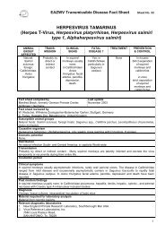
ANIMAL<br />
GROUP<br />
AFFECTED<br />
Ruminants<br />
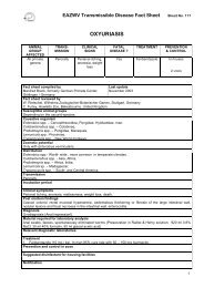
<strong>EAZWV</strong> <strong>Transmissible</strong> <strong>Disease</strong> <strong>Fact</strong> <strong>Sheet</strong> <strong>Sheet</strong> No. 91<br />
PARATUBERCULOSIS OR JOHNE’S DISEASE<br />
TRANS-<br />
MISSION<br />
Faecal-oral<br />
CLINICAL<br />
SIGNS<br />
Chronic or<br />
intermittent<br />
diarrhoea,<br />
wasting away<br />
FATAL<br />
DISEASE ?<br />
Yes<br />
TREATMENT PREVENTION<br />
& CONTROL<br />
None available In houses<br />
Good hygiene<br />
in zoos<br />
Quarantine and<br />
regular screening<br />
<strong>Fact</strong> sheet compiled by Last update<br />
F. Vercammen, Royal Zoological Society of December 2008<br />
Antwerp, Belgium<br />
<strong>Fact</strong> sheet reviewed by<br />
J. Kaandorp, Safari Beekse Bergen, Hilvarenbeek, The Netherlands<br />
D. Geysen, Animal Health, Institute of Tropical Medicine, Antwerp, Belgium<br />
Susceptible animal groups<br />
Although most information from clinical and experimental infections concerns domestic animal species,<br />
presumably all domestic and wild ruminants are susceptible. Reports exist of infection in water buffalo, whitetailed<br />
deer, red deer, roe deer, elk, bison, bighorn sheep, Rocky Mountain goat, aoudad, mouflon sheep,<br />
camel, mountain goat, reindeer, antelopes, yak, moose.The tylopods llama and alpaca can also be affected by<br />
this disease. But also monogastric animals can be receptive. Horses and pigs can be infected experimentally<br />
with development of intestinal lesions. Rabbits, mice, hamsters and guinea pigs are used as laboratory<br />
animals. Wild rabbits in Scotland have been shown to harbour these mycobacteria in their lymph nodes and<br />
intestines. Other nonruminant wildlife species infected in Scotland include small carnivorous mammals as well<br />
as carrion-eating birds and rodents. Also, two reports of infection in nonhuman primates (mandrill and<br />
stumptail macaque) exist.<br />
Causative organism<br />
The etiological agent of paratuberculosis is Mycobacterium avium subspecies paratuberculosis (MAP). In the<br />
past the organism was known as Mycobacterium paratuberculosis or Mycobacterium johnei. It is an acid-fast<br />
small bacillus that can appear gram-positive. It is closely related with, but distinctly different from<br />
Mycobacterium avium, which causes avian tuberculosis. MAP requires mycobactin for its growth, which is<br />
relatively slow in culture media. In the environment MAP is relatively resistant to adverse influences and can<br />
survive for many months in faeces, soil and water.<br />
Zoonotic potential<br />
The discussion about involvement of MAP in human Crohn’s <strong>Disease</strong> is still going on. The isolation of MAP<br />
from Crohn’ <strong>Disease</strong> patients is rare. Evidence to support an etiological role for MAP in Crohn’s <strong>Disease</strong> is for<br />
the moment still lacking. It is clear that if MAP is involved in human disease it is only one of the factors and that<br />
genetic and immunological elements play a significant role.<br />
Distribution<br />
This disease has a worldwide distribution. It appears to be more prevalent in temperate and wetter areas than<br />
in hotter and drier ones and usually occurs in regions with high cattle density.<br />
Transmission<br />
Both vertical and horizontal transmission exists. Although intrauterine infection can occur either because of an<br />
infected mother or because of infected semen or male genitalia, the faecal-oral route is most important.<br />
Horizontal transmission by ingestion of contaminated feed and water by the faeces of an infected animal that is<br />
excreting MAP remains most important.<br />
Incubation period<br />
In cattle it can take up to 2 - 5 years for first symptoms to appear. However, in captive wild animals it can occur<br />
also in young animals in which the incubation period can be much shorter (4 – 5 months).<br />
Clinical symptoms<br />
Chronic or intermittent diarrhoea that is unresponsive to treatment is the main clinical symptom in cattle. When<br />
diarrhoea becomes less prominent the animal appears to recover. In the case of prolonged protein-losing<br />
enteropathy the animal loses weight and wastes away. Sheep, goats and many exotic ruminants may not show<br />
real diarrhoea, but only softer faeces. However, they can also appear unhealthy with chronic weight loss.<br />
Post mortem findings<br />
In advanced cases macroscopic thickening of the intestinal mucosa is present in the lower part of the small<br />
intestine (especially in ileum) along with enlargement of the associated mesenteric lymph nodes. The intestinal
<strong>EAZWV</strong> <strong>Transmissible</strong> <strong>Disease</strong> <strong>Fact</strong> <strong>Sheet</strong> <strong>Sheet</strong> No. 91<br />
wall is diffusely thickened and the mucosa shows transverse folds or corrugations. These lesions can also be<br />
found in the jejunum, caecum or colon. Pathological changes of intestine and / or lymph nodes can vary<br />
between animal species and individuals.<br />
Diagnosis<br />
Anamnesis and clinical signs are only suggestive for paratuberculosis and differential diagnosis includes other<br />
chronic wasting diseases. A definitive diagnosis can be made by demonstrating MAP. Serology can also be<br />
very helpful.<br />
1. Direct methods: detection of the organism.<br />
a) Smears<br />
Mucosal scrapings or faecal smears can be examined by Ziehl-Neelsen staining, but only in the case of<br />
heavy shedders clumps of organisms can be found.<br />
b) Culture<br />
MAP is a very slow grower and culturing can take up to six to eight months. A radiometric culture with<br />
BACTEC can detect infection in 4 - 8 weeks and has a sensitivity of about 50 %, but is 100 % specific.<br />
c) Histology<br />
Histopathological examination of intestine and lymph nodes can reveal acid-fast bacilli in macrophages or<br />
giant cells.<br />
d) Polymerase Chain Reaction (PCR)<br />
By far the most rapid test is the polymerase chain reaction to detect the specific insertion sequence IS900<br />
of MAP. PCR diagnosis takes just a few days, but although 100 % specific, the test has a low 8 – 33 %<br />
sensitivity. Recently, IS900-like sequences have been reported in other mycobacteria. Therefore, a second<br />
assay on the f57 sequence was developed. The combination of the two PCR assays has proven to be<br />
useful for the identification of MAP, but validation on a large range of clinical samples still needs to be<br />
done.<br />
2. Indirect methods: detection of reaction against the organism.<br />
a) Serology<br />
Different types of serological tests for the detection of antibodies are used with varying sensitivity and<br />
specificity, but they all score low in subclinical cases. Complement fixation (CF), agar gel immunodiffusion<br />
(AGID) and enzyme-linked immunosorbent assay (ELISA) are the most known techniques.<br />
b) Cell Mediated Immunity<br />
The delayed-type hypersensitivity skin test with intradermal Johnin is unreliable because of minimal skin<br />
reactivity in most animals. A gamma-interferon stimulation test is helpful for identification of animals with<br />
early infection, but becomes unreliable when animals progress towards an anergic state. A lymphocyte<br />
stimulation test in deer has a sensitivity of 95 % and a specificity of 92 %, but in other species these<br />
figures are unknown.<br />
c) Histology<br />
Localized or diffuse granulomatous changes with accumulation of macrophages in the intestinal laminar<br />
propria and submucosa or lymph node cortex are suggestive of infection with MAP.<br />
Material required for laboratory analysis<br />
Faeces, serum, lymph node, intestine (ileum).<br />
OIE Reference Laboratories<br />
• Dr Jacek Gwozdz<br />
Johne's <strong>Disease</strong> Laboratory, Research and Development Division, Department of Primary Industries<br />
475 Mickleham Road, Attwood, Victoria 3049<br />
AUSTRALIA<br />
Tel: (61.3) 92.17.42.00 Fax: (61.3) 92.17.42.99<br />
Email: jacek.gwozdz@dpi.vic.gov.au<br />
• Dr Bernardo Alonso<br />
Gerencia de Laboratorios (GELAB) del Servicio Nacional de Sanidad y Calidad, Agroalimentaria<br />
(SENASA)<br />
Av. Alexander Fleming 1653, 1640 Martinez - Pcia de Buenos Aires<br />
ARGENTINA<br />
Tel: (54.11) 48.36.00.36 Fax: (54.11) 48.36.00.36<br />
Email: balonso@senasa.gov.ar<br />
Email: dilab@inea.com.ar<br />
• Dr I. Pavlik<br />
Veterinary Research Institute<br />
Hudcova 70, 62132 Brno<br />
CZECH (Rep.)<br />
Tel: (420.5) 33.33.16.01 Fax: (420.5) 33.33.12.29
Email: pavlik@vri.cz<br />
<strong>EAZWV</strong> <strong>Transmissible</strong> <strong>Disease</strong> <strong>Fact</strong> <strong>Sheet</strong> <strong>Sheet</strong> No. 91<br />
• Mme María Laura Boschiroli-Cara<br />
AFSSA Alfort, Unité Zoonoses Bactériennes, Laboratoire d'études et de recherches en pathologie animale<br />
et zoonoses<br />
23 avenue du Général de Gaulle, 94706 Maisons-Alfort Cedex<br />
FRANCE<br />
Tel: (33 (0)1) 49.77.13.00 Fax: (33 (0)1) 49.77.13.44<br />
Email: ml.boschiroli@afssa.fr<br />
Relevant diagnostic laboratories<br />
1. CODA, Groeselenberg 99, 1180 Brussel, Belgium<br />
2. ID/DLO Lelystad, P.O. Box 65, 8200 AB Lelystad, the Netherlands<br />
Treatment<br />
Today an effective etiological treatment is not available. Only supportive and symptomatic treatment can be<br />
initiated.<br />
Prevention and control in zoos<br />
Routine screening of all susceptible animals with radiometric culture and an ameliorated PCR assay in the<br />
future will be important to assess the real prevalence in a zoo population. Identifying subclinical carriers before<br />
entering the population will be most important. Isolation of infected animals with strict hygienic and<br />
zootechnical measures is necessary to have a chance of eradication of this disease. Contaminated pastures<br />
should be kept free of susceptible animals for at least one year. Contaminated enclosures should have the<br />
topsoil replaced.<br />
Suggested disinfectant for housing facilities<br />
MAP is resistant to many disinfectants, but among those that are effective belong formaldehyde, phenol,<br />
cresylic disinfectants and calcium hypochlorite. Generally, any disinfectant with tuberculocidal activity can be<br />
used.<br />
Notification<br />
Guarantees required under EU Legislation<br />
Guarantees required by EAZA Zoos<br />
Measures required under the Animal <strong>Disease</strong> Surveillance Plan<br />
Measures required for introducing animals from non-approved sources<br />
Measures to be taken in case of disease outbreak or positive laboratory findings<br />
Conditions for restoring disease-free status after an outbreak<br />
Contacts for further information<br />
References<br />
1. Beard PM, Daniels MJ, Henderson D, Pirie A, Rudge K, Buxton D, Rhind S, Greig A, Hutchings MR,<br />
McKendrick I, Stevenson K, Sharp JM. (2001) Paratuberculosis infection in nonruminant wildlife in<br />
Scotland. J Clin Microbiol 39 (4): 1517-1521.<br />
2. Graig A, Stevenson K, Perez V, Pirie AA, Grant JM, Sharp JM. (1997) Paratuberculosis in wild rabbits<br />
(Oryctolagus cuniculus). Vet Rec 140: 141-143.<br />
3. Kaandorp J. (1998) Diagnosis in mycobacteriosis. Proc 2 nd Scient Meeting <strong>EAZWV</strong>, Chester, 85-92.<br />
4. Kaandorp J. (2000) Paratuberculosis in zoo animal medicine. Proc 3 rd Scient Meeting <strong>EAZWV</strong>, Paris, 43-<br />
46.<br />
5. McClure HM, Chiodini RJ, Anderson DC, Swenson RB, Thayer WR, Coutu JA. (1987) Mycobacterium<br />
paratuberculosis infection in a colony of stumptail macaques (Macaca arctoides). J Infect Dis 155: 1011-<br />
1019.<br />
6. Morrow AN. Johne’s <strong>Disease</strong>. In: Sewell MMH, Brocklesby DW (eds.). Handbook on Animal <strong>Disease</strong>s in<br />
the Tropics. Baillière Tindall, 1990, 77-80.<br />
7. Payeur JB. (1998) Overview of Johne’s <strong>Disease</strong> Diagnostics. Proceedings of the Workshop on Diagnosis,<br />
Prevention, and Control of Johne’s <strong>Disease</strong> in Non-Domestic Hoofstock, White Oak Conservation Center,<br />
Florida, 21-26.<br />
8. Radostits OM, Blood DC, Gay CC (eds.). Veterinary Medicine. A Textbook of the <strong>Disease</strong>s of Cattle,
<strong>EAZWV</strong> <strong>Transmissible</strong> <strong>Disease</strong> <strong>Fact</strong> <strong>Sheet</strong> <strong>Sheet</strong> No. 91<br />
Sheep, Pigs, Goats and Horses. Baillière Tindall, 1994, 841-850.<br />
9. Rubery E. (2001) A Review of the Evidence for a Link between Exposure to Mycobacterium<br />
Paratuberculosis (MAP) and Crohn’s <strong>Disease</strong> (CD) in Humans. A Report for the Food Standard Agency.<br />
Conclusions and Recommendations, University of Cambridge, 30-32.<br />
10. Thoen CO. Tuberculosis and other Mycobacterial <strong>Disease</strong>s in captive wild animals. In: Fowler ME (ed).<br />
Zoo & Wild Animal Medicine: Current Therapy 3. WB Saunders Company, 1993, 45-49.<br />
11. Vansnick E., De Rijk P., Vercammen F., Geysen D., Rigouts L., Portaels F. (2004) Newly developed<br />
primers for the detection of Mycobacterium avium subspecies paratuberculosis. Vet. Microbiol. 100 (3-4):<br />
197-204.<br />
12. Walsh TF, Murnane RD, Barbiers R, Collins M. (1995) Enteric Mycobacterium paratuberculosis in a<br />
mandrill (Papio sphinx). Proc Jt Conf AAZV/WDA/AAWV: 262-264.<br />
13. Williams ES, Snyder SP, Martin KL. (1983) Experimental infection of some North American wild ruminants<br />
and domestic sheep with Mycobacterium paratuberculosis: clinical and bacteriological findings. J Wildlife<br />
Dis 19 (3): 185-191.