Clinical evaluation of refraction using a handheld wavefront ...
Clinical evaluation of refraction using a handheld wavefront ...
Clinical evaluation of refraction using a handheld wavefront ...

Create successful ePaper yourself
Turn your PDF publications into a flip-book with our unique Google optimized e-Paper software.
<strong>handheld</strong> without a table and chin rest. These features<br />
make it suitable for measuring very young children and<br />
disabled or uncooperative patients. In this respect, it is<br />
comparable to the Nikon Retinomax <strong>handheld</strong><br />
autorefractor. 11<br />
An interesting feature <strong>of</strong> the <strong>wavefront</strong> autorefractor<br />
is its fairly large working distance <strong>of</strong> 0.35 m. Although<br />
this distance is smaller than that used by<br />
photo<strong>refraction</strong> devices, 12–14 it is large enough to perform<br />
a valid <strong>refraction</strong> in children who would normally<br />
show strong resistance when an examiner comes into<br />
close range with a huge conventional autorefractor.<br />
In the present study, we evaluated the accuracy <strong>of</strong><br />
the <strong>wavefront</strong> autorefractor in ametropic patients with<br />
and without cycloplegia. The influence <strong>of</strong> accommodation<br />
when measuring children and adults without cycloplegia<br />
is also discussed.<br />
Patients and Methods<br />
Patients<br />
The accuracy <strong>of</strong> the <strong>wavefront</strong> autorefractor was studied<br />
in 2 groups <strong>of</strong> patients recruited from the outpatient department<br />
and ward <strong>of</strong> the University Eye Hospital, Hamburg-<br />
Eppendorf. Group 1 consisted <strong>of</strong> 195 eyes (108 patients, aged<br />
1 to 81 years) measured under cycloplegia. The median spherical<br />
equivalent (SE) was 0.75 diopter (D) 2.04 (SD)<br />
(range 8.13 to 5.75 D; 51 eyes with myopia, 138 with<br />
hyperopia, and 6 with emmetropia). The mean cylinder<br />
power was 0.70 0.62 D (range 0 to 3.75 D).<br />
Group 2 consisted <strong>of</strong> 96 eyes (51 patients, aged 2 to<br />
76 years) from Group 1 who were also measured without<br />
cycloplegia. The median SE <strong>of</strong> the 96 eyes was 1.13 <br />
2.24 D (range 7.75 to 5.75 D). The mean cylinder power<br />
was 0.76 0.76 D (range 0 to 3.75 D). A subgroup <strong>of</strong><br />
these patients consisted <strong>of</strong> 27 children (57 eyes, aged 2 to 17<br />
years). The median SE was 1.25 2.07 D (range 6.50 to<br />
5.75 D). The mean cylinder power was 0.71 0.71 D<br />
(range 0 to 3.00 D).<br />
All patients were first screened by an ophthalmologist.<br />
Patients with heterotropia, eccentric fixation, suppression,<br />
opacities <strong>of</strong> the optical media, or any disease <strong>of</strong> the retina were<br />
excluded. Cycloplegia was carried out by application <strong>of</strong> 1 drop<br />
<strong>of</strong> cyclopentolate 1% and a second drop 10 minutes later.<br />
After a waiting period <strong>of</strong> 20 minutes, retinoscopy and the<br />
measurement with the SureSight was performed. In children<br />
younger than 3 years, cyclopentolate 0.5% or tropicamide 5%<br />
was used instead <strong>of</strong> cyclopentolate 1%. Patients with dark<br />
irides were given an additional dose if it was judged that cycloplegia<br />
was incomplete on the basis <strong>of</strong> pupil activity and<br />
variability in the retinoscopic neutral point.<br />
1656<br />
HANDHELD WAVEFRONT AUTOREFRACTOR IN YOUNG AND ADULT PATIENTS<br />
J CATARACT REFRACT SURG—VOL 28, SEPTEMBER 2002<br />
Working Principle <strong>of</strong> the Wavefront Autorefractor<br />
Wavefront analysis does not require moving optical components.<br />
The data acquisition is based solely on optical imaging<br />
and electronic calculations. The patient must look straight<br />
at the device. A circle <strong>of</strong> 8 green blinking diodes is provided to<br />
attract the patient’s attention. The optical setup is shown in<br />
Figures 1A and 1B. An infrared beam from a laser diode is<br />
directed into 1 eye. When it emerges from the instrument, the<br />
beam can be seen by the patient as a red spot. This serves as an<br />
additional fixation target. The beam has a small diameter and<br />
is thus imaged on the retina with a large depth <strong>of</strong> focus. The<br />
diameter <strong>of</strong> the laser focus on the retina is small and almost<br />
independent <strong>of</strong> the degree <strong>of</strong> ametropia. The beam is reflected<br />
at the retina and propagates back to the autorefractor, where it<br />
enters the instrument, passes the beam-splitter, and reaches<br />
the essential part <strong>of</strong> the optical construction—the so-called<br />
Figure 1A. (Schimitzek) An infrared laser beam is reflected by a<br />
beam splitter and sent into the eye. The beam is focused at the<br />
macula and diffusely reflected from the fundus. Some <strong>of</strong> the reflected<br />
light rays leave the eye, enter the autorefractor, and fall on a Hartmann-Shack<br />
sensor that consists <strong>of</strong> numerous lenses arranged as a<br />
matrix. The multiple images are recorded with a CCD camera.<br />
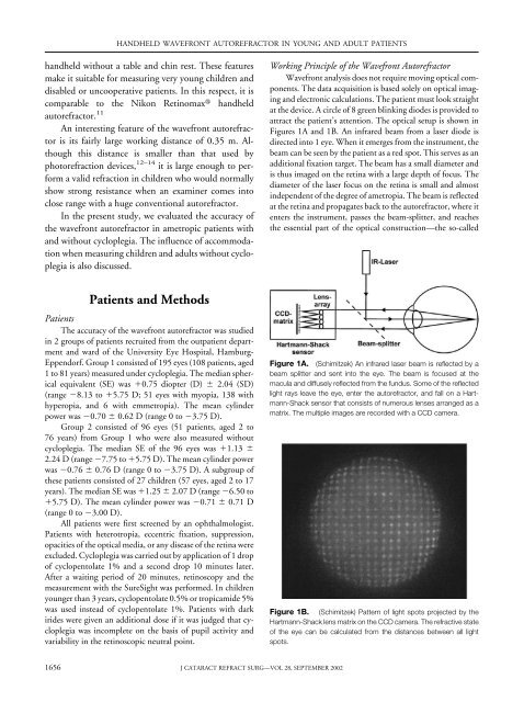
Figure 1B. (Schimitzek) Pattern <strong>of</strong> light spots projected by the<br />
Hartmann-Shack lens matrix on the CCD camera. The refractive state<br />
<strong>of</strong> the eye can be calculated from the distances between all light<br />
spots.