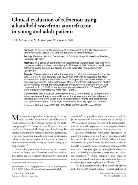
Clinical evaluation of refraction using a handheld wavefront ...
Clinical evaluation of refraction using a handheld wavefront ...
Clinical evaluation of refraction using a handheld wavefront ...
Create successful ePaper yourself
Turn your PDF publications into a flip-book with our unique Google optimized e-Paper software.
<strong>Clinical</strong> <strong>evaluation</strong> <strong>of</strong> <strong>refraction</strong> <strong>using</strong><br />
a <strong>handheld</strong> <strong>wavefront</strong> autorefractor<br />
in young and adult patients<br />
Thilo Schimitzek, MD, Wolfgang Wesemann, PhD<br />
Purpose: To determine the accuracy <strong>of</strong> measurement by the SureSight autorefractor<br />
(s<strong>of</strong>tware version 2.0) and the influence <strong>of</strong> accommodation.<br />
Setting: Pediatric Section, Department <strong>of</strong> Ophthalmology, University <strong>of</strong> Hamburg,<br />
Hamburg, Germany.<br />
Methods: In a series <strong>of</strong> comparative measurements, autorefractor readings were<br />
compared with cycloplegic retinoscopy in 195 eyes <strong>of</strong> 108 patients (1 to 81 years)<br />
measured under cycloplegia. Ninety-six eyes were also measured without<br />
cycloplegia.<br />
Results: The <strong>wavefront</strong> autorefractor was able to refract human eyes from a distance<br />
<strong>of</strong> 0.35 m. The accuracy was lower than that with conventional tabletop<br />
autorefractors. A difference <strong>of</strong> less than 0.51 diopter (D) was found in 68% <strong>of</strong> the<br />
spherical equivalents under cycloplegia. Many emmetropic and hyperopic children<br />
accommodated during the noncycloplegic measurements and were minus-overcorrected<br />
up to 6.13 D. In our group <strong>of</strong> young patients (2 to 17 years), 47%<br />
were minus-overcorrected by more than 2.00 D.<br />
Conclusions: The <strong>wavefront</strong> autorefractor uses a new method to determine the<br />
refractive state <strong>of</strong> the eye from a distance. It was less accurate than other conventional<br />
autorefractors. A benefit is its application in infants and disabled and<br />
uncooperative subjects. Cycloplegia is necessary in young hyperopic patients.<br />
J Cataract Refract Surg 2002; 28:1655–1666 © 2002 ASCRS and ESCRS<br />
Most automatic eye refractors currently in use are<br />
based on well-known optical principles such as<br />
streak retinoscopy, the Scheiner method, or the knifeedge<br />
principle. 1–3 During the past 30 years, these autorefractors<br />
have reached a high state <strong>of</strong> perfection. By<br />
incorporating modern computer and video technology,<br />
it has been possible to simplify the optical construction,<br />
reduce the measurement time, and improve the accuracy<br />
<strong>of</strong> measurement without changing the underlying optical<br />
principles. 4,5<br />
A completely new way <strong>of</strong> measuring the refractive<br />
state <strong>of</strong> the human eye is based on <strong>wavefront</strong> analysis.<br />
This technique was originally developed by Liang and<br />
Accepted for publication March 6, 2002.<br />
Reprint requests to Dr. Wolfgang Wesemann, HFAK, Bayenthalguertel<br />
6-8, D-50968 Cologne, Germany. E-mail: wesemann@hfak.de.<br />
coauthors 6 and provides a rapid, noninvasive, and objective<br />
measure <strong>of</strong> the wave aberration <strong>of</strong> the eye. In<br />
1997, Liang and Williams 7 developed the technique further<br />
and were able to present the most complete data on<br />
the various optical errors <strong>of</strong> the human eye to date.<br />
Another promising ophthalmic application <strong>of</strong><br />
<strong>wavefront</strong> analysis is in the field <strong>of</strong> <strong>wavefront</strong>-guided<br />
laser in situ keratomileusis (LASIK). When the wave<br />
aberrations <strong>of</strong> a given human eye are known, it may be<br />
possible to calculate a customized ideal ablation pr<strong>of</strong>ile<br />
that corrects not only spherical and cylindrical components<br />
but also high-order refractive errors such as spherical<br />
aberration. 8,9 Several companies have begun to<br />
incorporate <strong>wavefront</strong> aberrometers into their laser systems<br />
to support topography-guided ablation. 9,10<br />
The first autorefractor based on <strong>wavefront</strong> analysis<br />
is the SureSight (Welch Allyn). It is small and can be<br />
© 2002 ASCRS and ESCRS 0886-3350/02/$–see front matter<br />
Published by Elsevier Science Inc. PII S0886-3350(02)01426-8
<strong>handheld</strong> without a table and chin rest. These features<br />
make it suitable for measuring very young children and<br />
disabled or uncooperative patients. In this respect, it is<br />
comparable to the Nikon Retinomax <strong>handheld</strong><br />
autorefractor. 11<br />
An interesting feature <strong>of</strong> the <strong>wavefront</strong> autorefractor<br />
is its fairly large working distance <strong>of</strong> 0.35 m. Although<br />
this distance is smaller than that used by<br />
photo<strong>refraction</strong> devices, 12–14 it is large enough to perform<br />
a valid <strong>refraction</strong> in children who would normally<br />
show strong resistance when an examiner comes into<br />
close range with a huge conventional autorefractor.<br />
In the present study, we evaluated the accuracy <strong>of</strong><br />
the <strong>wavefront</strong> autorefractor in ametropic patients with<br />
and without cycloplegia. The influence <strong>of</strong> accommodation<br />
when measuring children and adults without cycloplegia<br />
is also discussed.<br />
Patients and Methods<br />
Patients<br />
The accuracy <strong>of</strong> the <strong>wavefront</strong> autorefractor was studied<br />
in 2 groups <strong>of</strong> patients recruited from the outpatient department<br />
and ward <strong>of</strong> the University Eye Hospital, Hamburg-<br />
Eppendorf. Group 1 consisted <strong>of</strong> 195 eyes (108 patients, aged<br />
1 to 81 years) measured under cycloplegia. The median spherical<br />
equivalent (SE) was 0.75 diopter (D) 2.04 (SD)<br />
(range 8.13 to 5.75 D; 51 eyes with myopia, 138 with<br />
hyperopia, and 6 with emmetropia). The mean cylinder<br />
power was 0.70 0.62 D (range 0 to 3.75 D).<br />
Group 2 consisted <strong>of</strong> 96 eyes (51 patients, aged 2 to<br />
76 years) from Group 1 who were also measured without<br />
cycloplegia. The median SE <strong>of</strong> the 96 eyes was 1.13 <br />
2.24 D (range 7.75 to 5.75 D). The mean cylinder power<br />
was 0.76 0.76 D (range 0 to 3.75 D). A subgroup <strong>of</strong><br />
these patients consisted <strong>of</strong> 27 children (57 eyes, aged 2 to 17<br />
years). The median SE was 1.25 2.07 D (range 6.50 to<br />
5.75 D). The mean cylinder power was 0.71 0.71 D<br />
(range 0 to 3.00 D).<br />
All patients were first screened by an ophthalmologist.<br />
Patients with heterotropia, eccentric fixation, suppression,<br />
opacities <strong>of</strong> the optical media, or any disease <strong>of</strong> the retina were<br />
excluded. Cycloplegia was carried out by application <strong>of</strong> 1 drop<br />
<strong>of</strong> cyclopentolate 1% and a second drop 10 minutes later.<br />
After a waiting period <strong>of</strong> 20 minutes, retinoscopy and the<br />
measurement with the SureSight was performed. In children<br />
younger than 3 years, cyclopentolate 0.5% or tropicamide 5%<br />
was used instead <strong>of</strong> cyclopentolate 1%. Patients with dark<br />
irides were given an additional dose if it was judged that cycloplegia<br />
was incomplete on the basis <strong>of</strong> pupil activity and<br />
variability in the retinoscopic neutral point.<br />
1656<br />
HANDHELD WAVEFRONT AUTOREFRACTOR IN YOUNG AND ADULT PATIENTS<br />
J CATARACT REFRACT SURG—VOL 28, SEPTEMBER 2002<br />
Working Principle <strong>of</strong> the Wavefront Autorefractor<br />
Wavefront analysis does not require moving optical components.<br />
The data acquisition is based solely on optical imaging<br />
and electronic calculations. The patient must look straight<br />
at the device. A circle <strong>of</strong> 8 green blinking diodes is provided to<br />
attract the patient’s attention. The optical setup is shown in<br />
Figures 1A and 1B. An infrared beam from a laser diode is<br />
directed into 1 eye. When it emerges from the instrument, the<br />
beam can be seen by the patient as a red spot. This serves as an<br />
additional fixation target. The beam has a small diameter and<br />
is thus imaged on the retina with a large depth <strong>of</strong> focus. The<br />
diameter <strong>of</strong> the laser focus on the retina is small and almost<br />
independent <strong>of</strong> the degree <strong>of</strong> ametropia. The beam is reflected<br />
at the retina and propagates back to the autorefractor, where it<br />
enters the instrument, passes the beam-splitter, and reaches<br />
the essential part <strong>of</strong> the optical construction—the so-called<br />
Figure 1A. (Schimitzek) An infrared laser beam is reflected by a<br />
beam splitter and sent into the eye. The beam is focused at the<br />
macula and diffusely reflected from the fundus. Some <strong>of</strong> the reflected<br />
light rays leave the eye, enter the autorefractor, and fall on a Hartmann-Shack<br />
sensor that consists <strong>of</strong> numerous lenses arranged as a<br />
matrix. The multiple images are recorded with a CCD camera.<br />
Figure 1B. (Schimitzek) Pattern <strong>of</strong> light spots projected by the<br />
Hartmann-Shack lens matrix on the CCD camera. The refractive state<br />
<strong>of</strong> the eye can be calculated from the distances between all light<br />
spots.
HANDHELD WAVEFRONT AUTOREFRACTOR IN YOUNG AND ADULT PATIENTS<br />
Hartmann-Shack sensor. This sensor consists <strong>of</strong> a matrix <strong>of</strong><br />
tiny lenses arranged in many rows and columns. These lenses<br />
form many small light spots on the active surface <strong>of</strong> a chargedcouple-device<br />
(CCD) matrix camera.<br />
The refractive power is calculated from the positions <strong>of</strong><br />
the light spots according to the following general concept. If<br />
the patient has an emmetropic eye, parallel light rays leave the<br />
eye. These rays produce light spots on the CCD camera that<br />
are uniformly spaced. The distance between the light spots<br />
(Figure 1B) is typical in an emmetropic eye. In the case <strong>of</strong> pure<br />
hyperopia or myopia, the light spots are also uniformly<br />
spaced. The spot pattern, however, is expanded or contracted<br />
compared to that in emmetropia because the light rays that are<br />
reflected from the fundus leave the eye divergent or convergent.<br />
If the patient has an astigmatic ametropia, a light bundle<br />
that is no longer rotationally symmetric leaves the eye. In this<br />
case, the distance between the light spots on the CCD matrix<br />
varies in a way that depends on cylinder power and axis.<br />
Method <strong>of</strong> Comparative Measurements<br />
All autorefractor measurements without cycloplegia were<br />
taken immediately before the eyedrops were dispensed. After<br />
the waiting period, streak retinoscopy was performed under<br />
cycloplegia by 1 <strong>of</strong> us (T.S.) <strong>using</strong> <strong>handheld</strong> corrective lenses.<br />
The investigator was unaware <strong>of</strong> the results with the <strong>wavefront</strong><br />
autorefractor. The cycloplegic measurement with the Sure-<br />
Sight followed immediately. The “child mode” explained in<br />
the discussion section was not used.<br />
Criteria for the Accuracy <strong>of</strong> Measurement<br />
To obtain information about the accuracy <strong>of</strong> the <strong>wavefront</strong><br />
autorefractor, all readings were compared to the results<br />
<strong>of</strong> cycloplegic retinoscopy. All comparison criteria, with the<br />
exception <strong>of</strong> the J-vector analysis, were used in our earlier<br />
studies 1,2,5,11 and allow direct comparison <strong>of</strong> the <strong>wavefront</strong><br />
autorefractor described here with autorefractors tested in earlier<br />
years.<br />
The difference <strong>of</strong> the spherical equivalent (DSE) was calculated<br />
by<br />
DSE (S t 0.5 * C t) S c 0.5 * C c<br />
where S and C denote the spherical and the cylinder powers;<br />
the subscripts “t” (test) and “c” (comparison) denote the instrument<br />
under test (SureSight) and the comparison technique<br />
(cycloplegic retinoscopy). A negative DSE indicates a<br />
minus-overcorrection by the instrument under test.<br />
The difference <strong>of</strong> the cylindrical powers (DC) was calculated<br />
by<br />
DC Ct – Cc The weighted axes difference (DA) is a quality criterion<br />
based on the power-vector approach. It is evaluated by a mathematical<br />
expression in which the difference between the<br />
2 cylinder axes (test and comparison, measured in degrees) is<br />
weighted with the cylinder power measured with the comparison<br />
method.<br />
DA 2C c sin ( t – c)<br />
The formula makes it possible to compare axis values,<br />
even when the actual cylinder powers are different. C c is taken<br />
as a weighting factor since it is assumed to be more accurate<br />
then the cylinder power <strong>of</strong> the instrument under test. Geometrically,<br />
DA is the length <strong>of</strong> the difference vector between<br />
both methods given that the cylinder power found with the<br />
SureSight is equal to the cylinder power found with retinoscopy.<br />
The resulting number has the dimension “diopter.” A<br />
value <strong>of</strong> DA 0.5 D is equivalent to an axes difference <strong>of</strong><br />
14.5 degrees given a cylinder power <strong>of</strong> 1.0 D.<br />
The J-vector difference ( J ) is a measure <strong>of</strong> the difference<br />
between the cylindrical components. J describes the<br />
cylindrical difference in terms <strong>of</strong> 2 Jackson crossed cylinders<br />
with orientations <strong>of</strong> 0 degrees and 45 degrees, respectively. J<br />
is determined <strong>using</strong> the method <strong>of</strong> Raasch and coauthors. 15 At<br />
first, the J 0 and J 45 components <strong>of</strong> the (M, J 0, J 45) space<br />
described by Thibos and Horner 16 are calculated from the<br />
results <strong>of</strong> the SureSight and cycloplegic retinoscopy.<br />
J 0t –C t/2 cos 2 t<br />
J 45t –C t/2 sin 2 t<br />
and<br />
J 0c –C c/2 cos 2 c<br />
J 45c –C c/2 sin 2 c<br />
Then the J-vector difference ( J) between the cylindrical<br />
results <strong>of</strong> the test and the comparison instrument can be written<br />
as<br />
J J 0, J 45<br />
where J 0 and J 45 are defined by<br />
J 0 J 0t – J 0c<br />
J 45t J 45t – J 45c<br />
Our last quality criterion is the total cylindrical difference<br />
(TCD). It is the length <strong>of</strong> the vector J. The TCD is a summarizing<br />
measure for the cylindrical accuracy <strong>of</strong> the measurement.<br />
When the normal cylinder notation is used instead <strong>of</strong><br />
the Jackson crossed-cylinder notation, TCD is obtained by<br />
TCD C t 2 Cc 2 2CtC ccos 2 t c<br />
The terminology for this criterion differs in the literature.<br />
The term total cylindrical difference was introduced by 1 <strong>of</strong> us<br />
in 1987. 5 In our earlier papers, 2 the term combined cylindrical<br />
error was used. Wesemann and Dick 11 called the above expression<br />
the difference <strong>of</strong> the cylindrical corrections. In 2000, Raasch<br />
and coauthors 15 proposed the name astigmatic difference (see<br />
Appendix).<br />
J CATARACT REFRACT SURG—VOL 28, SEPTEMBER 2002 1657
HANDHELD WAVEFRONT AUTOREFRACTOR IN YOUNG AND ADULT PATIENTS<br />
Results<br />
Accuracy <strong>of</strong> Measurement in Ametropic Patients<br />
Under Cycloplegia<br />
Difference <strong>of</strong> the spherical equivalents. The difference<br />
in the SE between <strong>wavefront</strong> refractor and cycloplegic<br />
retinoscopy in 195 eyes is shown in Figure 2a. The<br />
frequency distribution is almost normally distributed<br />
about the central column. This central column indicates<br />
that 45% <strong>of</strong> the SEs differed by less than 0.26 D. The<br />
other columns show how <strong>of</strong>ten larger errors occurred.<br />
The maximal differences were 2.38 D and 3.00 D.<br />
On average, the SureSight delivered an SE that was in<br />
exact agreement with retinoscopy under cycloplegia<br />
(DSE mean 0.01 0.65 D).<br />
Difference <strong>of</strong> the cylindrical powers. The cylinder<br />
powers determined by the SureSight and retinoscopy<br />
were very similar. The mean difference was 0.01 <br />
0.49 D. The distribution in Figure 2b is almost symmetric<br />
and shows few cylinder power differences larger than<br />
0.50 D. In all eyes that showed no significant astigmatism<br />
with cycloplegic retinoscopy, the SureSight displayed<br />
cylinder powers 1.0 D.<br />
Weighted axes difference. The accuracy <strong>of</strong> the axis<br />
was evaluated in the cases in which a cylinder power <strong>of</strong><br />
0.25 D or greater had been determined with the autorefractor<br />
and with retinoscopy. This occurred in 167 <strong>of</strong><br />
195 eyes. The mean DA was 0.55 0.48 D (Figure<br />
2c). In a few cases, large differences were found; the<br />
largest was 2.88 D (cylinder power 1.50 D, axis<br />
deviation 74 degrees).<br />
Accuracy <strong>of</strong> Measurement in Ametropic Patients<br />
Without Cycloplegia<br />
Difference <strong>of</strong> the spherical equivalents. The DSE distribution<br />
in Figure 3a is not symmetric. It exhibits a shift<br />
toward negative values. The SE measured with the <strong>wavefront</strong><br />
autorefractor was accurate to 0.50 D in only<br />
33% <strong>of</strong> cases. In 47%, the DSE was not larger than<br />
1.00 D. The mean DSE was 1.59 1.95 D (range<br />
2.75 to 6.13 D).<br />
Difference <strong>of</strong> the cylindrical powers. The accuracy in<br />
measuring cylinder power was similar to the measurement<br />
under cycloplegia (Figure 3b). The mean difference<br />
was 0.06 0.47 D.<br />
Weighted axes difference. Eighty-one percent <strong>of</strong> eyes<br />
showed a cylinder power <strong>of</strong> 0.25 D or greater and were<br />
included in the analysis. The mean DA was 0.45 <br />
0.49 D (Figure 3c). The largest difference was 3.15 D<br />
(cylinder power 2.50 D, axis deviation 39 degrees).<br />
Accuracy Indices<br />
Table 1 summarizes the data presented in the histograms<br />
(Figures 2 and 3). It indicates how <strong>of</strong>ten the differences<br />
were smaller than a selected criterion value.<br />
This is a measure <strong>of</strong> the percentage <strong>of</strong> correct or almost<br />
correct results. Data found with 7 table-top refractors <strong>of</strong><br />
an earlier generation 5 and with the Nikon Retinomax 11<br />
are presented for comparison.<br />
Accuracy <strong>of</strong> Cycloplegic Measurements. The percentage<br />
<strong>of</strong> correct or almost correct results with the Sure-<br />
Sight in cycloplegic eyes (rows 1 and 2) lie below the<br />
range found with 7 different table-top autorefractors 5<br />
Figure 2. (Schimitzek) Frequency distribution <strong>of</strong> the differences between SureSight and retinoscopy (Group 1); both measurements under<br />
cycloplegia. 2a: difference <strong>of</strong> the spherical equivalents; 2b: difference <strong>of</strong> the cylinder powers; 2c: weighted axes difference.<br />
1658<br />
J CATARACT REFRACT SURG—VOL 28, SEPTEMBER 2002
Figure 3. (Schimitzek) Frequency distribution <strong>of</strong> the differences between SureSight and retinoscopy (Group 2, SureSight without cycloplegia).<br />
3a: difference <strong>of</strong> the spherical equivalents; 3b: difference <strong>of</strong> the cylinder powers; 3c: weighted axes difference. The percentage <strong>of</strong> minusovercorrected<br />
cases (DSE 0in3a) is much larger than in Figure 2a. The distributions in 3b and 3c are similar to those in Figures 2b and 2c.<br />
Table 1. Frequency <strong>of</strong> “correct or almost correct” results obtained in eyes under and without cycloplegia with the SureSight. The<br />
percentages indicate how <strong>of</strong>ten the result <strong>of</strong> the SureSight differed by less than 0.51 D or 0.63 D from cycloplegic retinoscopy. Data from 2<br />
other studies that used the same criteria on 7 table-top autorefractors and the <strong>handheld</strong> Retinomax refractor are presented for comparison.<br />
Measurement<br />
SureSight under cycloplegia vs. cycloplegic<br />
retinoscopy (Group 1, n 195)<br />
SureSight under cycloplegia vs. cycloplegic<br />
retinoscopy (Group 2, n 96)<br />
SureSight without cycloplegia vs. cycloplegic<br />
retinoscopy (Group 2, n 96)<br />
Retinomax K-Plus vs. subjective <strong>refraction</strong><br />
(adult patients without cycloplegia) 11<br />
Cycloplegic Retinomax K-Plus vs. cycloplegic<br />
retinoscopy (children) 11<br />
Range <strong>of</strong> 7 table-top autorefractors (adult<br />
patients without cycloplegia) 5<br />
HANDHELD WAVEFRONT AUTOREFRACTOR IN YOUNG AND ADULT PATIENTS<br />
DSE<br />
Figure 4. (Schimitzek) Two-dimensional polar plot <strong>of</strong> the vector<br />
difference 2 J between the cylindrical corrections obtained with cycloplegic<br />
retinoscopy and cycloplegic SureSight in Group 1. a: All<br />
patients (N 195). b: Patients with astigmatism 0.5D(n 136). J<br />
was calculated for each eye. All vectors start at the origin. For clarity,<br />
only the endpoint <strong>of</strong> each vector is shown. The distance <strong>of</strong> each<br />
diamond from the origin indicates the discrepancy between the 2<br />
methods. All data points inside the circle denote measurements in<br />
which the TCD was smaller than our criterion difference <strong>of</strong> 0.63 D.<br />
Further Analysis <strong>of</strong> the Cylindrical Differences<br />
Results <strong>of</strong> the J-vector analysis <strong>of</strong> all patients in<br />
Group 1 are shown in Figure 4. The 2-dimensional scatterplots<br />
visualize the distribution <strong>of</strong> the J vectors calculated<br />
for all eyes. All difference-vectors start at the<br />
origin. For clarity, only the endpoint <strong>of</strong> each vector (the<br />
tip <strong>of</strong> the vector) is denoted by a black diamond; the<br />
entire vector is not shown. (As cylindrical differences<br />
were measured in the conventional way and not in Jackson<br />
crossed-cylinder units, both axes in Figure 4 are<br />
scaled in units <strong>of</strong> 2J [see Appendix].)<br />
The distance <strong>of</strong> each diamond from the origin characterizes<br />
the discrepancy between SureSight and retinos-<br />
1660<br />
HANDHELD WAVEFRONT AUTOREFRACTOR IN YOUNG AND ADULT PATIENTS<br />
J CATARACT REFRACT SURG—VOL 28, SEPTEMBER 2002<br />
copy. Diamonds lying exactly at the origin indicate<br />
measurements in which cylinder power and cylinder axis<br />
obtained with both measurement techniques were identical.<br />
All vectors whose endpoints lie within the circle<br />
have a length <strong>of</strong> TCD 0.63 D and were listed in Table<br />
1as“correct or almost correct” cylindrical results.<br />
The scatterplot depicted in the upper panel <strong>of</strong> Figure<br />
4 indicates an almost random distribution <strong>of</strong> the<br />
cylindrical differences. This indicates that the cylindrical<br />
results found with the SureSight were, on average, equal<br />
to the results found with retinoscopy. A thorough statistical<br />
analysis with the Kolmogorov-Smirnov normality<br />
test reveals, however, that the data are normally distributed<br />
(P .05) along the horizontal J 0 axis only. The<br />
distribution along the vertical J 45 axis has a marked<br />
kurtosis (k 1.77), indicating an excess <strong>of</strong> data points<br />
inside the circle, and so it fails the normality test.<br />
The lower panel (Figure 4b) shows the same data as<br />
in Figure 4a, but all patients with a cylinder power less<br />
than 0.5 D were omitted; 136 eyes were plotted. The<br />
excess in the center disappeared. Now the data pass the<br />
normality test in both directions. The number <strong>of</strong> large<br />
differences (points outside the circle) is almost<br />
unchanged.<br />
Our quality criterion (TCD 0.63 D) is met by<br />
61% <strong>of</strong> all measurements in Figure 4a and 51% in Figure<br />
4b. The decreasing percentage indicates that the discrepancy<br />
between SureSight and retinoscopy increases<br />
with increasing cylinder power.<br />
Influence <strong>of</strong> Accommodation in Patients Under<br />
and Without Cycloplegia<br />
The individual SEs measured by the SureSight in<br />
Group 2 are plotted against results obtained by cycloplegic<br />
retinoscopy in Figure 5. The upper panel shows<br />
the data points obtained under cycloplegia and the lower<br />
panel, the results without cycloplegia. To learn more<br />
about the changes with age, all patients in Group 2 were<br />
divided into 3 subgroups. The first subgroup (triangles)<br />
comprised “children” (aged 2 to 6 years [n 32] and 8<br />
to 17 years [n 25]). Squares denote “young adults”<br />
(aged 20 to 40 years [n 18]). Diamonds denote “old<br />
adults” (older than 40 years [n 21]). The data points<br />
<strong>of</strong> 2 patients with a myopia 4 D lie outside the<br />
plotted area.<br />
All data points in the upper panel <strong>of</strong> Figure 5 lie<br />
close to the diagonal line that represents ideal agree-
HANDHELD WAVEFRONT AUTOREFRACTOR IN YOUNG AND ADULT PATIENTS<br />
Figure 5. (Schimitzek) Individual SEs measured by the SureSight<br />
versus cycloplegic retinoscopy (Group 2). Upper panel: Both measurements<br />
under cycloplegia. Lower panel: SureSight without cycloplegia.<br />
The cycloplegic results agree well with retinoscopy. All data<br />
points lie close to the diagonal line that indicates perfect agreement.<br />
Without cycloplegia, many data points lie below the diagonal DSE <br />
0 D line, indicating a minus-overcorrection. The accommodating patients<br />
are mainly children (triangles) and young adults (squares). The<br />
additional dotted line at 2 D shows the autorefractor reading that<br />
would be expected when patients accommodate at a distance <strong>of</strong><br />
0.5 m.<br />
ment. This illustrates the fact that the SEs found with<br />
the <strong>wavefront</strong> autorefractor under cycloplegia were similar<br />
to the values obtained with cycloplegic retinoscopy.<br />
Without cycloplegia (lower panel), many data<br />
points obtained by the SureSight fell below the diagonal<br />
line. These data points indicate a minus-overcorrection<br />
by the <strong>wavefront</strong> autorefractor. It is obvious from Figure<br />
5 that minus-overcorrected results were more frequent<br />
in emmetropic and hyperopic patients. More than 50%<br />
<strong>of</strong> all children with hyperopia 1.0 D were minus-overcorrected<br />
by more than 2.0 D. Ten <strong>of</strong> 57 children eyes<br />
showed a minus-overcorrection <strong>of</strong> 2.0Dto4.0 D.<br />
An even larger minus-overcorrection <strong>of</strong> 4.13 D to<br />
6.13 D was seen in 15 eyes. The median <strong>of</strong> the difference<br />
between the SEs under and without cycloplegia in<br />
the subgroup <strong>of</strong> children was 2.22 D.<br />
The data <strong>of</strong> the emmetropic and hyperopic young<br />
adults (squares) also lie consistently below the diagonal<br />
line. The old adults, however, show no significant differences<br />
under and without cycloplegia.<br />
Discussion<br />
Analysis <strong>of</strong> Autorefractor Problems Caused<br />
by Accommodation<br />
In the present study, a large number <strong>of</strong> children and<br />
young adults accommodated during the noncycloplegic<br />
auto<strong>refraction</strong>. What was the reason for this undesired<br />
accommodation?<br />
Conventional autorefractors suffer from “instrument<br />
myopia.” Conventional autorefractors measure at close<br />
range and use special fogging techniques to relax accommodation.<br />
These fogging techniques work well on<br />
adults and reduce the so-called instrument myopia substantially.<br />
In children, however, these fogging techniques<br />
are less effective. 11 A physiological explanation<br />
for the undesired instrument myopia is that the children<br />
“feel” the fixation target very close to their eyes.<br />
Distant autorefractors suffer from “fixation myopia.”<br />
Several companies have tried to develop computerized<br />
eye <strong>refraction</strong> devices that operate from a longer distance.<br />
The first fully operational instrument <strong>of</strong> this kind<br />
was the Topcon pediatric refractor PR1100. 17 Modern<br />
alternatives are the PowerRefractor 14 and the SureSight.<br />
In addition, a new open-field autorefractor has been<br />
recently introduced by Shin-Nippon. 18,19 This autorefractor<br />
is a successor to the well-known Canon R1 and<br />
allows a binocular field <strong>of</strong> view through a large beam<br />
splitter. However, all these refractors have a common<br />
disadvantage: They do not have a fogging system.<br />
Distant autorefractors use real fixation targets that<br />
are mounted close to the front end <strong>of</strong> the instrument or<br />
to the wall <strong>of</strong> the examination room. When a hyperopic<br />
patient is asked to look at the fixation target, he or she<br />
may start to accommodate. As a result, the autorefractor<br />
will measure the eye not in its relaxed position but in a<br />
state <strong>of</strong> pseudo-myopia.<br />
When a patient accommodates exactly at the front<br />
end <strong>of</strong> the SureSight (0.35 m), the instrument should<br />
J CATARACT REFRACT SURG—VOL 28, SEPTEMBER 2002 1661
detect an SE <strong>of</strong> 2.86 D regardless <strong>of</strong> the actual<br />
ametropia <strong>of</strong> the patient. When the patient accommodates<br />
to an object at a greater distance, the spherical<br />
autorefractor result will lie between 2.86 and 0 D. We<br />
denote this special type <strong>of</strong> pseudomyopia caused by accommodation<br />
at a real object by the physiological term<br />
fixation myopia.<br />
A closer inspection <strong>of</strong> Figure 5, lower panel, reveals<br />
that the behavior <strong>of</strong> our young hyperopic patients can be<br />
explained by fixation myopia. Most <strong>of</strong> our hyperopic<br />
children did not appear as hyperopes but as myopes. A<br />
large percentage appeared as 1.50 to 2.25 D<br />
myopes, indicating that these young patients accommodated<br />
at distances close to 0.5 m. Almost all other hyperopic<br />
children showed up as myopes with SEs from<br />
1.5 to 0 D.<br />
Figure 6 illustrates the general properties <strong>of</strong> fixation<br />
myopia from a different perspective. The graph plots the<br />
degree <strong>of</strong> undesired accommodation against the SEs <strong>of</strong><br />
the distance <strong>refraction</strong> determined with retinoscopy.<br />
When the patient fixates and accommodates at a real<br />
point somewhere in the space between the distance <strong>of</strong><br />
the autorefractor and infinity, his or her amount <strong>of</strong> accommodation<br />
will fall somewhere within the triangular<br />
hatched area shown. The horizontal boundary (accommodation<br />
0 D) denotes that the patient does not<br />
accommodate at all. The diagonal line [accommodation<br />
Figure 6. (Schimitzek) Accommodation in children without cycloplegia<br />
during the automatic measurement as a function <strong>of</strong> the spherical<br />
distance <strong>refraction</strong>. The 2 boundary lines define the possible<br />
range <strong>of</strong> accommodation caused by fixation myopia. The accommodation<br />
increases with the degree <strong>of</strong> hyperopia. Most data points lie<br />
within a narrow band.<br />
1662<br />
HANDHELD WAVEFRONT AUTOREFRACTOR IN YOUNG AND ADULT PATIENTS<br />
J CATARACT REFRACT SURG—VOL 28, SEPTEMBER 2002<br />
1/(distance <strong>of</strong> eye refractor)] reflects the situation in<br />
which the patient accommodates exactly at the autorefractor.<br />
The vertical extent <strong>of</strong> the hatched area in Figure<br />
6 depends on the spherical distance <strong>refraction</strong> <strong>of</strong> the eye<br />
and increases with the degree <strong>of</strong> hyperopia. All values<br />
within the hatched area are possible.<br />
The accommodation found with the <strong>wavefront</strong> autorefractor<br />
in our group <strong>of</strong> children without cycloplegia<br />
is included in Figure 6 as black squares. The data points<br />
form a narrow channel inside the hatched region.<br />
All children with spherical distance <strong>refraction</strong>s<br />
larger than 2.0 D accommodated between 3.0 and<br />
6.0 D. The mean accommodation in children with an<br />
SE between 2.0 and 3.0 D was 3.5 D and between<br />
4.0 and 5.0 D, 5.0 D. The mean accommodation<br />
in children with spherical distance <strong>refraction</strong>s <strong>of</strong> less<br />
than 2.0 D was unpredictable. Some children accommodated<br />
exactly at the instrument, others accommodated<br />
at a greater distance. Cases with 3.0 D<br />
accommodation as well as cases with no accommodation<br />
were found.<br />
The scatter <strong>of</strong> the data in Figure 6 indicates that the<br />
amount <strong>of</strong> accommodation that occurs with the Sure-<br />
Sight in hyperopic patients cannot be predicted before<br />
the measurement. This is a serious handicap in young<br />
patients because they are mostly hyperopic (at least in<br />
our white population) and have a large range <strong>of</strong><br />
accommodation.<br />
The fact that most data points lie below the upper<br />
boundary <strong>of</strong> the hatched area indicates further that our<br />
young patients did not accommodate exactly at the<br />
SureSight but at greater distances. Children with spherical<br />
distance <strong>refraction</strong>s larger than 2.0 D accommodated<br />
predominantly at distances <strong>of</strong> about 0.5 to 1.2 m.<br />
The accommodation at a greater distance than the distance<br />
<strong>of</strong> the SureSight may be explained by the dim<br />
room illumination or by the well-known lag <strong>of</strong> accommodation<br />
in hyperopic children. 20<br />
The observed increase in accommodation with the<br />
degree <strong>of</strong> hyperopia appears to be typical for a distance<br />
eye refractor without a fogging system. In a conventional<br />
autorefractor, the amount <strong>of</strong> accommodation does not<br />
normally increase with ametropia. With the Retinomax,<br />
11 for example, about 60% <strong>of</strong> all hyperopic children<br />
with SEs between 2.0 and 8.0 D did not accommodate<br />
more than 1.0 D. In these cases, the fogging system<br />
worked well.
HANDHELD WAVEFRONT AUTOREFRACTOR IN YOUNG AND ADULT PATIENTS<br />
In conclusion, we can state that minus-overcorrected<br />
results occur in table-top autorefractors as well as<br />
in autorefractors that operate from a distance. The reasons<br />
for the minus-overcorrection are different. In tabletop<br />
autorefractors, it is mainly the subjective feeling <strong>of</strong><br />
the close distance to the eye. Both hyperopes and<br />
myopes are affected. In autorefractors that operate from<br />
a greater distance, the accommodation at or close to a<br />
real fixation target is the main cause for the spherical<br />
measurement error. Hyperopes and emmetropes are primarily<br />
affected.<br />
Child Mode<br />
The manufacturer <strong>of</strong> the SureSight recommends<br />
a special “child mode” when children younger than<br />
7 years are to be measured without cycloplegia. In this<br />
mode, a constant value <strong>of</strong> 2.5 D is added to the spherical<br />
result. This constant correction factor is supposed to<br />
compensate for a child foc<strong>using</strong> at the instrument that is<br />
only 0.35 m away.<br />
One main goal <strong>of</strong> vision screening is to detect all<br />
hyperopic and anisometropic children at risk for amblyopia<br />
and strabismus. Our findings indicate that this goal<br />
cannot be reached with the SureSight without cycloplegia.<br />
The SE differed by more than 2.0 D from cycloplegic<br />
retinoscopy in 47% <strong>of</strong> the patients under 18 years<br />
<strong>of</strong> age. Adding a constant factor does not solve the problem<br />
because the individual degree <strong>of</strong> accommodation<br />
apparent in Figure 5 is unpredictable.<br />
Results in Other Studies <strong>of</strong> Handheld Autorefractors<br />
A comparison <strong>of</strong> the present study with others is<br />
difficult because <strong>of</strong> different aims and different ways <strong>of</strong><br />
analyzing and presenting the results. Harvey et al. 21<br />
evaluated the accuracy <strong>of</strong> cycloplegic auto<strong>refraction</strong> by<br />
comparing autorefractor results measured with the Nikon<br />
Retinomax with cycloplegic retinoscopy. The mean<br />
difference and standard deviation <strong>of</strong> the SE was 0.02 <br />
0.37 D and 0.02 0.38 D for the cylinder power.<br />
The mean axis deviation was 6.97 13.87 degrees.<br />
Analysis <strong>of</strong> the axis deviations <strong>using</strong> the vector dioptric<br />
distance (VDD) method showed a mean deviation <strong>of</strong><br />
0.57 0.28 VD.<br />
El-Defrawy and coauthors 22 compared the Nikon<br />
Retinomax and retinoscopy in a group <strong>of</strong> children<br />
younger than 6 years. The mean difference in the SE was<br />
0.09 0.71 D and 0.23 0.13 D for the cylinder<br />
power. The mean VDD was 0.97 0.76 VD. These<br />
results showed larger variations than those in the study<br />
by Harvey et al., 21 probably because <strong>of</strong> the prevalence <strong>of</strong><br />
high ametropia in the group and the age <strong>of</strong> the children.<br />
Shoemaker 23 tested the agreement between 2 repeated<br />
refractive measurements with the SureSight in 21<br />
cooperative, healthy adults. The correlation coefficients<br />
between the repeated measures were 0.99 for sphere and<br />
0.94 for cylinder.<br />
Harvey and coauthors 24 compared noncycloplegic<br />
results obtained with the SureSight and the Retinomax<br />
with those <strong>of</strong> cycloplegic <strong>refraction</strong> in preschool children<br />
with a high prevalence <strong>of</strong> astigmatism. The mean<br />
differences between cycloplegic <strong>refraction</strong> and auto<strong>refraction</strong><br />
for sphere and cylinder, respectively, were 2.27<br />
1.45 D and 0.13 0.44 D for the SureSight and 1.29<br />
0.87 D and 0.15 0.32 D for the Retinomax.<br />
A comparative study <strong>of</strong> Retinomax, SureSight,<br />
PowerRefractor, and retinoscopy without cycloplegia<br />
was carried out by Bobier and coauthors. 25 The mean<br />
differences in the SEs between auto<strong>refraction</strong> and cycloplegic<br />
retinoscopy in a sample <strong>of</strong> 43 preschool children<br />
were 1.15 1.47 D (Retinomax), 0.49 1.06 D<br />
(SureSight), 0.85 0.77 D (PowerRefractor), and 0.64<br />
0.49 D (retinoscopy). In both studies, 24,25 a minusovercorrection<br />
is indicated by a positive sign.<br />
The average minus-overcorrection <strong>of</strong> 2.27 D found<br />
by Harvey and coauthors 24 appears to agree with our<br />
findings, whereas Bobier and coauthors 25 found a small<br />
plus-overcorrection <strong>of</strong> 0.49 D with the SureSight. This<br />
discrepancy can be explained. Bobier and coauthors<br />
used the child mode—an instrument setting that adds a<br />
constant values <strong>of</strong> 2.5 D to all spherical results. This<br />
constant correction factor disguises the actual minusovercorrection<br />
in emmetropic or nearly emmetropic patients.<br />
It is not effective, however, when patients with<br />
ametropias larger than 2.0 D are to be measured, as we<br />
have shown in Figures 5 (lower panel) and 6.<br />
Influence <strong>of</strong> Patient Selection<br />
Chat and Edwards 18 applied the Shin-Nippon<br />
SRW-5000 in children. This autorefractor uses a fixation<br />
star at 20 feet. Following our arguments, the star<br />
should also stimulate accommodation. If the subjects<br />
would focus at the star, all hyperopes should appear as<br />
emmetropes [1/(distance SRW-5000) 0.2 D].<br />
However, they found a high accuracy in children with-<br />
J CATARACT REFRACT SURG—VOL 28, SEPTEMBER 2002 1663
out cycloplegia. Closer inspection <strong>of</strong> their data, however,<br />
revealed their children (primarily Asian) had<br />
myopia or moderate hyperopia up to 1.75 D. In such<br />
a group, the influence <strong>of</strong> fixation myopia is weak; it<br />
occurs chiefly in patients with greater hyperopia. It<br />
would be interesting to evaluate the performance <strong>of</strong> the<br />
Shin-Nippon in young patients with ametropias ranging<br />
from 1.0 to 6.0 D.<br />
Applicability <strong>of</strong> Cyclopentolate and Tropicamide<br />
Cyclopentolate and tropicamide have been shown<br />
to be effective cycloplegic agents for the measurement <strong>of</strong><br />
refractive errors in most healthy infants. 26 Cycloplegic<br />
agents, however, are normally not used in routine vision<br />
screening tests <strong>of</strong> young children. Nevertheless, a current<br />
survey <strong>of</strong> pediatric eye centers reports a rising acceptability<br />
<strong>of</strong> cycloplegic drops. In particular, Barry and<br />
Loewen 27 discuss the advantages <strong>of</strong> topical cyclopentolate.<br />
It has a sufficient but short effect, a low risk <strong>of</strong><br />
severe and light complications, and extensive follow-up<br />
since its introduction in 1972.<br />
Weighted Axes Difference<br />
The DA used in this study is not an easy concept to<br />
understand because it is expressed in diopters and not in<br />
degrees. The multiplication with the cylinder power,<br />
however, makes it possible to compare the results in<br />
different patients even when the actual cylinder powers<br />
are different. Other authors 28–31 calculate the DA regardless<br />
<strong>of</strong> the actual cylinder power. We believe that<br />
such a simple DA is not a valid comparison criterion<br />
since the results <strong>of</strong> such an analysis depend strongly on<br />
the cylinder power distribution in the selected group <strong>of</strong><br />
patients. It is well known that the tolerable axes error in<br />
eyes with a large cylinder power is small, whereas larger<br />
axes error can be tolerated in eyes with a small cylinder<br />
power. Our analysis method avoids these problems as it<br />
normalizes the axis difference according to the actual<br />
cylinder power.<br />
Limitations <strong>of</strong> Our Patient Selection<br />
Since our group <strong>of</strong> patients was recruited from an<br />
eye clinic, it is not representative <strong>of</strong> the general public;<br />
eg, there was a preponderance <strong>of</strong> higher ametropias. The<br />
percentage <strong>of</strong> larger measurement errors usually increases<br />
with the degree <strong>of</strong> ametropia. Better agreement<br />
can be expected in subjects with small refractive errors.<br />
1664<br />
HANDHELD WAVEFRONT AUTOREFRACTOR IN YOUNG AND ADULT PATIENTS<br />
J CATARACT REFRACT SURG—VOL 28, SEPTEMBER 2002<br />
Limitations <strong>of</strong> Our Comparison Method<br />
The ideal comparison method (the gold standard)<br />
against which the results <strong>of</strong> a <strong>refraction</strong> technique can be<br />
checked does not exist. This problem has been addressed.<br />
11 In the present study, the accuracy <strong>of</strong> the Sure-<br />
Sight was assessed by comparing its results to cycloplegic<br />
retinoscopy. Cycloplegic retinoscopy has also been applied<br />
in other comparative studies. 22,25,32,33 In addition,<br />
it has been proven by Littmann 34 that retinoscopy<br />
is one <strong>of</strong> the most accurate <strong>refraction</strong> techniques. Nevertheless,<br />
its results are prone to a bias and small errors.<br />
Thus, the results <strong>of</strong> our comparison method may differ<br />
from the result <strong>of</strong> an “ideal” <strong>refraction</strong> technique. It<br />
should by mentioned, however, that according to results<br />
by Zadnick et al. 35 both retinoscopy and subjective <strong>refraction</strong><br />
are suitable for a comparative study because the<br />
standard deviation <strong>of</strong> the results obtained with an accurate<br />
retinoscopy and subjective <strong>refraction</strong> under cycloplegia<br />
are almost equal.<br />
Concluding Remarks<br />
The present study has shown that the SureSight<br />
<strong>wavefront</strong> autorefractor can be applied to measure the<br />
refractive state <strong>of</strong> young and adult patients. The accuracy<br />
<strong>of</strong> the spherical <strong>refraction</strong> is very low under noncycloplegic<br />
conditions because many patients start to<br />
accommodate. This occurs mainly in young children<br />
and young hyperopic adults. The accuracy <strong>of</strong> cylinder<br />
power is not affected by accommodation.<br />
Under cycloplegia, the SureSight is an interesting<br />
new tool to assess the refractive state, although the accuracy<br />
is still lower than that <strong>of</strong> conventional autorefractors<br />
and the range <strong>of</strong> measurement is limited. The<br />
<strong>wavefront</strong> autorefractor could be used by ophthalmologists<br />
who already have a conventional autorefractor and<br />
seek an additional device for patients who cannot be<br />
measured with the conventional autorefractor; ie, infants<br />
and disabled patients.<br />
Appendix<br />
The terms total cylindrical difference (TCD) used in the<br />
present paper and astigmatic difference (AD) used by Raasch<br />
and coauthors 15 are mathematically equivalent.<br />
In many <strong>of</strong> our studies on <strong>refraction</strong> devices since<br />
1984, 1,2,4,5,11,36 we evaluated the length <strong>of</strong> the difference vector<br />
between the cylindrical corrections measured with the test<br />
and the comparison method. This difference served as a mea-
sure <strong>of</strong> the deviations between the 2 measurement techniques.<br />
In our earlier papers, this vector difference was called total<br />
cylindrical difference. 5,36 In 2000, we used the term difference<br />
<strong>of</strong> the cylindrical corrections (DCC) 11 and calculated it according<br />
to the formula<br />
TCD DCC <br />
HANDHELD WAVEFRONT AUTOREFRACTOR IN YOUNG AND ADULT PATIENTS<br />
C t 2 Cc 2 2CtC c cos2 t c (1)<br />
The subscripts “t” and “c” denote the results <strong>of</strong> the instrument<br />
under test and the comparison method, respectively.<br />
In a recent paper, Raasch and coauthors 15 proposed calculating<br />
the AD. Their approach was based on the (M, J 0, J 45)<br />
space <strong>of</strong> Thibos and Horner. 16 M refers to the spherical equivalent<br />
and J 0 and J 45 refer to Jackson crossed cylinders with<br />
orientations <strong>of</strong> 0° and 45°, respectively.<br />
J 0 –C/2 cos2<br />
J 45 C/2 sin(2)<br />
Raasch and coauthors evaluated the AD from the differences<br />
between the J 0 and J 45 components <strong>of</strong> 2 <strong>refraction</strong>s in<br />
the astigmatic plane.<br />
J 0 J 0t – J 0c<br />
J 45 J 45t – J 45c<br />
(2)<br />
(3)<br />
AD J 0 2 J 45 2 (4)<br />
When equations 2 and 3 are inserted, equation 4 can be<br />
simplified by well-known trigonometric relationships.<br />
AD 1<br />
2 C t 2 sin 2 2 t – 2C tC c cos2 t) cos2 c<br />
C c 2 cos 2 2c C t 2 sin 2 2t<br />
– 2C tC c sin2 t) sin2 c C c 2 sin 2 2c<br />
1<br />
2 C t 2 C c 2 – 2CtC c [cos2 t cos2 c<br />
sin2 t sin2 c]}<br />
AD 1<br />
2 C t 2 C c 2 2CtC c cos2 t – 2 c (5)<br />
AD 1<br />
TCD (6)<br />
2<br />
By comparing equations 1 and 5, it can be seen that the<br />
AD used by Raasch and coauthors is mathematically identical<br />
to one half <strong>of</strong> our TCD.<br />
The factor 1/2 in equation 6 derives from the use <strong>of</strong><br />
different units. Raasch and coauthors characterized the vector<br />
difference AD in terms <strong>of</strong> the power <strong>of</strong> the equivalent Jackson<br />
crossed cylinder, whereas we describe the vector difference<br />
TCD in terms <strong>of</strong> the cylinder power used in prescriptions.<br />
Thus, the approach by Raasch and coauthors gives exactly the<br />
same result as ours.<br />
References<br />
1. Rassow B, Wesemann W. Moderne Augenrefraktometer;<br />
Funktionsweise und vergleichende Untersuchungen.<br />
Buch Augenarzt 1984; 102<br />
2. Rassow B, Wesemann W. Automatic infrared refractors—1984.<br />
Ophthalmology 1984; 91(instrument &<br />
book suppl):10–26<br />
3. Bennett AG, Rabbetts RB. <strong>Clinical</strong> Visual Optics, 2nd<br />
ed. Boston, MA, Butterworth, 1989<br />
4. Rassow B, Wesemann W. Automatische Augenrefraktometer.<br />
Buch Augenarzt 1987; 111:42–65<br />
5. Wesemann W, Rassow B. Automatic infrared refractors—a<br />
comparative study. Am J Optom Physiol Opt<br />
1987; 64:627–638<br />
6. Liang J, Grimm B, Goelz S, Bille JF. Objective measurement<br />
<strong>of</strong> wave aberrations <strong>of</strong> the human eye with the use<br />
<strong>of</strong> a Hartmann-Shack wave-front sensor. J Opt Soc Am A<br />
1994; 11:1949–1957<br />
7. Liang J, Williams DR. Aberrations and retinal image<br />
quality <strong>of</strong> the normal human eye. J Opt Soc Am A 1997;<br />
14:2873–2883<br />
8. Schwiegerling J, Snyder RW. Corneal ablation patterns<br />
to correct for spherical aberration in photorefractive keratectomy.<br />
J Cataract Refract Surg 2000; 26:214–221<br />
9. MacRae SM, Krueger RR, Applegate RA. Customized<br />
Corneal Ablation; the Quest for Super Vision. Thor<strong>of</strong>are,<br />
NJ, Slack, 2001<br />
10. MacRae SM, Schwiegerling J, Snyder R. Customized<br />
corneal ablation and super vision. J Refract Surg 2000;<br />
16:S230–S235<br />
11. Wesemann W, Dick B. Accuracy and accommodation<br />
capability <strong>of</strong> a <strong>handheld</strong> autorefractor. J Cataract Refract<br />
Surg 2000; 26:62–70<br />
12. Norcia AM, Zadnik K, Day SH. Photo<strong>refraction</strong> with a<br />
catadioptric lens; improvement on the method <strong>of</strong> Kaakinen.<br />
Acta Ophththalmol 1986; 64:379–385<br />
13. Hsu-Winges C, Hamer RD, Norcia AM, et al. Polaroid<br />
photorefractive screening <strong>of</strong> infants. J Pediatr Ophthalmol<br />
Strabismus 1989; 26:254–260<br />
14. Choi M, Weiss S, Schaeffel F, et al. Laboratory, clinical<br />
and kindergarten test <strong>of</strong> a new eccentric infrared photorefractor<br />
(PowerRefractor). Optom Vis Sci 2000; 77:537–<br />
548<br />
15. Raasch TW, Schechtman KB, Davis LJ, Zadrik K. Repeatability<br />
<strong>of</strong> subjective <strong>refraction</strong> in myopic and kerato-<br />
J CATARACT REFRACT SURG—VOL 28, SEPTEMBER 2002 1665
conic subjects: results <strong>of</strong> vector analysis; CLEK Study<br />
Group. Ophthalmic Physiol Opt 2001; 21:376–383<br />
16. Thibos LN, Horner D. Power vector analysis <strong>of</strong> the optical<br />
outcome <strong>of</strong> refractive surgery. J Cataract Refract<br />
Surg 2001; 27:80–85<br />
17. Tsukamoto M, Nakajima T, Nishino J, et al. Accommodation<br />
causes with-the-rule astigmatism in emmetropes.<br />
Optom Vis Sci 2000; 77:150–155<br />
18. Chat SWS, Edwards MH. <strong>Clinical</strong> <strong>evaluation</strong> <strong>of</strong> the<br />
Shin-Nippon SRW-5000 autorefractor in children.<br />
Ophthalmic Physiol Opt 2001; 21:87–100<br />
19. Mallen EAH, Wolffsohn JS, Gilmartin B, Tsujimura S.<br />
<strong>Clinical</strong> <strong>evaluation</strong> <strong>of</strong> the Shin-Nippon SRW-5000 autorefractor<br />
in adults. Ophthalmic Physiol Opt 2001; 21:<br />
101–107<br />
20. Atkinson J, Braddick O, Bobier B, et al. Two infant vision<br />
screening programmes: prediction and prevention <strong>of</strong><br />
strabismus and amblyopia from photo- and videorefractive<br />
screening. Eye 1996; 10:189–198<br />
21. Harvey EM, Miller JM, Dobson V, et al. Measurement <strong>of</strong><br />
refractive error in native American preschoolers: validity<br />
and reproducibility <strong>of</strong> auto<strong>refraction</strong>. Optom Vis Sci<br />
2000; 77:140–149<br />
22. El Defrawy S, Clarke WN, Belec F, Pham B. Evaluation<br />
<strong>of</strong> a hand-held autorefractor in children younger than 6.<br />
J Pediatr Ophthalmol Strabismus 1998; 35:107–109<br />
23. Shoemaker JA. Repeatability <strong>of</strong> Welch Allyn SureSight<br />
auto<strong>refraction</strong> in adults. ARVO abstract 1590. Invest<br />
Ophthalmol Vis Sci 2000; 41(4):S301<br />
24. Harvey EM, Miller JM, Dobson V. Preschool vision<br />
screening with a new portable vision screener: effectiveness<br />
in a population with a high prevalence <strong>of</strong> astigmatism.<br />
ARVO abstract 1594. Invest Ophthalmol Vis Sci<br />
2000; 41(4):S302<br />
25. Bobier WR, Suryakumar R, Machan C. Reducing differences<br />
between wet and dry <strong>refraction</strong>s <strong>of</strong> pre-school children.<br />
ARVO abstract 2110. Invest Ophthalmol Vis Sci<br />
2000; 42(4):S391<br />
26. Twelker JD, Mutti DO. Retinoscopy in infants <strong>using</strong> a<br />
near noncycloplegic technique, cycloplegia with tropic-<br />
1666<br />
HANDHELD WAVEFRONT AUTOREFRACTOR IN YOUNG AND ADULT PATIENTS<br />
J CATARACT REFRACT SURG—VOL 28, SEPTEMBER 2002<br />
amide 1%, and cycloplegia with cyclopentolate 1%. Optom<br />
Vis Sci 2001; 78:215–222<br />
27. Barry J-C, Loewen N. Zykloplegie-Tropferfahrungen in<br />
deutschsprachigen Zentren für Kinderophthalmologie<br />
und Strabologie—Umfrageergebnisse 1999. Klin<br />
Monatsbl Augenheilkd 2001; 218:26–30<br />
28. McBrien NA, Millodot M. <strong>Clinical</strong> <strong>evaluation</strong> <strong>of</strong> the<br />
Canon Autoref R-1. Am J Optom Physiol Opt 1985;<br />
62:786–792<br />
29. Salvesen S, Køhler M. Automated <strong>refraction</strong>; a comparative<br />
study <strong>of</strong> automated <strong>refraction</strong> with the Nidek AR-<br />
1000 autorefractor and retinoscopy. Acta Ophthalmol<br />
1991; 69:342–346<br />
30. Cordonnier M, Dramaix M, Kallay O, de Bideran M.<br />
How accurate is the hand-held refractor Retinomax in<br />
measuring cycloplegic <strong>refraction</strong>: a further <strong>evaluation</strong>.<br />
Strabismus 1998; 6:133–142<br />
31. Cordonnier M, Dramaix M. Screening for refractive errors<br />
in children: accuracy <strong>of</strong> the hand held refractor Retinomax<br />
to screen for astigmatism. Br J Ophthalmol 1999;<br />
83:157–161<br />
32. Berman M, Nelson P, Caden B. Objective <strong>refraction</strong>:<br />
comparison <strong>of</strong> retinoscopy and automated techniques.<br />
Am J Optom Physiol Opt 1984; 61:204–209<br />
33. Hodi S, Wood ICJ. Comparison <strong>of</strong> the techniques <strong>of</strong><br />
video<strong>refraction</strong> and static retinoscopy in the measurement<br />
<strong>of</strong> refractive error in infants. Ophthalmic Physiol<br />
Opt 1994; 14:20–24<br />
34. Littmann H. Foveale Präzisionsskiaskopie. Albrecht von<br />
Graefes Arch Ophthalmol 1949; 149:520–539<br />
35. Zadnik K, Mutti DO, Adams AJ. The repeatability <strong>of</strong><br />
measurement <strong>of</strong> the ocular components. Invest Ophthalmol<br />
Vis Sci 1992; 33:2325–2333<br />
36. Wesemann W, Rassow B. Modern instruments for subjective<br />
<strong>refraction</strong>; a comparative study. Ophthalmology<br />
1986; 93(instrument & book suppl):52–60<br />
From the Department <strong>of</strong> Ophthalmology, University <strong>of</strong> Freiburg<br />
(Schmitzek), Freiburg, and the School <strong>of</strong> Optometry (Wesemann), Cologne,<br />
Germany.<br />
Neither author has a financial interest in any product mentioned.


